
When I listened to this video below, I found it initeresting to hear the woman talk about how surfactant is related to mucous membranes.
In 1955, a scientist named Richard Pattle worked as a scientific researcher in a top secret chemical defense experimental establishment in the West of London. and h e did a lot of research on Surfactants. He discovered a scientific method to estimate surface tension.
Richard Pattle was considered to be a "Boffin," which is a erson who is engaged in scientific research in an area of science that is completely and highly complex.

- a person engaged in scientific or technical research."a computer boffin"
- a person with knowledge or a skill considered to be complex, arcane, and difficult."he had a reputation as a tax boffin, a learned lawyer"

The Discovery of Surfactant
Surfactants are amazing.
7 Science Tricks with Surface Tension
7.2 Surfactants and Surface Tension
There are 4 types of surfactants with a brief review of each as follows. These classifications are based upon the composition of the polarity of the head group: nonionic, anionic, cationic, amphoteric. A non-ionic surfactant has no charge groups in its head. The head of an ionic surfactant carries a net charge
Surfactants are a BIG DEAL, for the human body. Your body actually produces surfactant.
This is how soap works against enveloped viruses, like the SARS-CoV2 virus.
Photo from this page:
https://www.bnl.gov/newsroom/news.php?a=213090
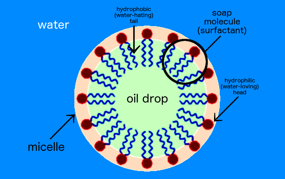
Photo from this page:
https://www.meritech.com/blog/how-soap-works

Surfactants are classified by their ionic (electrical charge) properties in water: anionic (negative charge), non-ionic (no charge), cationic (positive charge) and amphoteric (either positive or negative charge). Soap is an anionic surfactant. Information about Soaps and Detergents | Healthy Cleaning 101
About Micelles:
https://www.mpikg.mpg.de/886719/MicelleFormation.pdf
From this page: https://thehealthnexus.org/the-biology-of-coronavirus-preventative-measures/
Here we tap Dr. Schnell’s knowledge of viruses to understand some of the advice we’ve been getting about the coronavirus.
Why does something as simple as washing hands help against something so deadly?
Coronaviruses have a weakness that we can use to our advantage to prevent spread. They are covered in a rather fragile lipid bilayer, a sort of thin film of grease. When you drop soap on a film of grease in a pan, that grease separates. It’s the same idea with the layer of lipids covering this virus particle, except in the case of a virus, the film is so tiny that it can’t be seen by standard microscopes. By rubbing soap on your hands into a good lather, you help break this nano-scale lipid viral covering.
What about hand sanitizers, do they really work?
Hand sanitizers are effective against bacteria, but also have been tested to kill some, but not all viruses. We do know that they effectively destroy the coronavirus that causes COVID-19. It works a little differently than soap. Rather than breaking open the lipid bilayer, the alcohol mixed with a little bit of water (the small amount of water in sanitizer is important) destroys the virus by crumpling the surface proteins it uses to enter and infect cells. It’s like the sanitizer crushes the set of lock-picks that the coronavirus uses to break into our cells and infect them.

By our bad luck, the genetic code of the virus that causes COVID-19 has a lock-pick shaped perfectly for a lock or protein on human cells called ACE2. Unfortunately humans have ACE2 on many cell such as the cells that line the lungs, blood vessels, kidney, and intestine. Unlike the common cold or flu, this coronavirus has additional tools in its lock-pick set that allows it to unlock and enter cells with ACE2 more easily and quickly, making it more dangerous. This is probably also why older adults are at greater risk for complications. If the virus is infecting organs of people whose organs are already stressed and busy fighting pre-existing conditions like heart disease, hypertension, or lung disease, they are more likely to have difficulty fighting the disease.

https://thehealthnexus.org/the-biology-of-coronavirus-preventative-measures/
https://www.atsjournals.org/doi/full/10.1164/rccm.202004-1471LE
https://virologyj.biomedcentral.com/articles/10.1186/s12985-019-1182-0
Coronavirus envelope protein: current knowledge
Virology Journal 16, Article number: 69 (2019)
Abstract
Background
Coronaviruses (CoVs) primarily cause enzootic infections in birds and mammals but, in the last few decades, have shown to be capable of infecting humans as well. The outbreak of severe acute respiratory syndrome (SARS) in 2003 and, more recently, Middle-East respiratory syndrome (MERS) has demonstrated the lethality of CoVs when they cross the species barrier and infect humans. A renewed interest in coronaviral research has led to the discovery of several novel human CoVs and since then much progress has been made in understanding the CoV life cycle. The CoV envelope (E) protein is a small, integral membrane protein involved in several aspects of the virus’ life cycle, such as assembly, budding, envelope formation, and pathogenesis. Recent studies have expanded on its structural motifs and topology, its functions as an ion-channelling viroporin, and its interactions with both other CoV proteins and host cell proteins.
Main body
This review aims to establish the current knowledge on CoV E by highlighting the recent progress that has been made and comparing it to previous knowledge. It also compares E to other viral proteins of a similar nature to speculate the relevance of these new findings. Good progress has been made but much still remains unknown and this review has identified some gaps in the current knowledge and made suggestions for consideration in future research.
Conclusions
The most progress has been made on SARS-CoV E, highlighting specific structural requirements for its functions in the CoV life cycle as well as mechanisms behind its pathogenesis. Data shows that E is involved in critical aspects of the viral life cycle and that CoVs lacking E make promising vaccine candidates. The high mortality rate of certain CoVs, along with their ease of transmission, underpins the need for more research into CoV molecular biology which can aid in the production of effective anti-coronaviral agents for both human CoVs and enzootic CoVs.
Background
Coronaviruses (CoVs) (order Nidovirales, family Coronaviridae, subfamily Coronavirinae) are enveloped viruses with a positive sense, single-stranded RNA genome. With genome sizes ranging from 26 to 32 kilobases (kb) in length, CoVs have the largest genomes for RNA viruses. Based on genetic and antigenic criteria, CoVs have been organised into three groups: α-CoVs, β-CoVs, and γ-CoVs (Table 1) [1, 2]. Coronaviruses primarily infect birds and mammals, causing a variety of lethal diseases that particularly impact the farming industry [3, 4]. They can also infect humans and cause disease to varying degrees, from upper respiratory tract infections (URTIs) resembling the common cold, to lower respiratory tract infections (LRTIs) such as bronchitis, pneumonia, and even severe acute respiratory syndrome (SARS) [5,6,7,8,9,10,11,12,13,14]. In recent years, it has become increasingly evident that human CoVs (HCoVs) are implicated in both URTIs and LRTIs, validating the importance of coronaviral research as agents of severe respiratory illnesses [7, 9, 15,16,17].
Some CoVs were originally found as enzootic infections, limited only to their natural animal hosts, but have crossed the animal-human species barrier and progressed to establish zoonotic diseases in humans [19,20,21,22,23]. Accordingly, these cross-species barrier jumps allowed CoVs like the SARS-CoV and Middle Eastern respiratory syndrome (MERS)-CoV to manifest as virulent human viruses. The consequent outbreak of SARS in 2003 led to a near pandemic with 8096 cases and 774 deaths reported worldwide, resulting in a fatality rate of 9.6% [24]. Since the outbreak of MERS in April 2012 up until October 2018, 2229 laboratory-confirmed cases have been reported globally, including 791 associated deaths with a case-fatality rate of 35.5% [25]. Clearly, the seriousness of these infections and the lack of effective, licensed treatments for CoV infections underpin the need for a more detailed and comprehensive understanding of coronaviral molecular biology, with a specific focus on both their structural proteins as well as their accessory proteins [26,27,28,29,30]. Live, attenuated vaccines and fusion inhibitors have proven promising, but both also require an intimate knowledge of CoV molecular biology [29, 31,32,33,34,35,36].
The coronaviral genome encodes four major structural proteins: the spike (S) protein, nucleocapsid (N) protein, membrane (M) protein, and the envelope (E) protein, all of which are required to produce a structurally complete viral particle [29, 37, 38]. More recently, however, it has become clear that some CoVs do not require the full ensemble of structural proteins to form a complete, infectious virion, suggesting that some structural proteins might be dispensable or that these CoVs might encode additional proteins with overlapping compensatory functions [35, 37, 39,40,41,42]. Individually, each protein primarily plays a role in the structure of the virus particle, but they are also involved in other aspects of the replication cycle. The S protein mediates attachment of the virus to the host cell surface receptors and subsequent fusion between the viral and host cell membranes to facilitate viral entry into the host cell [42,43,44]. In some CoVs, the expression of S at the cell membrane can also mediate cell-cell fusion between infected and adjacent, uninfected cells. This formation of giant, multinucleated cells, or syncytia, has been proposed as a strategy to allow direct spreading of the virus between cells, subverting virus-neutralising antibodies [45,46,47].
Unlike the other major structural proteins, N is the only protein that functions primarily to bind to the CoV RNA genome, making up the nucleocapsid [48]. Although N is largely involved in processes relating to the viral genome, it is also involved in other aspects of the CoV replication cycle and the host cellular response to viral infection [49]. Interestingly, localisation of N to the endoplasmic reticulum (ER)-Golgi region has proposed a function for it in assembly and budding [50, 51]. However, transient expression of N was shown to substantially increase the production of virus-like particles (VLPs) in some CoVs, suggesting that it might not be required for envelope formation, but for complete virion formation instead [41, 42, 52, 53].
The envelope protein
Structure
The CoV E protein is a short, integral membrane protein of 76–109 amino acids, ranging from 8.4 to 12 kDa in size [69,70,71]. The primary and secondary structure reveals that E has a short, hydrophilic amino terminus consisting of 7–12 amino acids, followed by a large hydrophobic transmembrane domain (TMD) of 25 amino acids, and ends with a long, hydrophilic carboxyl terminus, which comprises the majority of the protein (Fig. 1) [1, 60, 72,73,74,75]. The hydrophobic region of the TMD contains at least one predicted amphipathic α-helix that oligomerizes to form an ion-conductive pore in membranes [76,77,78].
Release: Viroporin
While the accumulation of E at the ERGIC points largely to a role in assembly and budding, only a small portion is incorporated into the viral envelope, suggesting that E has additional functions centred around the ER and Golgi region [66, 92, 109, 159]. Viroporins are viral-encoded membrane pore-forming proteins that can modulate cellular ion channels and have been suggested to regulate and function in multiple stages of the viral life cycle, from viral entry to assembly and release, and even pathogenesis [184, 187,188,189,190,191,192,193,194,195,196]. Although viroporins are not essential to viral replication, their absence does weaken or attenuate the virus and diminishes its pathogenic effects [35, 197,198,199,200]. They tend to be small proteins (~ 60–120 amino acids) of a predominantly hydrophobic nature that oligomerise in the membranes of infected cells, forming hydrophilic pores. The hydrophobic residues line the outside of the structure, oriented toward the phospholipids, while the inside of the pore is made up of the hydrophilic resides [140, 159, 201,202,203,204]. Most viroporins share certain structural features such as an amphipathic α-helix in the hydrophobic domain (HD) along with a cluster of positively charged, basic amino acids (such as lysine or arginine) which anchor the pore to the membrane through electrostatic interactions with the negatively charged phospholipids (Fig. 5) [187, 205,206,207].

Illustration of a typical viroporin structure and motifs. The pore of the viroporin (brown) is created by the amphipathic α-helix and the viroporin is anchored to a lipid bilayer by terminal positively charged residues (lysine or arginine). Conformational changes in the structure regulate the flow ions through the viroporin by opening (left) and closing (right) the pore [208]
Viroporins can transport different ions but appear to be largely selective for the positively charged ions hydrogen (H+), K+, Na+, and calcium (Ca2+) [209, 210]. Although preferentially selective for cations, viroporins can also transport anions. The preference simply appears to be for cations over anions [211,212,213]. It is, however, interesting to note that, at a neutral pH, the ion selectivity of the respiratory syncytial virus (RSV) small hydrophobic (SH) protein can change from cationic to anionic [214]. This suggests that viroporins are sensitive to changes in the cellular environment, a property that could be of therapeutic value. After all, the influenza A virus M2 protein is pH-gated and activates upon acidification of the endosome following receptor-mediated endocytosis of the virus [215]. In the same study, Schnell and Chou [215] showed that the anti-viral drug rimantadine exerts its anti-viral property by stabilising the M2 viroporin in its closed conformation and in doing so inhibits viral replication [209, 216]. Similarly, the E protein of several CoVs possesses ion channel activity, though the only structural data of the CoV viroporin has been derived from SARS-CoV using synthetic peptides [75, 135, 136, 138, 217, 218].
Synthetic peptides of SARS-CoV E demonstrate that the TMD is responsible for its ion-conductive properties [135, 136, 138]. Computational predictions and spectroscopic studies show that the SARS-CoV E TMD undergoes oligomerisation, characteristic of ion-channelling proteins, to form a stable pentamer [75, 135,136,137]. Viroporin formation appears to be mediated by ionic interactions rather than disulphide bonds as mutation of the porcine reproductive and respiratory syndrome virus (PRRSV) E protein cysteine residues appears to be dispensable for oligomerisation [219]. Research into the mechanism of viroporin formation is hampered by the hydrophobic nature of the TMD and has thus far been limited largely to mutational studies and the use of ion channel inhibitors such as amantadine and hexamethylene amiloride.
The CoV E viroporin is equally cation-selective when it comes to its ion-channelling properties, demonstrating a preference for the monovalent cations Na+ and K+ [217, 218]. Synthetic peptides of SARS-CoV E, that resemble the CoV E viroporin, are able to transport Na+, K+, and chloride ions (Cl−) but are more selective of Na+ over K+ and least selective of Cl− [217]. Synthetic peptides that correspond to E from HCoV-229E, MHV, and IBV exhibit a similar cation-selectivity for MHV and IBV E as for SARS-CoV E. However, it is interesting that although the E viroporin synthetic peptides of HCoV-229E were still cation-selective, it exhibits a slightly higher selectivity for K+ than for Na+ [218]. The SARS-CoV E synthetic peptide findings were corroborated using a full-length SARS-CoV E protein [76]. More recently, purified full-length MERS-CoV E has also demonstrated limited ion-channelling properties and would benefit from a more comprehensive characterisation to establish whether it has ion-channelling properties similar to that of the other CoVs [140].
It should be cautioned that the charge on the lipid head group of membranes used can modulate the ion-selectivity of the viroporin. Neutral lipids appear to negate the selectivity of the viroporin as the channels formed did not seem to differentiate cations from anions. In contrast, negatively charged lipids were more cation-selective than neutral lipids, being more permeable to cations [76]. This suggests that the lipid head group of the membranes in use should be taken into consideration when interpreting the results as it might skew the results and inaccurate conclusions may be drawn. At times, the ion channels were only marginally more selective of cations, bringing into question the ion-selectivity of the CoV E viroporin for one cation over another. In fact, an ion channel is only considered ion-specific when its permeability is nearly exclusive to one ion while extremely low to others [220]. Synthetic peptides corresponding to the full-length SARS-CoV E viroporin have also recently been shown to be capable of transporting Ca2+ and was linked to the inflammatory response often observed in ARDS [221]. This is the only study so far to have shown that the E viroporin of any CoV is capable of Ca2+ transport.
Recent efforts have been directed toward understanding how mutant CoV E viruses carrying ion channel-inactivating mutations revert to their original pathogenic state. Mutants of SARS-CoV E carrying mutations N15A and V25F in the TMD restored ion channel activity by incorporating compensatory mutations in both in vitro and in vivo systems [77]. Mutant N15A reverted by incorporating a single mutation that led to an amino acid change at the same position (A15D), creating a more stable mutant. Conversely, mutant V25F reverted to mutants with amino acid substitutions at either the same position (F25D) or positions relatively close to the original mutation (L19A, F20 L, F26 L, L27S, T30I, L37R). Intriguingly, the V25F mutants appeared as early as 2 days after mice were infected where revertant mutant T30I surpassed the growth of the original virus by day two. This suggests that while some of these mutations appear to merely restore the loss of ion channel activity, it is not entirely inconceivable that revertant viruses would acquire gain of function mutations that can render it more virulent [77]. Similar results were recently reported for IBV E TMD residues analogous to N15A and V25F (T16A and A26F) [222]. It is interesting to note that in both cases SARS-CoV E and IBV E followed a similar trend in their reversion: mutations at N15A and T16A both reverted by substitution of a single residue, whereas mutations at V25F and A26F produced revertants by acquisition of multiple residues.
Some viroporins have been implicated in the release of viruses, but it is not yet known whether the release is mediated by the ion channel activity of the proteins [187, 223,224,225,226]. An intriguing study recently reported that both IBV infected and IBV E transfected cells exhibited a marked increase in the pH of the Golgi lumen [227]. These findings suggest that the IBV E viroporin could channel H+ and possibly mediate viral release by its ion channel activity. However, this increase in pH was found only in cells expressing a monomeric form of IBV E and not the oligomeric form as required for viroporin formation. The authors proposed that the change in pH could be attributed to an interaction between the monomeric form of E and a host protein. Although possible, only a very small number of host proteins have been shown to interact with CoV E. The monomeric and oligomeric forms were produced by transfection of mutated IBV E A26 to F26 (EA26F) and T16 to A16 (ET16A), respectively. In an earlier study, the same authors demonstrated that these two forms were present in IBV E-infected cells but that the monomeric form was much less (~ 10%) in infected cells than in transfected cells (~ 50%). The oligomeric form, however, was the dominant form in infected cells [90]. This suggests that other viral proteins might affect or modulate the oligomerisation of IBV E. It is interesting to note that the M2 protein amphipathic helix motif was required for release of influenza A virus (IAV) particles, perhaps indicating that this motif might be required for the processes budding, scission, and for viroporin activity [181]. It might be worth investigating whether ion-channel inhibitors, such as amantadine, or proton pump inhibitors specifically are able to inhibit this increase in Golgi pH. For now, though, it still remains to be seen whether CoV release is mediated by viroporin ion channel activity or through PPIs with host proteins of the secretory pathway.
Pathogenesis: ER stress response/unfolded-protein response (UPR) and apoptosis
The ER can sustain a high load of protein content without being overwhelmed [228]. However, when the ER’s capacity for folding and processing proteins is exceeded, unfolded or misfolded proteins rapidly accumulate in the lumen and the ER stress response, or unfolded-protein response (UPR), is activated. The various signalling pathways that make up the UPR collectively function by enhancing the folding of proteins, chaperoning, and ER-assisted degradation (ERAD) [229]. If, however, the UPR is prolonged and irreversible, apoptosis will be initiated [230]. By increasing the protein content, folding, and processing of the ER, viral infections can also trigger the UPR and this pathway can be used by the host cell as an antiviral response [231]. Very few studies have looked at the role of CoV E in the ER stress response and its ability to induce apoptosis. In cultured cell lines, overexpressed MHV E and epitope-tagged SARS-CoV E induces apoptosis [87, 232]. However, cells infected with rSARS-CoV and rSARS-CoVΔE, a more biologically relevant system, demonstrated that SARS-CoV E may regulate the UPR as part of its pathogenesis [233]. Cells infected with SARS-CoVΔE exhibit a stronger stress response compared to cells infected with the wild-type virus. Moreover, a higher degree of apoptosis was observed in SARS-CoVΔE-infected cells than in those infected with the wild-type virus.
This study demonstrates the risk of interpreting data from overexpression and epitope-tagged studies. Results generated by such studies might offer some insight into the putative functions of viral proteins but should be interpreted with great care as they can be misleading. Findings can only be more conclusive when supported by results from studies in more biologically relevant systems. The study also shows that CoV E has an anti-apoptotic function in infected cells by suppressing the UPR during infection, likely as a survival mechanism and to continue viral propagation. This function of E has only been demonstrated in SARS-CoV so far, one of the most virulent HCoVs. It would be interesting to see whether E of the other CoVs, as well as the less virulent HCoVs, are also able to contribute to pathogenesis by regulating the host cell stress response.
Immune response: Inflammasome activation
Viruses often encode proteins that interfere with the immune system to either inhibit a response or enhance one as part of their pathogenicity. Some viral proteins disrupt components of the immune response pathways to disrupt the immune system and promote their viral evasion and pathogenesis [234,235,236,237]. Alternatively, viral proteins can modulate other cellular factors that could also disrupt the immune response to promote pathogenesis. Coxsackievirus 2B protein promotes the internalisation of major histocompatibility complex class I (MHC-I) proteins and, in doing so, prevents their transport to the cell surface for immune recognition [238]. This protein also delays the transport of proteins along the secretory pathway by altering the Ca2+ and H+ concentrations of the Golgi and ER compartments and has been proposed to be a mechanism of immune evasion as well [239]. Influenza virus M2 protein triggers activation of the NOD-like receptor family, pyrin domain containing 3 (NLRP3) inflammasome by creating ionic imbalances through its ion-channel activity [240]. Other viruses use viroporins to stimulate an immune response as part of their pathogenicity, including the E protein of PRRSV [241,242,243].
Inflammasome activation by CoV E was first reported in PRRSV [242]. Blocking ion channel activity with amantadine significantly inhibited activation of the inflammasome, demonstrating an association between E viroporin activity and inflammation. Recently, the transport of Ca2+ by SARS-CoV E was shown to trigger inflammasome activation [221]. This establishes the link between inflammasome induction by SARS-CoV E and the inflammatory-mediated lung damage seen in SARS-CoV-infected mice [77]. Interestingly, despite attempts to inhibit ion channel activity in SARS-CoV E, by mutating N15A and V25F, viruses restored ion channel activity by incorporating additional mutations after several passages. The authors concluded that this ion-channelling function confers a selective advantage to the virus [77]. The reduction of inflammatory cytokines in the absence of CoV E ion channel activity suggests that inhibition of the CoV E viroporin limits CoV pathogenicity and could be of therapeutic value to CoV infections.
Future perspectives and conclusion
While most CoV infections, such as those caused by HCoV-229E, HCoV-OC43, HCoV-NL63, and HCoV-HKU1, are mild and self-limiting, SARS-CoV and MERS-CoV cause severe infections that lead to high mortality rates [244,245,246]. There are currently no effective, licensed therapies for HCoV infections and existing treatment strategies are generally limited to symptomatic treatment and supportive care [26,27,28, 247]. While an extensive amount of research has gone into identifying potential treatment options, most have only shown promise in vitro and will likely not progress further as they often have one or more limitations. Anti-viral candidates either exhibit only a narrow spectrum of activity, are only effective at unusually high therapeutic dosages or cause serious side effects or immune suppression [248]. A few studies have investigated the potential of rCoVs with a mutated E or lacking E, specifically focussing on SARS- and MERS-CoV, as live attenuated vaccine candidates with some promising results [34, 36, 165, 249, 250]. Vaccinated animal models developed robust immune responses, both cellular and humoral, and were protected against infective challenges. This shows that CoV vaccines with mutated or deficient in E can potentially be used for prophylactic treatment, but the duration of immunity does not seem to have been established yet.
Viruses exploit the extensive network of their host cell’s signalling pathways to promote viral replication and propagation [251, 252]. This dependence on PPIs offers the unique opportunity to target both viral-host and intraviral PPIs and, thereby, stop viral replication and propagation. Therapies that use small-molecule drugs have the advantage of small size, which allows the drugs to cross cell membranes efficiently, but it also severely limits the selectivity and targeting capabilities of the drug, which often leads to undesired side-effects [253]. Interactions between proteins take place over large, flat surface areas that feature shallow interaction sites. Small-molecule drugs, however, tend to bind to deep grooves or hydrophobic pockets not always found on the surface of target proteins, making it difficult for such drugs to disrupt PPIs (Fig. 6) [253,254,255]. Larger, protein-based therapies, on the other hand, make use of insulin, growth factors, and engineered antibodies, that form many more, and much stronger, interactions, making these therapies more potent and selective for their targets. Such properties result in fewer side-effects but the size of these agents also restricts their ability to cross the membranes of target cells [253]. This calls for therapeutic agents that can bridge the gap between molecules that are large enough to be specific and potent for their targets but still small enough to be able to cross target cell membranes efficiently and can also be manufactured easily.

Mechanisms of interaction between small molecules and proteins, and protein-protein interactions. Left: The binding of biotin to avidin occurs in a deep groove, while the interaction between the human growth hormone (hGH) and the hGH receptor (hGHR) occurs over a larger, flatter area [254]
Stapled peptides fulfil these criteria to a large extent and have been applied to various human diseases and fields such as cancer, infections, metabolism, neurology, and endocrinology [256,257,258,259,260]. In fact, Aileron Therapeutics have already developed two stapled peptides, ALRN-5281 and ATSP-7041. The company has already completed the first-in-human trail with ALRN-5281 for the treatment of rare endocrine diseases, such as adult growth hormone deficiency. Moreover, ATSP-7041 was designed to target intracellular PPIs, specifically murine double minute 2 (MDM2) and murine double minute X (MDMX) [261]. To the best of the author’s knowledge, only a few studies so far have investigated the potential of stapled peptides as antiviral agents, with promising results for both intracellular and extracellular targets. The focus so far has only been on HIV-1, RSV, and HCV [260, 262,263,264,265].
Granted, the therapeutic application of stapled peptides, particularly regarding viral infections, is still relatively new, but their numerous advantages give them tremendous potential as antiviral agents. Stapled peptides (1) can inhibit PPIs; (2) are more specific for their targets than small-molecule drugs, which also decreases the risk of unwanted side-effects; (3) can target diseases that are otherwise difficult to treat, referred to as “undruggable”; (4) can be modified easily to enhance membrane permeability, potency, and half-life; (5) have a short market time [253, 266, 267]. As more viral PPIs for CoV E are identified, the repertoire of stapled peptide targets also expands making it easier to limit viral replication, propagation, and even pathogenesis. Stapled peptides have the potential to be used as antiviral agents that can work effectively at multiple levels.
Autophagy is a cellular process that recycles excess or damaged cellular material to maintain the energy levels of the cell and ensure its survival. The material is removed from the cytoplasm by forming enclosed DMVs known as autophagosomes and then fused with lysosomes to be degraded [268, 269]. Recent studies have increasingly pointed to the involvement of autophagy components in viral infections [270]. Some suggest that it might have an antiviral function by inhibiting viral replication [271,272,273]. Others reported inhibition or subversion of autophagy as a defence mechanism to promote viral propagation [274,275,276]. Others still, notably RNA viruses, appear to exploit autophagy for the purpose of viral propagation [277, 278]. Regarding CoVs, replication of TGEV is negatively regulated by autophagy [279]. Interestingly, PRRSV activates autophagy machinery, possibly to enhance viral replication as certain components of autophagy are required for MHV replication [280, 281]. These studies suggest the possibility of CoVs exploiting autophagy for replicative purposes. It has even been proposed that the DMVs formed in CoV-infected cells might be the result of autophagy and derived from the rough ER [281]. Recently, an increase in cytosolic Ca2+, presumably from the ER lumen, has been implicated in autophagy induction by protein 2B (P2B) of the foot and mouth disease virus (FMDV) [282]. The rotavirus non-structural protein 4 (NSP4) reportedly induces autophagy by a similar mechanism [283]. Considering these studies, along with the ability of SARS-CoV to channel Ca2+, it is not inconceivable that CoV E viroporin could induce autophagy in CoV-infected cells by increasing cytosolic Ca2+. However, experimental evidence would be required to support the possibility of such a mechanism in CoVs.
The multifunctional role CoV E protein: A central role in assembly, release, and pathogenesis?
From studies, it appears that some viral proteins do not have unique, definitive functions. Despite the deletion of some viral genes, the viral life cycle continues, suggesting that other viral genes can compensate for this loss. It was recently shown to be the case for the vaccinia virus [284]. This is also evident in the varied requirements of the E protein for different CoVs and the reason(s) for this is not understood. Trafficking and maturation of TGEV virions is arrested without E [40]. Virions of MHV ΔE are capable of producing viable, replicating progeny [39]. Deletion of E from SARS-CoV attenuates the virus whereas, in the case of MERS-CoV, virions are propagation deficient [35, 165]. Certain CoV accessory proteins appear to be able to complement, or sometimes even compensate for, the absence of E in processes such as assembly, release, and the pathogenesis of some CoVs [30]. It is particularly noteworthy that SARS-CoV encodes two accessory proteins, 3a and 8a, that might exhibit relative compensatory functions in the absence of E [285, 286]. In terms of viral replication in vivo and in vitro, 3a could partially compensate for the loss of E. Moreover, 3a also contains a PBM and might be able to compensate for the loss of E to an extent but utilises different signalling pathways [285]. Although the study demonstrated that even the accessory proteins demonstrate some measure of dispensability, the virus still encodes these additional proteins with overlapping functions. The dynamics between these proteins, however, are not quite clear yet and warrants further investigation. What is clear, though, is that viroporin proteins, case in point IAV M2, can exhibit a multitude of different functions independent of their ion-channel properties [153, 184]. The studies in this review have shown that CoV E could be involved in multiple aspects of the viral replication cycle: from assembly and induction of membrane curvature to scission or budding and release to apoptosis, inflammation and even autophagy. Although a lot of progress has been made on CoV E, there is still much to be discovered about this small, enigmatic protein.
Abbreviations
- A15D:
Alanine residue 15 mutated to aspartic acid
- A26F:
Alanine residue 26 mutated to phenylalanine
- altPBM:
alanine mutated PBM
- ARDS:
Acute respiratory distress syndrome
- Bcl-xL:
B-cell lymphoma-extra-large
- BCoV:
Bat coronavirus
- Ca2+ :
Calcium ion
- CCoV:
Canine coronavirus
- Cl− :
Chloride ion
- CMs:
Convoluted membranes
- CoV(s):
Coronavirus (es)
- C-terminus:
Carboxy terminus
- Dlg1:
Drosophila disc large tumour \ressor
- DMVs:
Double-membrane vesicles
- E:
Envelope protein
- EM:
Electron microscopy
- Env:
Envelope glycoprotein gp160
- ER:
Endoplasmic reticulum
- ERAD:
ER-assisted degradation
- ERGIC:
Endoplasmic reticulum Golgi intermediate compartment
- ESCRT:
Endosomal sorting complex required for transport
- F13 L:
vaccinia virus envelope phospholipase F13 protein
- F20 L:
phenylalanine residue 20 mutated to leucine
- F25D:
phenylalanine residue 20 mutated to aspartic acid
- F26 L:
phenylalanine residue 26 mutated to leucine
- FeCoV:
feline coronavirus
- FMDV:
foot and mouth disease virus
- GFP:
Green fluorescent protein
- GST:
Glutathione-S-transferase
- H+ :
Hydrogen ion
- HA:
Haemagglutinin
- HBV:
Hepatitis B virus
- HCoV(s):
Human coronavirus (es)
- HCoV-229E:
Human coronavirus 229E
- HCoV-4408:
Human coronavirus 4408
- HCoV-HKU1:
Human coronavirus HKU1
- HCoV-NL63:
Human coronavirus NL63
- HCoV-OC43:
Human coronavirus OC43
- HCV:
Hepatitis C virus
- HD:
Hydrophobic domain
- HEV:
porcine hemagglutinating encephalomyelitis virus
- hGH:
human growth hormone
- hGHR:
human growth hormone receptor
- HIV:
human immunodeficiency virus
- IAV:
Influenza A virus
- IBV:
avian infectious bronchitis virus
- K+ :
potassium ion
- kb:
kilobases
- kDa:
kilodalton
- L19A:
Leucine residue 19 mutated to alanine
- L27S:
Leucine residue 27 mutated to serine
- L37R:
Leucine residue 37 mutated to arginine
- LRTIs:
Lower respiratory tract infections
- M:
Membrane protein
- M2:
Matrix-2 protein
- MAPK:
Mitogen-activated protein kinase
- MDM2:
Murine double minute 2
- MDMX:
Murine double minute X
- MERS:
Middle-East respiratory syndrome
- MERS-CoV:
Middle-East respiratory syndrome coronavirus
- MHC-I:
major histocompatibility complex I
- MHV:
Murine hepatitis virus
- MS:
Mass spectrometry
- mutPBM:
glycine mutated PBM
- N:
Nucleocapsid protein
- N15A:
asparagine residue 15 mutated to alanine
- N5, 15, 48, 66:
asparagine residues 5, 15, 48, 66
- Na+ :
sodium ion
- Nef:
negative regulatory factor
- NLRP3:
NOD-like receptor family, pyrin domain containing 3
- Nsp(s) 3, 4, 6:
non-structural protein(s) 3, 4, 6
- NSP4:
Non-structural protein 4
- N-terminus:
amino terminus
- P2B:
protein 2B
- PALS1:
Protein associated with Caenorhabditis elegans lin-7 protein 1
- PBM:
PDZ-binding motif
- PDZ:
Postsynaptic density protein 95 (PSD95)/Drosophila disc large tumour suppressor (Dlg1)/zonula occludens-1 protein (zo-1)
- PEDV:
Porcine epidemic diarrhoea coronavirus
- PPI(s):
Protein-protein interaction(s)
- PRCoV:
Porcine respiratory coronavirus
- PRRSV:
Porcine reproductive and respiratory syndrome virus
- PSD95:
Postsynaptic density protein 95
- rCoVs:
recombinant coronaviruses
- RNA:
Ribonucleic acid
- RSV:
Respiratory syncytial virus
- S:
Spike protein
- SARS:
Severe acute respiratory syndrome
- SARS-CoV:
severe acute respiratory syndrome coronavirus
- Sf9:
Spodoptera frugiperda cell line
- SH:
Small hydrophobic
- SIV:
Simian immunodeficiency virus
- T16A:
Threonine residue 16 mutated to alanine
- T30I:
Threonine residue 30 mutated to isoleucine
- TAP:
Tandem affinity purification
- TAP-MS:
Tandem affinity purification coupled with mass spectrometry
- TCoV:
Turkey coronavirus
- TGEV:
Transmissible gastroenteritis coronavirus
- TMD:
Transmembrane domain
- UPR:
Unfolded-protein response
- URTIs:
Upper respiratory tract infections
- V25:
Valine residue 25
- V25F:
Valine residue 25 mutated to phenylalanine
- VLP(s):
Virus-like particle(s)
- zo-1:
zonula occludens-1 protein
- α:
alpha
- β:
beta
- γ:
gamma
- Δ6:
recombinant SARS-CoV deletion mutant number 6
- ΔE:
deleted E gene
- ΔPBM:
recombinant SARS-CoV mutant with deleted PBM
I've only seen ONE study about surfactants in the treatment of COFVID-19, and it's really just a proposal.
https://www.sciencedirect.com/science/article/pii/S0306987720308197
Surfactant-based prophylaxis and therapy against COVID-19: A possibility
Keywords
Introduction
The pandemic COVID-19 is spreading rapidly all over the world. At present, the search for a therapy to this viral disease is in a ‘to leave no stone unturned’ situation. Washing with soap is recommended for hand hygiene for preventing the spread of COVID-19. Interestingly, the damage to coronavirus spike proteins leads to the virus inactivation [1]. Soaps and detergents are classified under surfactants and are widely explained as damaging peplomers or spike proteins. Inactivation of the Ebola virus, a virus showing spike-protein mediated host attachment, by surfactant nanoemulsion is ideal support for this hypothesis [2]. Surfactants are approved and widely used as pharmaceutical inactive ingredients. Surfactants, including biosurfactants, have been known to have antiviral properties. But their potential in prophylaxis and therapy against COVID-19 is still not explored. Therefore, this hypothesis analyzes the possibility and speculates the use of surfactants against SARS-CoV-2 peplomers for the prophylaxis and treatment of COVID-19.
Hypothesis
Here we postulate that a simple surfactant-based gargle would be enough to stop the spread of this pandemic. In addition to gargle, surfactant-loaded throat paint, mouthwash, nasal drops, and eye drops could be used for prophylaxis. This would be particularly useful among high-risk categories and health workers. In addition to prophylaxis, we postulate that surfactant therapy would be beneficial in the treatment of COVID-19. Further, the use of surfactant against COVID-19 would prevent the spread of this disease.
Surfactant-based prophylaxis against COVID-19
The major route of entry of SARS-CoV-2 is through nose and mouth, and to some extent through eyes. Throat (pharynx) is a common point of entry to the lungs from both mouth and nose. In the case of eyes too, it is described that the virus travels through the lacrimal and nasolacrimal ducts (the tear ducts) and then into the nose, throat, trachea, and finally lung [3]. Thus, any virus accidentally entered through mouth, nose, and/or eyes could be stopped/inactivated at the pharynx or nasopharynx with the use of a surfactant-based gargle (Fig. 1a). Entrapment of viruses by surfactant micelles could also occur as a mechanism of inactivation. This would be possible when the critical micelle concentration (CMC) for a surfactant is achieved. Most surfactants have low CMC values. A high residence time of the virus in the pharynx would further enhance this prophylactic effect.

Fig. 1. The diagrammatic presentations of the prophylactic and therapeutic applications of surfactants against COVID-19 (a) how the surfactant-based gargle prevents COVID-19; (b) entry of SARS-CoV-2 virus into lung alveoli; (c) action of surfactant on the SARS-CoV-2 virus. The figure shows the inactivation of the virus by the action of surfactant on viral spike glycoprotein. The surfactant present in the interstitial fluid could also render the virus non-infective. The insert shows the internalization of the virus at low surfactant concentrations; (d) action of surfactant in circulation. The surfactant either inactivates the virus or coats the virus and renders it inactive.
A gargle is a widely used dosage form for throat infections and pain. Most importantly, gargles are considered as external preparations which render a high level of flexibility in choosing the excipients and actives for their preparation. In addition to gargle, similar effects could be expected from surfactant-loaded throat paint, mouthwash, nasal drops, and eye drops. Throat paints even contain chemicals such as phenol and iodine. So a surfactant-based gargle or throat paint would not be of any concern. Most of the aspects mentioned for gargles apply to throat paints and mouthwash too. Among these, surfactant-loaded throat paints also provide good promise for the prevention of infection. Based on the convenience of using gargle compared to throat paint, the former would be most suitable for prophylaxis.
Inclusion of antiviral agents, proteases, astringents (protein precipitants), etc are some of the possible modifications to achieve additional benefits in gargles and mouthwashes. The addition of some protein precipitants such as tannic acid would have added benefit in preventing viral infection. Nevertheless, their interactions should be studied with the surfactant. Their interaction should not interfere with the ability of surfactants to inactivate the virus by interacting with the spike glycoprotein.
The inhalation route would also prove to be useful in the prophylaxis of COVID-19. Inhalation drug delivery devices are helpful in such delivery of surfactants [4]. Surfactants are already reported for pulmonary drug delivery [5], [6]. The use of surfactant in lung and tracheal infections without causing any damage to the cilia is also reported [7]. On systemic delivery, the surfactant reaches both interstitial fluid and lung alveoli. Now when a virus enters the lungs, it has to encounter these surfactant molecules (Fig. 1b). We believe that surfactant therapy will prevent the attachment of the virus through its spike glycoproteins. The surfactant present at the alveolar surface could interfere with the spike glycoproteins resulting in their destruction. This would help avoid the infection when in contact with the virus (Fig. 1c).
Surfactant therapy against COVID-19
Surfactant therapy is already in use for respiratory problems. It is reported that inhalation of lung surfactant improves respiratory diseases. The tremendous benefit of an artificial surfactant against the HINI virus has been proved in mice [8]. Also, surfactants have some role in innate host defense during infections [9]. Thus, it could be reasonably hypothesized that surfactant therapy would be promising in COVID-19 cases. A list of approved lung surfactants is presented in Table 1 [10], [11], [12].
Table 1. Potential surfactants used in the pharmaceutical industry and approved for clinical use.
| Sl. No. | Name/class of surfactant | Present approved use | Approval details | [Ref.] |
|---|---|---|---|---|
| 1 | Lung surfactants – natural and synthetic [E.g.: colfosceril palmitate (Exosurf®), beractant (Alveofact®), poractant alpha (Curosurf®), calfactant (Infasurf®), lucinactant (KL4®), pumactant (Artificial Lung Expanding Compound or ALEC), and recombinant human surfactant protein C (Lusupultide, Venticute®)] | Respiratory distress syndrome in neonates. | USFDA approved lung surfactants | [10], [11], [12] |
| 2 | Polysorbate (non-ionic) | Approved inactive ingredient in intravenous, inhalation (respiratory), oral, nasal, ophthalmic, topical, and other formulations. Useful for external preparations too. | Included in the inactive ingredients database of USFDA. Included in Pharmacopoeia. | [22], [23], [24], [25] |
| 3 | Poloxamer (non-ionic) | Approved inactive ingredient in intravenous, inhalation (respiratory), oral, nasal, ophthalmic, topical, and other formulations. Useful for external preparations too. | Included in the inactive ingredients database of USFDA. Included in Pharmacopoeia. | [22], [24], [25] |
| 4 | Sodium lauryl sulfate (anionic) | Approved inactive ingredient in inhalation (respiratory), oral, topical, and other formulations. Useful for external preparations too. | Included in the inactive ingredients database of USFDA. Included in Pharmacopoeia. | [22], [24] |
| 5 | Cetrimide (cationic) | Pharmaceutical aid and bactericide. | Included in Pharmacopoeia. | [24], [25], [26] |
| 6 | Bile salt surfactants | Emulsifier and solubilizer. Used in combination with phospholipids in mixed micelles formulation. | Used in marketed formulations | [27], [28] |
| 7 | Phospholipids (zwitterionic) | Emulsifier and solubilizer. | Used in marketed formulations worldwide | [28], [29] |
| 8 | Vitamin E TPGS (semi-synthetic) | Solubilizer, absorption and permeation enhancer, and emulsifier. | Approved by USFDA. Included in Pharmacopoeia. | [24], [25], [30] |
| 9 | Polyoxyl 35 castor oil (semi-synthetic) | Emulsifying, solubilizing, and wetting agents. | Included in Pharmacopoeia. | [24], [25], [26] |
Lung surfactant is considered as a physiological barrier to viral infections. The lipid portion is mainly responsible for the antiviral activity [13], [14]. Inhibition of the H1N1 influenza virus is also possible with the lipid part [15]. Yet another advantage of lung surfactant phospholipids is their inhibition of virus-mediated inflammation and infection. Palmitoyl-oleoyl-phosphatidylglycerol has shown such activity [16]. Meanwhile, the antiviral activity of the protein part is also established [17]. Moreover, it is noteworthy that the lung surfactant protein has the ability for selective recognition of SARS coronavirus spike glycoprotein and subsequent macrophage activation [18].
Recently, the use of bear bile has been proposed against COVID-19 [19]. Bile contains bile salts that can function as surfactants [20]. We believe that these bio-surfactants present in the bear bile would have made a major contribution to the observed activity.
Now about the role of surfactant inside a cell, we postulate that the surfactant may reach the cells and the extracellular fluid. The surfactant would interfere with one or more steps of viral replication. Moreover, the spread of viruses from one cell to the adjacent one might be interrupted by the presence of a surfactant in the extracellular or interstitial fluid. This effect would be most important with the target site of SARS-CoV-2, the alveolar epithelial cell.
Surfactants against the spread of COVID-19
The next aspect or advantage of surfactant therapy is the prevention of virus spread. At first, let us consider the most common method of spread, the droplet contact transmission method. The presence of a surfactant near the cell membrane could interfere with the viral budding process by affecting the viral envelope formation. Each drop of the saliva of a patient could host millions of infective viruses which could be simply inactivated by the use of a surfactant-based gargle or mouthwash [21]. Now assuming the possibility of viral spread through blood, and possibly other body fluids too, the surfactant molecules in circulation would either coat the virus or destroy its peplomer and this renders it non-infective (Fig. 1d). Most importantly, these mechanisms could result in the loss of contagiousness of a patient. Therefore, we believe that surfactant-based methods could prevent the spread of this pandemic. Moreover, if we could demonstrate the utility of this method, it would be a solution to stop such viral infections in the future too.
A description on selected surfactants useful for the prophylaxis and/or therapy of COVID-19 is provided in the supplementary material along with other details regarding the validity of the proposed hypothesis.
Testing of the hypothesis
Two aspects should be studied to identify the potential of surfactant-based strategies against COVID-19. The first one is to identify the surfactant and its concentration or dose for its use. The critical micelle concentration would be the best point to start with for every surfactant. The second one is to check its safety profile for the intended use and route of administration. The in vitro studies would be sufficient for the primary screening of the surfactant and the required concentration or dose. A detailed study of the effect of surfactant on viral spike protein would be appropriate at this stage. Many surfactants are approved for pharmaceutical use as inactive ingredients and therefore issues related to safety will not be a concern (Table 1) [22]. The hypothesis may be tested under two categories. In one type of testing, the prophylactic action should be studied. In the other, therapeutic effect should be studied.
Conclusion
We believe that our hypothesis would stimulate debate or new research exploring the possibility of surfactant-based prophylaxis and therapy against COVID-19. The future and required clinical trials on surfactant-based therapeutics against COVID-19 would be further dependent on how this pandemic emerges in the future. It is presumed that this hypothesis would trigger some research in this unconventional approach against COVID-19.
https://www.sciencedirect.com/science/article/pii/S0306987720308197
I did a search for "covid" and "surfactant" and these are some things that came up, but it shocking to see how ethere are no studies on this. It seems pretty absurd given the fact that the influenza virus is also an enveloped virus, and we've had decades to study that.
Microbial Surfactants: The Next Generation Multifunctional Biomolecules for Applications in the Petroleum Industry and Its Associated Environmental Remediation
1. Introduction
Surfactants are a class of chemical compounds possessing amphiphilic (both hydrophobic and hydrophilic) moieties that distribute themselves between two immiscible fluids, with the effect of reducing the surface/interfacial tensions and causing the solubility of polar compounds in non-polar solvents [1]. They display properties, such as detergency, solubilization, and lubrication; have stabilizing and foaming capacity; and form phase dispersion [2]. Surfactants are either derived synthetically or biologically. Naturally derived surfactants are denominated biosurfactants since they are produced from biological entities, especially microorganisms. Fungi, bacteria, and yeast belonging to different species and strains are known for producing biosurfactants of a diverse variety of molecular structures [3]. Amongst the bacteria domain, genera of Pseudomonas, Bacillus, and Acinetobacter dominate the literature space as excellent producers of biosurfactants [2]. The species among these genera that have been extensively studied are Pseudomonas aeruginosa, Bacillus subtilis, and Acinetobacter calcoaceticus, amongst other species [1,4,5]. Bhardwaj et al. [6] and Morita et al. [7] respectively studied Candida bombicola and Pseudozyma rugulosa representing fungi and yeast. Biosurfactants are broadly grouped into low molecular weight (LMW) and high molecular weight (HMW) biosurfactants based on their biochemical natures. The former efficiently lowers surface and interfacial tensions while the latter is more of an emulsion-stabilizing agent. On the basis of chemical composition, biosurfactants are grouped into glycolipids (rhamnolipids, sophorolipids, trehalolipids, mannosylerithritol lipids), lipopeptides (surfactin, lichenysin, iturin, fengycin, serrwettin), fatty acids/phospholipids/neutral lipids (phosphatidylethanolamine, spiculisporic acid), polymeric biosurfactants (emulsan, alasan, biodispesan, liposan), and particulate biosurfactants (vesicles, whole-cell) [8,9,10]. Lipopeptides, glycolipids, and phospholipids belong to the LMW biosurfactants while the HMW biosurfactants include polymeric and particulate biosurfactants [11]. Microbial and synthetic surfactants are employed in diverse industries, including the cosmetics, food, and pharmaceutical sector.
Surfactants have a versatile phase character and diversity of colloidal structures, thus finding application in many industrial processes, especially where modification of the interface activity or stability of the colloidal systems is required [12]. There are four categories of surfactants: Anionic, cationic, nonionic, and zwitterionic [13] based on the composition of the polarity of the head group. The anionic surfactants carry a negative charge, which is the most commonly available surfactants chemically and naturally [14,15]. They have prominent application in personal care products and soaps because they are very effective in cleansing systems [16]. Further, they are also used in the oil industry, agriculture, health, cosmetics, remediation, and bioprospecting because of their wide range of hydrophilic–hydrophobic balance (HLB) values, emulsification property, and their excellent ability to reduce surface tension. The positively charged surfactants (cationic) are well suited for surfaces with a negative charge, thus they are used as anti-corrosion/antistatic agents, flotation collectors, fabric softeners, hair conditioners, and bactericides [17]. The nonionics are surfactants with uncharged hydrophilic head groups, which are good in low-temperature detergents and emulsifiers probably because of their low irritating effects [18]. The zwitterionics are amphoteric surfactants with poor cleansing and emulsifying properties [14] but have excellent dermatological properties and skin compatibility [19]. Also, they are used in manufacturing shampoos and cosmetics. So, surfactants can be used in the petroleum industry, health, pharmaceuticals, agriculture, detergents, cosmetics, bioprocessing, environmental remediation, textiles, paint, leader, papermaking, and other industries and activities where water could serve as an interactive medium [16,20,21,22,23,24,25].
Washing Your Hands Is Important Because Soap “Absolutely Annihilates” Coronavirus
The number one recommendation on the list of protective measures for COVID-19 from both WHO and the CDC is to regularly wash your hands. The CDC in particular recommends hand-washing over using hand sanitizer.
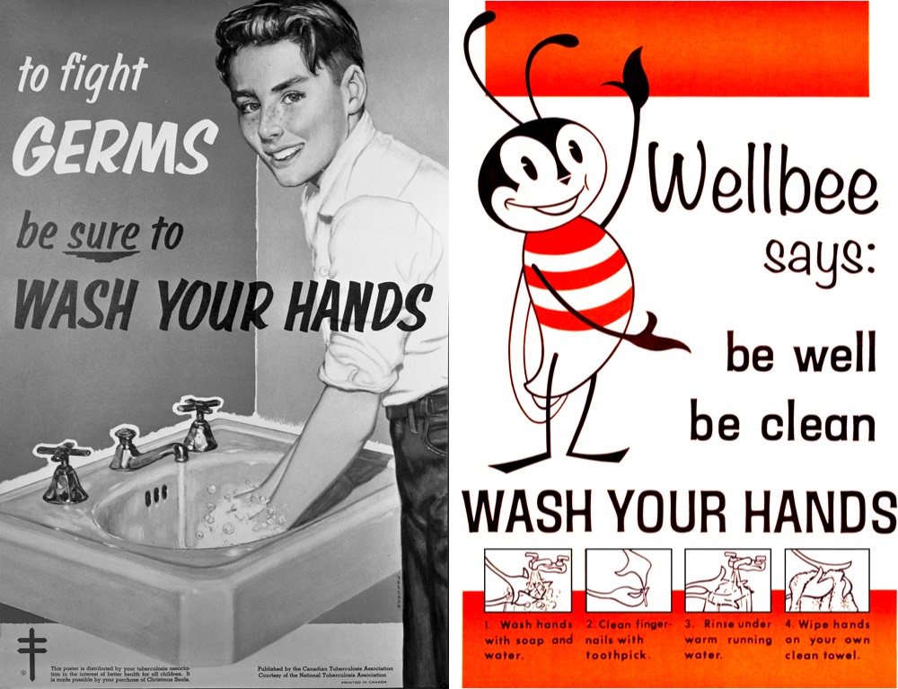
Vox recently talked with chemistry professor Palli Thordarson about why washing with soap is so effective when dealing with coronaviruses.
The soap takes care of the virus much like it takes care of the oil in the water. “It’s almost like a crowbar; it starts to pull all the things apart,” Thordarson says.
One side of the soap molecule (the one that’s attracted to fat and repelled by water) buries its way into the virus’s fat and protein shell. Fortunately, the chemical bonds holding the virus together aren’t very strong, so this intrusion is enough to break the virus’s coat. “You pull the virus apart, you make it soluble in water, and it disintegrates,” he says.
Then the harmless shards of virus get flushed down the drain. (And even if it the soap doesn’t destroy every virus, you’ll still rid them from your hands with soap and water, as well as any grease they may be clinging to.)
And why do you need to wash for 20 seconds? Because that gives soap time to do its work.
First off, your skin is wrinkly, and it takes time for soap to penetrate into all the tiny folds and demolish the viruses that lurk within. Then the soap needs a few moments to do its chemical work. “You do need a bit of time for all the soap to interact back and forth with the virus particle,” he says. Twenty seconds should do the trick just fine.
See also Why Soap Works from the NY Times, which explains why soap & water is better than hand sanitizer in these cases:
On the whole, hand sanitizers are not as reliable as soap. Sanitizers with at least 60 percent ethanol do act similarly, defeating bacteria and viruses by destabilizing their lipid membranes. But they cannot easily remove microorganisms from the skin. There are also viruses that do not depend on lipid membranes to infect cells, as well as bacteria that protect their delicate membranes with sturdy shields of protein and sugar. Examples include bacteria that can cause meningitis, pneumonia, diarrhea and skin infections, as well as the hepatitis A virus, poliovirus, rhinoviruses and adenoviruses (frequent causes of the common cold).
UPDATEThordarson also wrote an article for The Guardian on how effective soap is at killing coronavirus.


How does Soap Work?
https://www.livescience.com/57044-science-of-soap.html
Organelles of the Cell (updated)
https://www.livescience.com/57044-science-of-soap.html
What is soap?
Soap is a mixture of fat or oil, water, and an alkali, or basic salt.
The ancient Babylonians are credited with being the first people to make soap. Their recipe for animal fats, wood ash and water has been found carved into clay containers dating back to 2800 B.C., according to soaphistory.net. They likely used the concoction for washing wool and cotton so the materials could be woven into cloth and not so much for cleaning their bodies.
The ancient Egyptians developed a similar recipe for soap, which they used for treating sores, skin diseases and personal washing. The Romans also made soap, but it wasn't until the later centuries of the Roman era that soap was used for personal hygiene; prior to that, soap was a physician's tool for treating diseases.
The basic recipe for soap hasn't changed for thousands of years. It's still a combination of fat or oils with an alkali — basic ionic salt — and water. When those ingredients combine in the proper proportions, they go through a chemical process called saponification, which results in soap. Today, there are two techniques that people use to make soap: the cold process and the hot process.
In the cold process, a room-temperature lye solution (sodium hydroxide in water) is mixed with animal or vegetable oil. As the ingredients react with one another, the mixture thickens and heats up. Before it gets too thick, the mixture is poured into a mold where it solidifies, and the saponification process is complete. The last step is to let the soap sit, or cure for a few weeks, which allows excess water in the mixture to evaporate. This makes a harder soap, according to the Handcrafted Soap and Cosmetic Guild.
The hot process is the more traditional and ancient way to make soap and requires an outside source of heat. The ingredients are heated as they're mixed, which increases the speed of the saponification process. The soap is in a liquid form when it's poured into molds and it's ready for use as soon as it's solidified. Hot-process soap can be cured in a way that's similar to the cold-process soap, but it's not usually needed, according to the Handcrafted Soap and Cosmetic Guild.
How soap works
Soap doesn't kill germs on our hands, it removes them.
Germs stick to the oils and grease on our hands (sounds yucky, but it's totally normal). Water alone won't remove much of the germs on our hands because water and oil don't like each other, so they won't mix. But soap likes both water and oil. That's because soap molecules are a type of surfactant, which means they have one end that's water loving, or hydrophilic, and one end that's oil loving, or hydrophobic.
When you wash your hands with soap, the soap molecules act as a mediator between the water and oil molecules, and bind with both of them at the same time. Then when you rinse everything off, the soap carries away the germs with the water.
For the most effective hand washing, you must use soap and you must be thorough. Work up a lather because the friction helps lift dirt and oils from your skin, according to the Centers for Disease Control and Prevention (CDC). How long you should scrub depends on how dirty your hands are, but most health authorities recommend at least 20 seconds, or as long as it takes to sing "Happy Birthday" twice. And don't forget to scrape underneath your fingernails. That area is prime real estate for germs.
Once you've washed, be sure to air-dry or towel-dry. There's no agreed-upon best practice for drying, but wet hands are more likely to spread germs than dry ones, the CDC says.
Is antibacterial soap even better? Nope.
Antibacterial soaps have added ingredients like triclosan or triclocarban, which are hydrophobic molecules that can penetrate bacterial cell membranes and kill the bacteria. Sounds impressive, but studies have shown that antibacterial soaps are no more effective than regular soaps at removing bacteria.
In 2016, the FDA issued a rule that antibacterial soaps were no longer allowed to be marketed to the public.
"Consumers may think antibacterial washes are more effective at preventing the spread of germs, but we have no scientific evidence that they are any better than plain soap and water," Dr. Janet Woodcock, the director of the FDA's Center for Drug Evaluation and Research (CDER), said in a statement. "In fact, some data suggests that antibacterial ingredients may do more harm than good over the long term."
What about hand sanitizer?
The CDC recommends cleaning hands with soap and water, but if that's not an option, then hand sanitizer is a good backup. Studies have found that hand sanitizers with alcohol concentrations of 60-95% are more effective at killing germs than nonalcohol or low-alcohol sanitizers.
Related: Hand sanitizer sold out? Here's how to make your own.
The alcohol kills some bacteria and viruses by breaking down their protective membranes, which basically makes them fall apart. But it doesn't work for all germs, such as norovirus, Clostridium difficile, which can cause life-threatening diarrhea, or Cryptosporidium, a parasite that causes a diarrheal disease called cryptosporidiosis, the CDC says. Hand sanitizers also likely don't remove harmful chemicals like pesticides or heavy metals, nor does hand sanitizer work well on super dirty or greasy hands.
Hand washing with soap is, by far, the most effective way to keep harmful germs at bay.
Additional resources:
- Hand washing do's and don'ts, according to the Mayo Clinic.
- Learn more about the importance of hand washing from the CDC.
- Here's a video showing how to hand wash, from the World Health Organization.
Why Did The FDA Ban Antibacterial Soap?
No comments:
Post a Comment
Note: Only a member of this blog may post a comment.