
Please also have a look at THIS PAGE that shows how strenuous exercise (which generates free radicals which lead to oxidative stress) can make a COVID infection worse.
Oxidative Stress Associated with SARS-Cov-2 (COVID-19) Increases the Severity of the Lung Disease - A Systematic Review
Samir Derouiche1,2*
Abstract
COVID-19 patients have a higher risk of developing inflammatory responses associated with serious and even fatal respiratory diseases. This review focuses on the relationship between oxidative stress and COVID-19. Coronaviruses are a family of common RNA viruses that can cause serious lower respiratory tract infections, followed by bronchitis and pneumonia. Pulmonary inflammation, fever and fibrosis are symptoms of COVID-19 mediated by cytokine pro-inflammatory. Oxidative stress affect repair mechanisms and the immune control system, which is one of the main events of the inflammatory response which allows us also to conclude that oxidative stress is a major factor increasing the severity of COVID-19 especially during chronic diseases associated with the fragility of the antioxidant system, suggesting to recommend antioxidants supplementation in therapeutic strategies against COVID-19.
Keywords
Inflammatory response, Oxidative stress, Antioxidant therapy, SARS-CoV-2
Background
SARS-CoV-2, the virus responsible for COVID-2019 for (Coronavirus disease 2019) is a new coronavirus discovered in the city of Wuhan in Hubei province in China in December 2019 ref. COVID-19 has been described as a pandemic by the WHO from the date March 11, 2020, the first triggered by a coronavirus [1]. Coronaviruses are enveloped RNA viruses belonging to the family of Coronaviridae, genus betacoronavirus [2]. In humans, SARS-CoV-2 (COVID-19) has identified as the seventh now pathogenic Cornonavirus for humans after other coronavirus species which are: seasonal HCoV, SARS-CoV, MERS-CoV [3]. Whereas coronavirus 2 (SARS-CoV-2) causes a severe acute respiratory syndrome that spreads worldwide [4]. According to the guidelines of the World Health Organization (WHO), the communicability, severity and impact of the disease are the criteria for assessing the severity of pandemic influenza [5]. Communicability reflects the movement of the virus, which is influenced by the dynamics of spread [6]. The lungs are the preferred target of COVID-19 by the large area exposed to viruses, they are among the most oxygenated organs in the human body [7]. Multiple lung disease including apnea causes alveolar hypoventilation, vasoconstriction of the pulmonary artery and cyclic changes in hypoxemia contribute to increased production of reactive oxygen species (ROS) characteristic of the condition oxidative stress [8]. Oxidative stress is an important factor causing metabolic and physiological alterations and various diseases in the body [9]. COVID-19 attack triggers inflammatory reaction which releases pro-inflammatory cytokines characteristic of acute lung damage [10]. A great association between the pro-inflammatory elements and the reactive oxygen species (ROS) in the different lung disease including Coronavirus infection which is associated with inflammation and oxidative stress [11]. The current review focuses on the relationship between COVID19 infection and inflammation on one side and between oxidative stress and inflammation on the other to identify the possible effect of oxidative stress on the progression of the state of health of the COVID-19 host.
Oxidative Stress and Lung Disease
In lung tissue and during pulmonary ischemia, alveolar oxygen helps maintain aerobic metabolism, delaying hypoxia [12] which results in decreased levels of adenosine triphosphate (ATP) and more intense breakdown of ATP, resulting in increased production of hypoxanthine [13]. When oxygen is reintroduced into the environment by ventilation, the superoxide radical is formed under the action of the enzyme xanthine oxidase on hypoxanthine [14]. In the absence of blood circulation in the lungs there is lipid peroxidation and oxidative damage due to the presence of oxygen [15]. Furthermore, NADP oxidase and NO oxidase in the endothelium seem to be one of the main causes of oxidation in pulmonary ischemia. Also immune cells such as macrophages and neutrophils can contribute to oxidative damage in the lungs by the same enzymatic mechanism (NADP Oxidase) [16].
Oxidative stress and chronic obstructive pulmonary disease
Oxidative stress plays a central role in the pathogenesis of chronic obstructive pulmonary disease (COPD). Exposure to the environment is the main source of oxidative stress, such as cigarette smoke (CS) and air pollutants, which may be example for the role of cigarette smoke [17]. The study of Sundar, et al. showed that chronic smokers release more free radicals from leukocytes, have high levels of lipid peroxidation products with a decrease in antioxidants (vitamin E) in the distal respiratory tract compared to non-smoking controls [18]. This can cause inflammation and higher release of protease. Also it has been shown that in smokers there is a lack of vitamin A depletion which protects lipids against peroxidation caused by free radicals under the effect of benzopyrene, a component of cigarette smoke [19]. In addition, leukocytes and macrophages are involved in the inflammatory process in the lungs of subjects with chronic obstructive pulmonary disease, which increases the ROS [20]. The latter are capable of causing oxidative damage to DNA, lipids, carbohydrates and proteins, which contribute to the development and progression of COPD [21]. Reactive oxygen species also activate epithelial cells and alveolar macrophages, to generate chemotactic molecules that recruit neutrophils, monocytes and lymphocytes into the lung [22], which develops persistent inflammation and chronic oxidative stress in the lungs, and also develops defects in tissue repair mechanisms, accelerated apoptosis and increased autophagy in lung cells, all of which have been linked to the severity and progression of chronic obstructive pulmonary disease [23].
Oxidative stress and obstructive sleep apnea and asthma
During obstructive sleep apnea, circulating neutrophils increases free radical release, lipid peroxidation and reduce nitric oxide, which is an endothelial vasodilator [24]. In addition, reactive oxygen species cause an increase in platelet aggregation and can increase the expression of various endothelial genes, such as those responsible for the synthesis of adhesion molecules, endothelin and vascular endothelial growth factor [25]. On the other hand, Asthma is a disease characterized by chronic inflammation of the airways. Several pathophysiological changes in asthma are associated with the production of free radicals by inflammatory cells [26]. In asthma, an increased oxidative charge can lead to the release of nitric oxide which interact with the superoxide anions to form peroxynitrite (ONOO-), which has considerable oxidative capacity [27].
Oxidative stress and acute respiratory distress syndrome
Acute lung injury (ALI) and its most serious form, acute respiratory distress syndrome (ARDS) are common complications in critically ill patients and are responsible for significant morbidity and mortality [28]. Following a bacterial or virus infection, the pulmonary macrophages and the endothelium are activated and regulate the surface expression of the adhesion molecules [29]. This leads to the activation of neutrophils and to the subsequent transmigration of the intravascular space into the socket [30]. This produces a plethora of inflammatory mediators that include reactive oxygen species (ROS) such as hydroxyl radical and nitric oxide (NO), cytokines and chemokines which are the source of oxidative stress associated with acute lung injury [31].
COVID-19 Induced Inflammatory Reaction
Covid19 and host targets in lung cells
The large volume and area of the lung is an important factor in susceptibility to inhaled viruses, but there are also other biological factors [32]. More recently, the first and complete sequence of the COVID-19 genome has been deposited in NCBI (GenBank: MN908947.3) [33] which makes it possible to identify the key to the potential structure, the viral protein binding model and the model of interaction with target proteins of host cells (such as ACE2, cyclophilins and other cell adhesion factors) which important for cell adhesion and virulence [34]. The identification of COVID-19 structure makes it possible to know the nature of interaction protein with respect to the structure of SARS virus which shows an identity of 91% in the region of domain S2, but it lacks similarity in three other regions [35]. A greater sequence difference (55% identity) was found in the S1 domain which is known for its target host cell interaction underlying cell adhesion and virulence [36]. This suggests that COVID-19 may interact with some of the host targets previously described (ACE2, cyclophilins), but via slightly varied molecular interactions [37]. In the lower respiratory tract, angiotensin 2 converting enzyme (ACE2) has been shown to be the major receptor for glycoprotein S of SARS-CoV suggest that COVID-19 may also infect cells of the lower respiratory tract via the same ACE2 enzyme [38]. It has been shown that 83% of ACE2 is expressed in epithelial cells alveolar type II suggesting that these cells can serve as a reservoir for the virus and that these cells expressing ACE2 facilitates invasion coronavirale entry and replication, as well as serious lung damage [39]. On the other hand, Extracellular cyclophilins (eCyPs), one of the interactive targets of COVID-19 in lung cells, are also pro-inflammatory factors playing an important role in the pathogenesis of a number of inflammatory diseases through interaction with the CD147 receptor and the initiation of a poorly characterized signal transduction process leading to chemotaxis and the production of pro-inflammatory factors [40].
Covid19 and pro-inflammatory production
The data so far available seem to indicate that the COVID-19 infection is capable of producing an excessive immune reaction in the host [41]. In the study by Dan Zhang, et al. 2020, performed on patients with COVID-19 they illustrated larger than normal monocytes, easily identifiable by forward scattering, with the presence of a distinct population monocytes with strong forward diffusion (FSC-high) [42]. On a more detailed analysis, these elevated FSC monocytes are CD11b+, CD14+, CD16+, CD68+, CD80+, CD163+, CD206+ and secrete IL-6, IL-10 and TNF-alpha, consistent with an inflammatory phenotype [43]. Infection with COVID-19 leads to excessive activation of monocytes/macrophages with the development of a cytokine storm and consequently, leading to the appearance of acute respiratory distress syndrome (ARDS) [44]. Pulmonary inflammation, fever and fibrosis are symptoms of COVID-19 mediated by the production of active IL1 under the action of toll like receptors (TLR) when it interacts with cytokine pro-inflammatories, including IL-1b and IL-6 via COVID-19 induced [45]. In addition, an increase in the interferon gamma of type 1 helper T lymphocytes (Th1) (IFN-γ), inflammatory cytokines IL-1β, IL-6 and IL-12 have been reported in patients with SARS-Cov in for at least two weeks after the onset of the disease [46]. IL-6 is produced by activated leukocytes, acts on a large number of cells and tissues, promotes the differentiation of B lymphocytes, the growth of certain categories of cells and inhibits the growth of others [47]. IL-6 increases during inflammatory diseases, infections, autoimmune disorders, cardiovascular diseases and some types of cancer [48]. An increase in pro-inflammatory cytokines linked by inflammatory reactions and acute lung damage induced by Protein N of SARS-CoV which proves the induction of pulmonary inflammation during COVID19 attack [49]. On the other hand, Extracellular cyclophilins (eCyPs), one of the interactive targets of SARS-Cov in lung cells, are also pro-inflammatory factors playing an important role in the pathogenesis of a number of inflammatory diseases [50] through interaction with the CD147 receptor and the initiation of a poorly characterized signal transduction process leading to chemotaxis and the production of pro-inflammatory factors [51].
Oxidative Stress and Inflammation of Lung Cell
Inflammatory response markers in the lungs
The cooperation between the functions of cytokines, chemokines and adhesion molecules controls the inflammatory response in the lungs [52]. Pulmonary edema, infiltration of inflammatory cells and thickening of the alveolar interval have been shown to promote pulmonary edema and the spread of hypoxia gradually worsens inflammation of the lung tissue [53]. The induction of cytokines such as TNF-α and IL-6 is involved in transcriptional reprogramming induced by CIH [54].
Oxidative stress and inflammatory response
Inflammation of the airways and oxidative stress have been implicated in the pathogenesis of COPD [55]. A high number of neutrophils, macrophages and lymphocytes (TCD8+) have been shown in the bronchoalveolar lavage fluid during inflammation, and an elevated level of TNF-α and IL-8 has been detected in the plasma of patients with of COPD, in the case of IL-8 which is a powerful chemoattractant of neutrophils, it initiates degranulation and the production of reactive oxygen species (ROS) which induce oxidative stress. This last plays a primary role in the pathogenesis of COPD [56]. Oxidative stress is a condition caused by an imbalance between oxidants and antioxidants. Oxidative stress may affect extra-cellular matrix remodeling, mitochondrial respiration, cell proliferation and lung defense mechanisms [57]. Neutrophils and macrophages are inflammatory cells responsible for producing the majority of oxidants in the lungs of COPD patients by releasing cytokines and regulating cell adhesion molecules [58]. Moreover, oxidative stress affect repair mechanisms and the immune control system, which is one of the main events of the inflammatory response [59].
Strategies to Improve Oxidative Stress in COVID-19 and Lung Disease
Categories of antioxidants approaches
Several clinical approaches have been tested in order to repair the state of oxidative stress. These strategic approaches can generally be classified according to the therapeutic target as agents blocking the production of NO• in the case of excess NO• [60]; increase or supplement deficient antioxidants, in particular GSH and non-enzymatic antioxidants, including vitamins and trace elements; or trap the ROS directly [61]. Each of these categories of therapeutic approaches, most of which have ultimately had little or no success in treating pulmonary or pulmonary vascular disease [62]. But in parallel with the recommendation of one of these approaches there are problems concerning the chosen dose, with the half-life of the antioxidants supplemented; targeting of the appropriate tissue, organ or cells; type of patient and disease and according to the type of physiological disturbance of the oxidants [63]. Oxidative stress plays an important role in the development and progress of lung disease either by increasing the production of oxidants or by reducing antioxidant resources [64]. Numerous laboratory studies demonstrate the protection of pulmonary vascular diseases when the production of ROS is suppressed, so this is another strategy that has also been considered in the field of clinical research [65].
Glutathione and its biosynthesis in lung cells
Glutathione (GSH) plays an important protective role in the air and intracellular spaces in epithelial cells and also plays a role in maintaining the integrity of the epithelial barrier of the pulmonary air space [66]. Harju and colleagues (2002) found in the airways of smokers a decrease in the immunoreactivity of glutamate cysteine ligase (GCL), the speed-limiting enzyme in GSH synthesis compared to non-smokers, which suggests that cigarette smoke predisposes lung cells to oxidant during stress [67]. Protection against chronic inflammation and oxidative-mediated lesions during respiratory disease through induction of the enzyme glutamate cysteine ligase by therapy to increase cellular levels of GSH is also very important and promising [68]. Therefore the direct increase in GSH levels in lung cells would be a logical approach to protection against chronic inflammation and oxidant-mediated injury in lung disease [69].
Dietary polyphenols supplementation
Phytochemicals in the diet can exert on different targets that can relieve multiple pathological processes, including oxidative damage, epigenetic alterations, chronic inflammation, active stimulators, inhibitors and growth terminators and prevention of various diseases associated with oxidative stress [70]. A typical example of the action of polyphenols is Curcumin, an active ingredient in the perennial herb Curcuma longa [71]. It inhibits the expression/activation of NF-κ B, the release of IL-8, cyclooxygenase (COX) -2, heme oxygenase-1, cytokines and the recruitment of neutrophils in the lungs [72]. Moreover, it acts as a scavenger of oxygen and hydroxyl radicals and induces the activation of GCL and therefore increases the glutathione level [73]. On the other hand, an effective flavonoid, resveratrol, inhibits macrophages from producing inflammatory cytokines in infected lungs. Polyphenols can induce phase II detoxifying genes by mechanisms dependent on Nrf-2 [74]. The catechins present in green tea (epigal-locatechin-3-gallate) in addition to theophylline have antioxidant and anti-inflammatory character [75] and also possibly effective in the increase of glucocorticoids in lungs diseases [76].
Prevention strategy
Preventive measures are the current effective strategy to limit the spread of COVID-19. The most important strategy for people is to wash their hands frequently and use a portable hand sanitizer and avoid contact with the face and mouth after interacting with a potentially contaminated environment. For the health sector, preventive strategies focus on isolating patients and carefully controlling infections, including appropriate measures to be taken during diagnosis and the provision of clinical care to an COVID-19 infected patient.
Conclusion
The rapidly progressing COVID-19 pandemic has led to difficult decision-making regarding the treatment of critically ill patients with the new viral infection. This systematic review seeks to provide guidance based on the relationship between the inflammatory responses induced by COVID-19 and the release of ROS, which generates a state of oxidative stress, which suggests recommending antioxidants in therapeutic strategies against COVID-19.
Conflicts of Interest
No funding was provided for this manuscript. Author has no conflicts of interest to report.
Scholarly articles for oxidative stress covid | |
… from neutrophil-induced oxidative stress in COVID-19 - Laforge - Cited by 34 … radical NETwork in COVID-19: Oxidative stress, … - Schönrich - Cited by 37 … system and reducing inflammation and oxidative stress … - Iddir - Cited by 105 | |
Featured snippet from the web
Web results
COVID-19 alert
Common questions
Web results
https://particleandfibretoxicology.biomedcentral.com/articles/10.1186/s12989-014-0057-1



Carbonated soft drinks induce oxidative stress and alter the expression of certain genes in the brains of Wistar rats
- Authors:
- Published online on: February 18, 2016 https://doi.org/10.3892/mmr.2016.4903
- Pages: 3147-3154
Abstract
Introduction
Consumption of carbonated soft drinks is high in Saudi Arabia, particularly in middle-aged individuals aged between 35–50 years old. The effects of these products on health are unclear, although epidemiological studies have suggested their association with obesity, kidney disease, liver disease and osteoporosis (1,2). They predominantly consist of water but also commonly contain phosphoric acid, caffeine, sugar and chemicals in the form of colorings, flavors, preservatives and sweeteners. The rate of consumption of these drinks is particularly high in affluent countries (1).
The majority of individuals view carbonated soft drink consumption as fairly innocuous (1). However, there are a number of serious health issues associated with regular consumption of carbonated soft drinks, for example, previous peer-reviewed studies have reported 25 separate harmful effects, including osteoporosis, and kidney and liver disease (2,3). Carbonated soft drinks contain several compounds including caffeine; which is the most widely consumed behaviorally active substance worldwide. Almost all caffeine comes from dietary sources (4). Acute and chronic caffeine intake appear to have only minor negative consequences on health (5). For this reason and because few caffeine users report loss of control over their caffeine intake, governmental regulatory agencies impose no restrictions on its use. In the majority of carbonated beverages, caffeine is deliberately added to make it addictive. However, caffeine in carbonated drinks is more readily absorbed than that from other non-carbonated beverages. The majority of carbonated soft drinks also contain phosphoric acid, caffeine, sugar or aspartame or saccharin, caramel coloring, carbon dioxide, and aluminum. Each of which have been demonstrated to have negative effects on human health (5).
Caffeine is known to be an addictive drug that has the ability to stimulate mental alertness, overcome fatigue and enhance endurance. Caffeine acts by blocking adenosine (neurotransmitter) receptor sites in the central nervous system, and adenosine generally exhibits a depressant action in the brain, heart and kidneys. The resultant stimulation is accompanied by constriction of the cerebral arteries, elevated heartbeat, high blood pressure and excessive excretion of urine. Cases of caffeine-associated fatalities and seizures have previously been identified (4,5) due to a combination of excess caffeine intake and cardiovascular disorders. Moreover, several studies have reported a weaker compensatory response after consumption of caloric liquids (6,7). Previous studies have examined the effect of energy intake on brain histology and activity (6–9). It has been reported that soda exhibited an adverse effect on the cerebellum, whereas non-diet soda exhibited harmful effects (10).
In the Middle East, particularly in Saudi Arabia, it is common for individuals to consume carbonated soft drinks 3 times per day with each meal. Therefore, the current study was conducted to examine the effect of chronic consumption of three common drinks in Saudi Arabia (Cola, Pepsi and 7-UP) on oxidative stress, antioxidant levels, aggression markers, and histopathology of the brain to outline their potential effects on the brains of Wistar rats. In addition, the effect of soft beverages on the expression and activity of certain genes associated with anxiety, violence and/or aggression, such as monoamine oxidase (MAO) and dopamine D2 receptors (DD2R) were examined.
Materials and methods
Chemicals and kits
Ethidium bromide, agarose, Mayer's hematoxylin and eosin (H&E) and Tris-Borate-EDTA (TBE) were purchased from Sigma-Aldrich (St. Louis, MO, USA). The Wistar albino rats were purchased from the King Fahd Center for Scientific Research, King Abdel-Aziz University (Jeddah, Saudi Arabia). Serologic kits for catalase, malondialdehyde (MDA), glutathione reductase (GR) and glutathione peroxidase (GPx) were purchased from Bio-diagnostic Co., (Giza, Egypt). Cola (Atlanta, GA, Pepsi (PepsiCo, Purchase, NY, USA) and 7-UP (Dr Pepper Snapple Group, Inc., Plano, TX, USA) were used. DNA 100 bp ladder was purchased from MBI, Fermentas, Thermo Fisher Scientific. Inc. (Waltham, MA, USA). Qiazol for RNA extraction and oligo dT primers were purchased from Qiagen, Inc., (Valencia, CA, USA).
Animals, experimental design and sampling
All animal procedures were approved by the Ethical Committee Office of Taif University (Taif, Saudi Arabia). Forty male Wistar rats (age, 3 months; weight, 200–280 g) were used for this study. For acclimatization, animals were handled daily and kept under observation for 1 week prior to the onset of the experiment. The animals were kept under a 12-h light-dark cycle and had ad libitum access to food and water. Animals were divided into the following 4 groups: Control group (CNT) without any treatment; Cola group; Pepsi group and 7-UP group. Groups 2–4 received free access to food and only carbonated soft drinks for 3 consecutive months. At the end of the 3 months, all rats were anesthetized using diethyl ether inhalation and sacrificed via decapitation. Blood was collected in vacuteiner tubes from retro-orbital venous plexuses following anesthetization. Brain tissues from the right hemisphere were harvested for gene expression and left hemisphere tissues were used for histopathological analyses. Serum was extracted after blood centrifugation for 10 min at 4,000 × g. For gene expression analysis, brain tissues were kept in QIAzol reagent at −80°C for RNA extraction and in 10% neutral buffered formalin (NBF) at room temperature for 24 h for histopathological and immunohistochemical analysis.
Serum chemistry assays
Catalase, GR, GP and MDA were measured using commercial spectrophotometric analysis kits (Bio-Diagnostic Company, Giza, Egypt). MAO and acetylcholine esterase (AChE) levels were measured using commercial enzyme-linked immunosorbent assay kits obtained from MyBioSource, Co. (San Diego, CA, USA). All procedures were conducted according to the manufacturer's protocol. mRNA expression levels of glutathione-S-transferase (GST) and 5-hydroxy tryptamine transporter (5-HTT) were assessed using reverse transcription-polymerase chain reaction (RT-PCR) analysis.
Gene expression analysis
Total RNA was extracted from the brain tissue samples as previously described (11). RNA concentration and purity were determined spectrophoto-metrically after measuring the optical density at 260 and 280 nm using a SmartSpec Plus spectrophotometer (Bio-Rad, Hercules, CA, USA). The RNA integrity was confirmed after running in 1.5% denatured agarose gel stained with ethidium bromide. A mixture of 3 µg total RNA and 0.5 ng oligo dT primer (Qiagen Inc., Valencia, CA, USA) were used for cDNA synthesis in a total volume of 11 µl sterilized diethylpyrocarbonate (DEPC) water and was incubated in the Bio-Rad T100 Thermal cycler (Bio-Rad) at 65°C for 10 min for denaturation. Then, 2 µl of 10X RT-buffer, 2 µl of 10 mM dNTPs and 100 units Moloney Murine Leukemia Virus Reverse Transcriptase (SibEnzyme. Ak, Novosibirsk, Russia) were added and made up to a total volume of 20 µl with DEPC water. The mixture was then re-incubated in the thermal cycler at 37°C for 1 h, then at 90°C for 10 min to inactivate the enzyme. For semi-quantitative RT-PCR analysis, specific primers for examined genes (Table I) were designed using the Oligo-4 computer program (version 7; Molecular Biology Insights, Colorado Springs, CO, USA) and synthesized by Macrogen (Macrogen Inc., Gasadong, Korea). PCR was conducted in a final volume of 25 µl consisting of 1 µl cDNA, 1 µl of 10 pM of each primer (forward and reverse), and 12.5 µl PCR master mix (Promega Corporation, Madison, WI, USA), the volume was made up to 25/µl using sterilized deionized water. PCR was conducted using the Bio-Rad T100 Thermal Cycler with the following cycle sequence: 94°C for 5 min for one cycle, followed by 27–31 cycles (Table I) each of which consisted of denaturation at 94°C for 1 min, annealing at the specific temperature corresponding to each primer (Table I) and extension at 72°C for 1 min with an additional final extension at 72°C for 7 min. As a reference, expression of glyceraldehyde-3-phosphate dehydrogenase (GAPDH) mRNA was examined (Table I). PCR products were visualized under UV light after electrophoresis on 1.5% agarose (Bio Basic Int., Markham, ON, Canada) gel stained with ethidium bromide in TBE buffer. PCR products were confirmed using a 100 bp DNA ladder and were subsequently photographed using an InGenius 3.0 gel documentation system (Syngene, Frederick, MD, USA). The intensities of the bands were quantified densitometrically using Image J software version 1.47 (http://imagej.en.softonic.com/).

Brain histopathology
Brain was removed following diethyl ether inhalation and sacrifice of the rats and fixed overnight in a 10% NBF solution. Fixed brain tissues were processed routinely, washed and preserved in 70% ethanol, dehydrated in ascending grades of ethanol solution, cleared in xylene, embedded in paraffin wax, pressed and cut into 5-µm sections. Subsequently, the sections were placed on top of glass slides. The slides were stained with Mayer's H&E (12). Tissue slides were visualized using a Wolfe S9–0982 microscope (Carolina Biological Supply Co., Burlington, NC, USA) and photos were captured using a Canon Power-Shot SX500 IS digital camera (Canon, Tokyo, Japan).
Statistical analysis
Results are presented as the mean ± standard error of mean. Data were analyzed using analysis of variance and Fisher post hoc descriptive tests using SPSS software version 11.5 (SPSS, Inc., Chicago, IL, USA). Regression analysis was performed using the same software. P<0.05 was considered to indicate a statistically significant difference.
Results
Effect of carbonated soft drink consumption for 3 months on serum levels of MDA, GR, GPx and catalase in Wistar rats
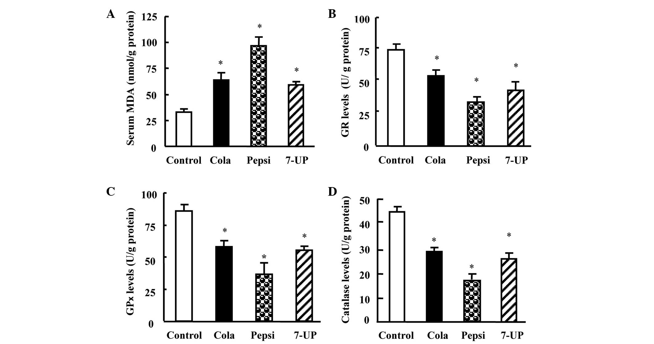
Consumption of Cola, Pepsi and 7-UP for 3 months showed a significant increase in MDA levels (Fig. 1A; P<0.05) with the greatest increase in the rats from the Pepsi group. In parallel, the levels of antioxidants GR, GPx and catalase in the rats were decreased significantly in all groups administered carbonated soft drinks compared with the control. Notably, the greatest changes were observed in the rats from the Pepsi group (Fig. 1B–D).
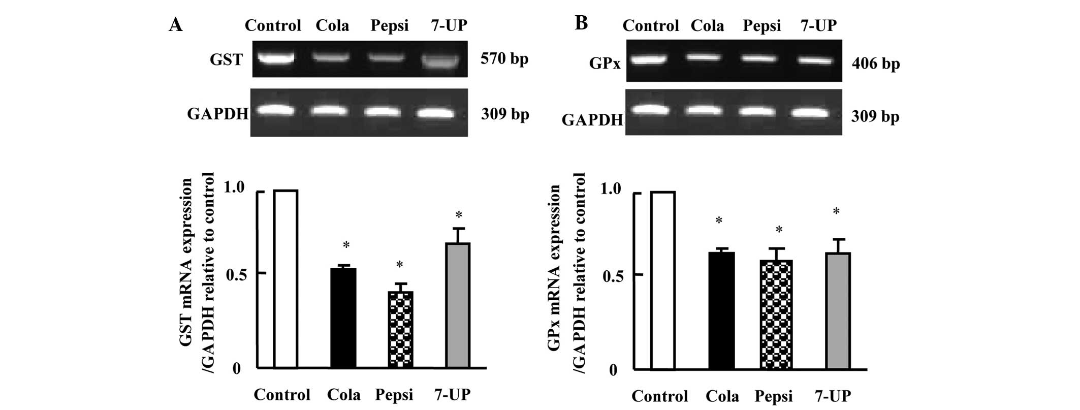
Effect of carbonated soft drink consumption for 3 months on mRNA expression of GST and GPx in the brain tissues of Wistar rats
As shown in Fig. 2, carbonated soft drink consumption for 3 months downregulated the mRNA expression of GST and GPx. Expression levels were significantly decreased by 50 and 40% in the Cola and Pepsi groups for GST and GPx, respectively (P<0.05). Although still significantly reduced when compared with the control (P<0.05), rats in the 7-UP group demonstrated increased levels of GST and GPx, as compared with the Cola and Pepsi groups.
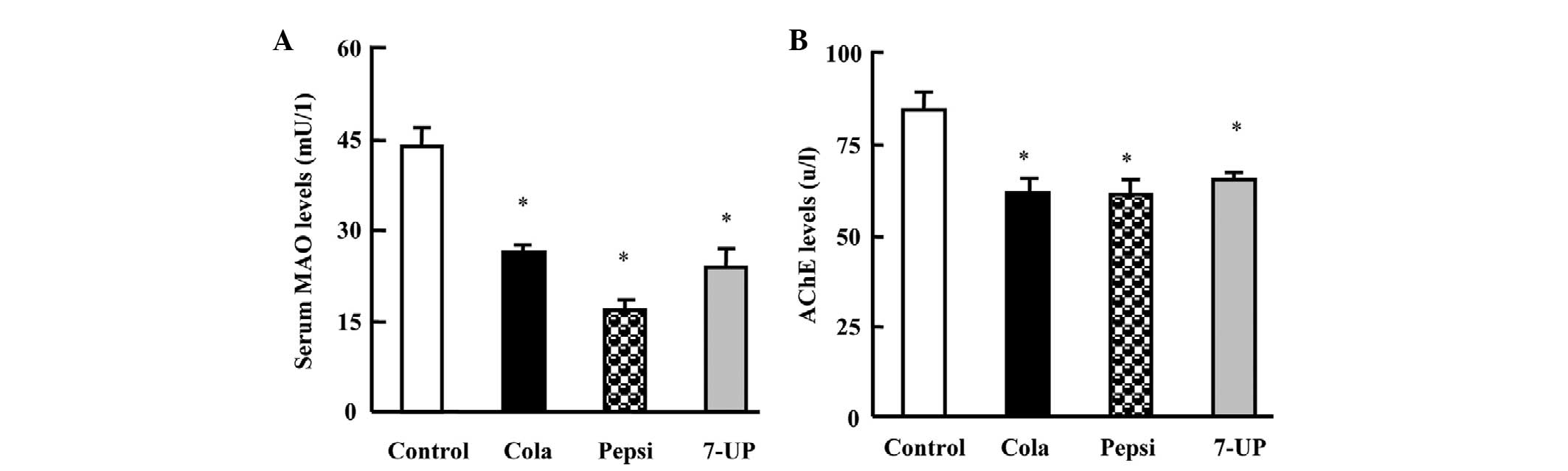
Effect of carbonated soft drink consumption for 3 months on serum levels of MAO-A and AChE in Wistar rats
Next, the changes in MAO-A and AChE levels (Fig. 3) were examined. Cola, Pepsi and 7-UP consumption for 3 months resulted in a significant decrease in MAO and AChE levels (P<0.05). Rats in the Pepsi group exhibited the greatest decreases in MAO-A and AChE levels, as compared with the other groups.
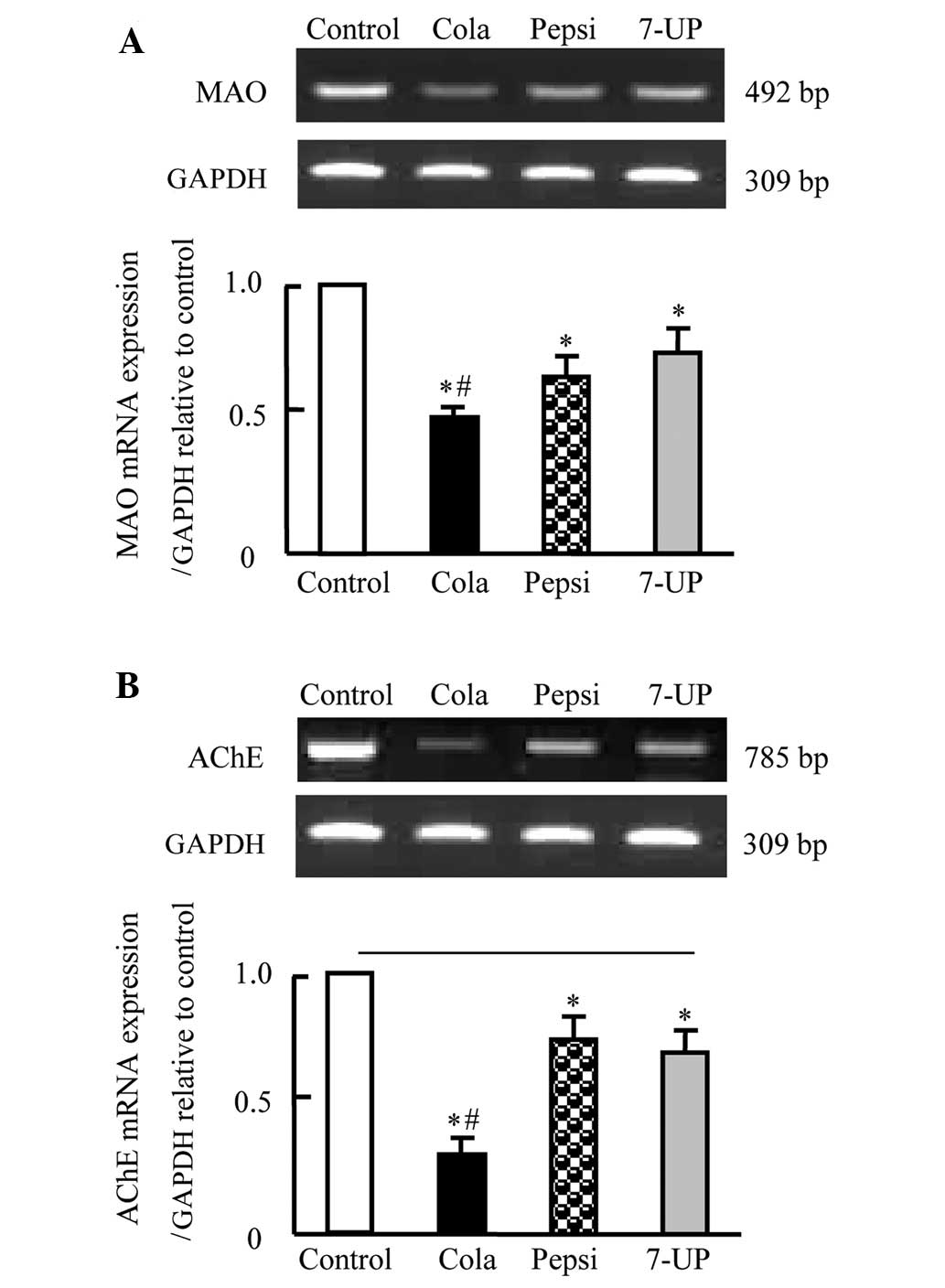
Effect of carbonated soft drink consumption for 3 months on mRNA expression of MAO-A and AChE in brain tissues of Wistar rats
Fig. 4 shows that, consistent with serum changes of MAO-A and AChE, the mRNA expression of MAO-A and AChE was significantly downregulated in the brain tissues of rats administered carbonated soft drinks (P<0.05). Rats in the Cola group exhibited the greatest decrease in MAO-A and AChE expression, followed by Pepsi and 7-UP, respectively. The decrease was not identified to be significantly different between the Pepsi and 7-UP groups (Fig. 4).
Effect of carbonated soft drink consumption for 3 months on mRNA expression of DD2R and 5-HTT in brain tissues of Wistar rats
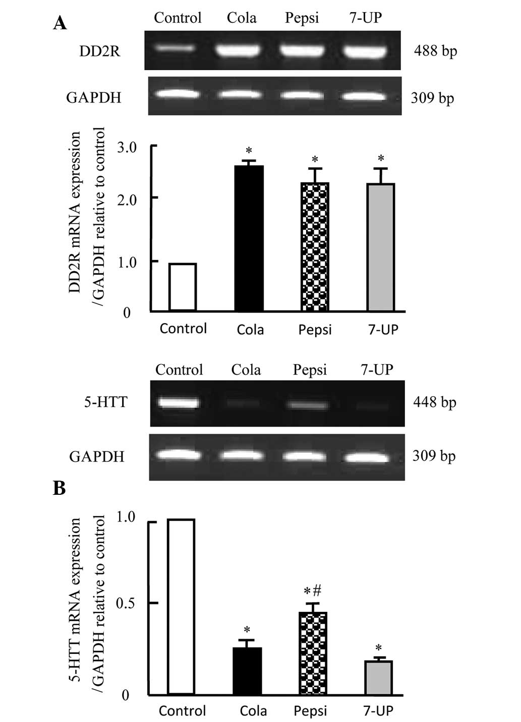
The effects of carbonated soft drink consumption on the expression of certain genes that have been shown to be associated with aggression were investigated. As shown in Fig. 5A, expression of DD2R was significantly upregulated in rats in the Cola, Pepsi and 7-UP groups (P<0.05). By contrast, the rats in these groups exhibited significant downregulation of 5-HTT mRNA expression (Fig. 5B; P<0.05). Furthermore, rats in the Cola and 7-UP groups exhibited significantly decreased expression of 5-HTT, as compared with rats in the Pepsi group (P<0.05).
Effect of carbonated soft drink consumption for 3 months on brain histopathology
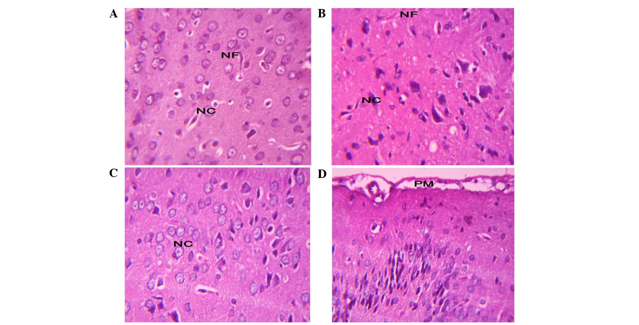
Although changes induced by carbonated soft drinks were observed at the biochemical and molecular levels, brain histopathology analysis showed normal brain architecture in all groups (Fig. 6). The gray matter of the rats appeared normal with its well-organized regularly arranged six layers and different size and shape nerve cells. The normal pattern of the white matter is formed of homogeneously stained nerve fibers running down the cortex was also identified.
Discussion
The results of current study confirmed that chronic consumption of carbonated soft drinks induced oxidative stress and changes in antioxidant expression in the brains of Wistar rats. Moreover, soft drink consumption decreased the serum levels and mRNA expression of MAO and AChE in the brain. Notably, it was also demonstrated that the levels of DD2R were downregulated and the levels of 5-HTT expression were upregulated in the brain, while brain histopathology remained unaffected.
Oxidative stress has been associated with the etiopatho-genesis of several chronic diseases and exhibits a key role in the aging process (13). One of the consequences of uncontrolled oxidative stress (imbalance between the prooxidant and antioxidant levels in favor of prooxidants) is injury to cells, tissues and organs by oxidative damage. It has long been recognized that high levels of free radicals or reactive oxygen species (ROS) can directly damage lipids. The primary sources of endogenous ROS production are the mitochondria, plasma membrane, endoplasmic reticulum and peroxisomes (14). ROS are produced through a variety of mechanisms, including enzymatic reactions and/or auto-oxidation of several compounds, such as catecholamines and hydroquinone. In this study, chronic carbonated soft drink consumption induced oxidative stress in the brain as indicated by the increase in MDA levels in addition to the decrease in the expression of antioxidants, GR, GPx and catalase.
Carbonated soft drinks contain caffeine and phosphoric acid. Caffeine causes the release of adrenaline and an accompanying increase in blood sugar levels to produce the required energy. Caffeine reaches its peak level in the blood within 1 h of consumption and remains in the body for 4–6 h (4,5). Caffeine in soft drinks causes an increases in the release of acid into the stomach, which may lead to an upset stomach or heartburn. Moreover, caffeine has been reported that chronic exposure to the various components of energy and soft drinks may result in significant alterations in the cardiovascular system and brain activity (15,16). The results of the present study demonstrated an alteration in MAO-A and AChE at the serum and mRNA levels. MAO has 2 isozymes, A and B. MAO-A in humans is encoded by the MAOA gene (17,18). It preferentially deaminates norepinephrine, epinephrine, serotonin and dopamine (all are equally deaminated by MAO-A and MAO-B). Inhibition of both MAO-A and MAO-B using MAO inhibitor is used in the treatment of clinical depression, erectile dysfunction and anxiety. MAO-A has been shown to be increased in patients with depression (19) and an association has been demonstrated between low-activity forms of the MAO-A gene and autism (20). A dysfunctional MAO-A gene has also been correlated with increased aggression levels in mice (21,22), as well as with heightened levels of aggression in humans (23). The results of the present study demonstrated that the consumption of carbonated soft drinks decreased MAO-A gene expression and were associated with oversensitivity, as MAO-A decrease is correlated with aggression and violence, this may suggest consumption of carbonated soft drinks is correlated with increased aggression and violence but this requires further investigation.
It is well-established that cholinergic neurons are involved in several neuropsychic functions, such as learning, memory and sleep. Acetylcholine exhibits a key role in modulating these functions (24). A central cholinergic deficit is strongly associated with certain neurodegenerative diseases, such as Alzheimer's disease and Parkinson's disease (25). Evidence of autism due to dysfunction of the cholinergic system has recently been reported (26). AChE is a specific cholinergic marker protein for the functional state of cholinergic neurons. It is key in the maintenance of acetylcholine levels at the cholinergic neurons (27) as it is responsible for degradation of acetylcholine to acetate and choline in the synaptic cleft. Notably, AChE was observed to be decreased in the serum and AChE mRNA expression was observed to be decreased in the brain. It has previously been suggested that acetylcholine disruption may be a primary cause of depression and/or aggression (28).
Dopamine is a neurotransmitter of the catecholamine and phenethylamine families that exhibits a number of roles in the human brain and body. There are 5 isoforms of the dopamine receptor, dopamine D1–5 receptors. DD2R is the most common receptor in the mammalian brain. DD2R antagonists have been used for decades to treat aggressive behavior in psychotic patients (29). In addition, in a preclinical study the role of dopamine D1, D2 and D3 receptors in the modulation of aggression has been documented (30). Several studies have indicated that the mesocorticolimbic dopamine system is involved in the preparation, execution and consequences of aggressive acts (31–33). Pharmacologically induced dopamine increases are associated with increased aggressive behavior under certain conditions (32,33). The results of the present study demonstrated that DD2R levels increased following chronic carbonated soft drink consumption. This was probably due to the increase in dopamine levels resulting from the downregulation of MAO-A expression.
Two major enzymes are responsible for catecholamine catabolism in the brain: Catechol-O-methyltransferase (COMT) and monoamine oxidase A (MAO-A). If aggressive behavior is enhanced by catecholaminergic activity, then decreased activity of COMT and MAO-A should indirectly increase levels of aggression (31,34).
5-HTT expression was shown to be downregulated following chronic carbonated soft drink consumption. A number of studies have shown that elevated serotonin levels lead to decreased aggression in a number of species (35), including humans (36). Blocking serotonin transporter molecules is effective in reducing and preventing aggressive behavior in humans and other animals, presumably due to increased brain 5-HT levels (35). Clinically, blocking 5-HTT with the administration of selective serotonin reuptake inhibitors (SSRIs), reduces aggressive outbursts and violent behavior in psychiatric patients (37–39). In addition, in animal models, acute and chronic treatment with SSRIs can dose-dependently reduce aggressive behavior (40). Acute administration of several SSRIs reduced aggression in different contexts and species, including rodents and non-human primates (40,41). 5-HTT is a type of monoamine transporter protein that transports serotonin from the synaptic cleft to the pre-synaptic neuron. This transport of serotonin by the 5-HTT protein terminates the action of serotonin and recycles it in a sodium-dependent manner. This protein is the target of numerous antidepressant agents, including those of the SSRI class (42). A repeat length polymorphism in the promoter of this gene has been shown to affect the rate of serotonin uptake and may exhibit a role in sudden infant death syndrome, aggressive behavior in Alzheimer disease patients, post-traumatic stress disorder and depression-susceptibility in individuals experiencing emotional trauma (43). It has also been suggested that alterations in 5-HTT expression levels following the consumption of carbonated soft drinks may be a predisposing factor for depression.
Histological examination of the brain revealed that the brain exhibited normal histology and cell distribution following chronic carbonated soft drink consumption. This finding was also reported in another study (10) for regular soft drinks; however, previously diet soft drinks were shown to exhibit adverse effects on the cerebellum of albino rats (10).
In conclusion, chronic term carbonated soft drink consumption induced oxidative stress and alterations in antioxidants and the expression levels of certain genes associated with brain function. Therefore, the results of the present study suggested that the consumption of carbonated soft drinks may induce adverse effects, thus these drinks must be consumed with caution.
Acknowledgments
The authors would like to thank Al-Saedan Research Chair for Genetic Behavioral Disorders, Taif University, Kingdom of Saudi Arabia, for their financial support.
References
Adjene JO, Ezeoke JC and Nwose EU: Histological effects of chronic consumption of soda pop drinks on kidney of adult Wister rats. N Am J Med Sci. 2:215–217. 2010.PubMed/NCBI | |
Amato D, Maravilla A, García-Contreras F and Paniagua R: Soft-drinks and health. Rev Invest Clin. 49:387–395. 1997. | |
Amato D, Maravilla A, Montoya C, Gaja O, Revilla C, Guerra R and Paniagua R: Acute effects of soft drink intake on calcium and phosphate metabolism in immature and adults rats. Rev Invest Clin. 50:185–189. 1998.PubMed/NCBI | |
Clauson KA, Shields KM, McQueen CE and Persad N: Safety issues associated with commercially available energy drinks. J Am Pharm Assoc (2003). 48:e55–e67. 2008. View Article : Google Scholar | |
Iyadurai SJ and Chung SS: New-onset seizures in adults: Possible association with consumption of popular energy drinks. Epilepsy Behav. 10:504–508. 2007. View Article : Google Scholar : PubMed/NCBI | |
DiMeglio DP and Mattes RD: Liquid versus solid carbohydrate: Effects on food intake and body weight. Int J Obes Relat Metab Disord. 24:794–800. 2000. View Article : Google Scholar : PubMed/NCBI | |
Mattes RD: Fluid energy-Where's the problem? J Am Diet Assoc. 106:1956–1961. 2006. View Article : Google Scholar : PubMed/NCBI | |
Frank GK, Oberndorfer TA, Simmons AN, Paulus MP, Fudge JL, Yang TT and Kaye WH: Sucrose activates human taste pathways differently from artificial sweetener. Neuroimage. 39:1559–1569. 2008. View Article : Google Scholar | |
Smeets PA, de Graaf C, Stafleu A, van Osch MJ and van der Grond J: Functional magnetic resonance imaging of human hypothalamic responses to sweet taste and calories. Am J Clin Nutr. 82:1011–1016. 2005.PubMed/NCBI | |
Eluwa MA, Inyangmme II, Akpantah AO, Ekanem TB, Ekong MB, Asuquo OR and Nwakanma AA: A comparative study of the effect of diet and soda carbonated drinks on the histology of the cerebellum of adult female albino Wistar rats. Afr Health Sci. 13:541–545. 2013.PubMed/NCBI | |
Soliman MM, Abdo Nassan M and Ismail TA: Immunohistochemical and molecular study on the protective effect of curcumin against hepatic toxicity induced by paracetamol in Wistar rats. BMC Complement Altern Med. 14:4572014. View Article : Google Scholar : PubMed/NCBI | |
Bancroft JD and Gamble M: Theory and practice of histological techniques. 6th ed. Churchill Livingstone Elsevier; Philadelphia: pp. 126–127. 2008 | |
Ceconi C, Boraso A, Cargnoni A and Ferrari R: Oxidative stress in cardiovascular disease: Myth or fact? Arch Biochem Biophys. 420:217–221. 2003. View Article : Google Scholar : PubMed/NCBI | |
Moldovan L and Moldovan NI: Oxygen free radicals and redox biology of organelles. Histochem Cell Biol. 122:395–412. 2004. View Article : Google Scholar : PubMed/NCBI | |
Higgins JP, Tuttle TD and Higgins CL: Energy beverages: Content and safety. Mayo Clin Proc. 85:1033–1041. 2010. View Article : Google Scholar : PubMed/NCBI | |
Usman A and Jawaid A: Hypertension in a young boy: An energy drink effect. BMC Res Notes. 5:5912012. View Article : Google Scholar : PubMed/NCBI | |
Hotamisligil GS and Breakefield XO: Human monoamine oxidase A gene determines levels of enzyme activity. Am J Hum Genet. 49:383–392. 1991.PubMed/NCBI | |
De Colibus L, Li M, Binda C, Lustig A, Edmondson DE and Mattevi A: Three-dimensional structure of human monoamine oxidase A (MAO A): Relation to the structures of rat MAO A and human MAO B. Proc Natl Acad Sci USA. 102:12684–12689. 2005. View Article : Google Scholar : PubMed/NCBI | |
Meyer JH, Ginovart N, Boovariwala A, Sagrati S, Hussey D, Garcia A, Young T, Praschak-Rieder N, Wilson AA and Houle S: Elevated monoamine oxidase a levels in the brain: An explanation for the monoamine imbalance of major depression. Arch Gen Psychiatry. 63:1209–1216. 2006. View Article : Google Scholar : PubMed/NCBI | |
Cohen IL, Liu X, Lewis ME, Chudley A, Forster-Gibson C, Gonzalez M, Jenkins EC, Brown WT and Holden JJ: Autism severity is associated with child and maternal MAOA genotypes. Clin Genet. 79:355–362. 2011. View Article : Google Scholar | |
Scott AL, Bortolato M, Chen K and Shih JC: Novel monoamine oxidase A knock out mice with human-like spontaneous mutation. Neuroreport. 19:739–743. 2008. View Article : Google Scholar : PubMed/NCBI | |
Vishnivetskaya GB, Skrinskaya JA, Seif I and Popova NK: Effect of MAO A deficiency on different kinds of aggression and social investigation in mice. Aggress Behav. 33:1–6. 2007. View Article : Google Scholar : PubMed/NCBI | |
Brunner HG, Nelen M, Breakefield XO, Ropers HH and van Oost BA: Abnormal behavior associated with a point mutation in the structural gene for monoamine oxidase A. Science. 262:578–580. 1993. View Article : Google Scholar : PubMed/NCBI | |
Mohapel P, Leanza G, Kokaia M and Lindvall O: Forebrain acetylcholine regulates adult hippocampal neurogenesis and learning. Neurobiol Aging. 26:939–946. 2005. View Article : Google Scholar : PubMed/NCBI | |
Oda Y: Choline acetyltransferase: The structure, distribution and pathologic changes in the central nervous system. Pathol Int. 49:921–937. 1999. View Article : Google Scholar : PubMed/NCBI | |
Lam KS, Aman MG and Arnold LE: Neurochemical correlates of autistic disorder: A review of the literature. Res Dev Disabil. 27:254–289. 2006. View Article : Google Scholar | |
Eckenstein F and Sofroniew MV: Identification of central cholinergic neurons containing both choline acetyltransferase and acetylcholinesterase and of central neurons containing only acetylcholinesterase. J Neurosci. 3:2286–2291. 1983.PubMed/NCBI | |
Shytle RD, Silver AA, Lukas RJ, Newman MB, Sheehan DV and Sanberg PR: Nicotinic acetylcholine receptors as targets for antidepressants. Mol Psychiatry. 7:525–535. 2002. View Article : Google Scholar : PubMed/NCBI | |
Glazer WM and Dickson RA: Clozapine reduces violence and persistent aggression in schizophrenia. J Clin Psychiatry. 59(Suppl 3): 8–14. 1998.PubMed/NCBI | |
Miczek KA, Covington HE III, Nikulina EM Jr and Hammer RP: Aggression and defeat: Persistent effects on cocaine self-administration and gene expression in peptidergic and aminergic mesocorticolimbic circuits. Neurosci Biobehav Rev. 27:787–802. 2004. View Article : Google Scholar : PubMed/NCBI | |
de Almeida RM, Ferrari PF, Parmigiani S and Miczek KA: Escalated aggressive behavior: Dopamine, serotonin and GABA. Eur J Pharmacol. 526:51–64. 2005. View Article : Google Scholar : PubMed/NCBI | |
Ferrari PF, van Erp AM, Tornatzky W and Miczek KA: Accumbal dopamine and serotonin in anticipation of the next aggressive episode in rats. Eur J Neurosci. 17:371–378. 2003. View Article : Google Scholar : PubMed/NCBI | |
Ferrari PF, Palanza P, Parmigiani S, de Almeida RM and Miczek KA: Serotonin and aggressive behavior in rodents and nonhuman primates: Predispositions and plasticity. Eur J Pharmacol. 526:259–273. 2005. View Article : Google Scholar : PubMed/NCBI | |
Gogos JA, Morgan M, Luine V, Santha M, Ogawa S, Pfaff D and Karayiorgou M: Catechol-O-methyltransferase-deficient mice exhibit sexually dimorphic changes in catecholamine levels and behavior. Proc Natl Acad Sci USA. 95:9991–9996. 1998. View Article : Google Scholar : PubMed/NCBI | |
Chiavegatto S and Nelson RJ: Interaction of nitric oxide and serotonin in aggressive behavior. Horm Behav. 44:233–241. 2003. View Article : Google Scholar : PubMed/NCBI | |
Coccaro EF and Kavoussi RJ: Neuropsychopharmacologic challenge in biological psychiatry. Clin Chem. 40:319–327. 1994.PubMed/NCBI | |
Barkan T, Peled A, Modai I, Barak P, Weizman A and Rehavi M: Serotonin transporter characteristics in lymphocytes and platelets of male aggressive schizophrenia patients compared to non-aggressive schizophrenia patients. Eur Neuropsychopharmacol. 16:572–579. 2006. View Article : Google Scholar : PubMed/NCBI | |
Blader JC: Pharmacotherapy and postdischarge outcomes of child inpatients admitted for aggressive behavior. J Clin Psychopharmacol. 26:419–425. 2006. View Article : Google Scholar : PubMed/NCBI | |
Bond AJ: Antidepressant treatments and human aggression. Eur J Pharmacol. 526:218–225. 2005. View Article : Google Scholar : PubMed/NCBI | |
Carrillo M, Ricci LA, Coppersmith GA and Melloni RH Jr: The effect of increased serotonergic neurotransmission on aggression: A critical meta-analytical review of preclinical studies. Psychopharmacology (Berl). 205:349–368. 2009. View Article : Google Scholar | |
Caldwell EE and Miczek KA: Long-term citalopram maintenance in mice: Selective reduction of alcohol-heightened aggression. Psychopharmacology (Berl). 196:407–416. 2008. View Article : Google Scholar | |
Deutch AY and Roth RH: Neurotransmitters. Fundamental Neuroscience. Squire L, Berg S, Bloom F, du Lac S, Ghosh A and Spitzer N: 3rd edition. Elsevier/Academic Press; Amsterdam: pp. 1432008 | |
Holmes A, Murphy DL and Crawley JN: Reduced aggression in mice lacking the serotonin transporter. Psychopharmacology (Berl). 161:160–167. 2002. View Article : Google Scholar |
Related Articles
- Chronic effects of soft drink consumption on the health state of Wistar rats: A biochemical, genetic and histopathological study
- Carbonated soft drinks alter hepatic cytochrome P450 isoform expression in Wistar rats
- Protective effect of grape seed extract against cadmium-induced testicular dysfunction
- Modulatory effects of levamisole and garlic oil on the immune response of Wistar rats: Biochemical, immunohistochemical, molecular and immunological study
- Magnesium sulfate ameliorates carbon monoxide‑induced cerebral injury in male rats
Impaired regulation of pH homeostasis by oxidative stress in rat brain capillary endothelial cells
- PMID: 15962511
- DOI: 10.1007/s10571-004-1379-6
Abstract
(1) Endothelial cells are permanently challenged by altering pH in the blood, and oxidative damage could also influence the intracellular pH (pH(i)) of the endothelium. Cerebral microvascular endothelial cells form the blood-brain barrier (BBB) and pH(i) regulation of brain capillary endothelial cells is important for the maintenance of BBB integrity. The aim of this study was to address the pH regulatory mechanisms and the effect of an acute exposure to hydrogen peroxide (H2O2) on the pH regulation in primary rat brain capillary endothelial (RBCE) cells The RBCE monolayers were loaded with the fluorescent pH indicator BCECF and pH(i) was monitored by detecting the fluorescent changes. (2) The steady-state pH(i) of RBCE cells in HEPES-buffer (6.83 +/- 0.1) did not differ significantly from that found in bicarbonate-buffered medium (6.90 +/- 0.08). Cells were exposed to NH4CI to induce intracellular acidification and then the recovery to resting pH was studied. Half-recovery time after NH4Cl prepulse-induced acid load was significantly less in the bicarbonate-buffered medium than in the HEPES-medium, suggesting that in addition to the Na+ / H+ exchanger, HCO3- / Cl- exchange mechanism is also involved in the restoration of pH(i) after an intracellular acid load in primary RBCE cells. We used RT-PCR-reactions to detect the isoforms of Na+ / H+ exchanger gene family (NHE). NHE-1 -2, -3 and -4 were equally present, and there was no significant difference in the relative abundance of the four transcripts in these cells. (3) No pH(i) recovery was detected when the washout after an intracellular acid load occurred in nominally Na+ -free HEPES-buffered medium or in the presence of 10 microM 5-(N-ethyl-N-isopropyl)amiloride (EIPA), a specific inhibitor of Na+ / H+ exchanger. The new steady-state pH(i) were 6.37 +/- 0.02 and 6.60 +/- 0.02, respectively. (4) No detectable change was observed in the steady-state pH(i) in the presence of 100 microM H2O2; however, recovery from NH4Cl prepulse-induced intracellular acid load was inhibited when H2O2 was present in 50 or 100 microM concentration in the HEPES-buffered medium during NH4Cl washout. These data suggest that H2O2 is without effect on the activity of Na+ / H+ exchanger at rest, but could inhibit the function of the exchanger after an intracellular acid load.
References
Publication types
MeSH terms
Substances
LinkOut - more resources
Full Text Sources

No comments:
Post a Comment
Note: Only a member of this blog may post a comment.