
https://www.ncbi.nlm.nih.gov/pmc/articles/PMC7112871/
How mRNA Vaccines Work - Simply Explained
How do RNA-based vaccines work?
https://www.quora.com/How-do-RNA-based-vaccines-work

Q. How does the new mRNA Covid-19 vaccine by Pfizer and Moderna actually work?
A. Mike, Mike, Mike! Wassup, Mike? 🐫🐪🐫
Pfizer’s BNT162 and Moderna’s mRNA-1273 have both been linked to preliminary phase 3 data showing 90% efficacy or greater among administered patients versus placebo arms at the first cutoff for total infection rates in their respective global, large-scale trials.
Vaccines train the immune system to recognize the disease-causing part of a virus. Vaccines traditionally contain either weakened viruses or purified signature proteins of the virus.
But an mRNA vaccine is different, because rather than having the viral protein injected, a person receives genetic material – mRNA – that encodes the viral protein. When these genetic instructions are injected into the upper arm, the muscle cells translate them to make the viral protein directly in the body.
This approach mimics what the SARS-CoV-2 does in nature – but the vaccine mRNA codes only for the critical fragment of the viral protein. This gives the immune system a preview of what the real virus looks like without causing disease. This preview gives the immune system time to design powerful antibodies that can neutralize the real virus if the individual is ever infected.
While this synthetic mRNA is genetic material, it cannot be transmitted to the next generation. After an mRNA injection, this molecule guides the protein production inside the muscle cells, which reaches peak levels for 24 to 48 hours and can last for a few more days. Thus, a second dose is required to achieve lasting protection.
ps://www.cdc.gov/coronavirus/2019-ncov/vaccines/different-vaccines/mrna.html
Understanding mRNA COVID-19 Vaccines
Messenger RNA vaccines—also called mRNA vaccines—are some of the first COVID-19 vaccines authorized for use in the United States.
New Approach to Vaccines
mRNA vaccines are a new type of vaccine to protect against infectious diseases. To trigger an immune response, many vaccines put a weakened or inactivated germ into our bodies. Not mRNA vaccines. Instead, they teach our cells how to make a protein—or even just a piece of a protein—that triggers an immune response inside our bodies. That immune response, which produces antibodies, is what protects us from getting infected if the real virus enters our bodies.
A Closer Look at How COVID-19 mRNA Vaccines Work
COVID-19 mRNA vaccines give instructions for our cells to make a harmless piece of what is called the “spike protein.” The spike protein is found on the surface of the virus that causes COVID-19.
COVID-19 mRNA vaccines are given in the upper arm muscle. Once the instructions (mRNA) are inside the immune cells, the cells use them to make the protein piece. After the protein piece is made, the cell breaks down the instructions and gets rid of them.
Next, the cell displays the protein piece on its surface. Our immune systems recognize that the protein doesn’t belong there and begin building an immune response and making antibodies, like what happens in natural infection against COVID-19.
At the end of the process, our bodies have learned how to protect against future infection. The benefit of mRNA vaccines, like all vaccines, is those vaccinated gain this protection without ever having to risk the serious consequences of getting sick with COVID-19.
Facts about COVID-19 mRNA Vaccines
They cannot give someone COVID-19.
- mRNA vaccines do not use the live virus that causes COVID-19.
They do not affect or interact with our DNA in any way.
- mRNA never enters the nucleus of the cell, which is where our DNA (genetic material) is kept.
- The cell breaks down and gets rid of the mRNA soon after it is finished using the instructions.
COVID-19 mRNA Vaccines Will Be Rigorously Evaluated for Safety
mRNA vaccines have been held to the same rigorous safety and effectiveness standards as all other types of vaccines in the United States. The only COVID-19 vaccines the Food and Drug Administration (FDA) will make available for use in the United States (by approval or emergency use authorization) are those that meet these standards.
mRNA Vaccines Are New, But Not Unknown
Researchers have been studying and working with mRNA vaccines for decades. Interest has grown in these vaccines because they can be developed in a laboratory using readily available materials. This means the process can be standardized and scaled up, making vaccine development faster than traditional methods of making vaccines.
mRNA vaccines have been studied before for flu, Zika, rabies, and cytomegalovirus (CMV). As soon as the necessary information about the virus that causes COVID-19 was available, scientists began designing the mRNA instructions for cells to build the unique spike protein into an mRNA vaccine.
Future mRNA vaccine technology may allow for one vaccine to provide protection for multiple diseases, thus decreasing the number of shots needed for protection against common vaccine-preventable diseases.
Beyond vaccines, cancer research has used mRNA to trigger the immune system to target specific cancer cells.
Related Links
2.8. Infectivity of viral RNA and detergent-disrupted virions
Infected Vero cells were prepared by inoculation with 20 μl of virus at a 106.37 TCID50 per ml of SARS-CoV in a final volume of 2 ml in a T25 flask for 1 h at 25 °C. DMEM with supplements was added to the flask and the cells were incubated at 37 °C for 3 days. The monolayer was washed with 1X phosphate buffered saline (PBS), cells were lysed with the addition of 2.5 ml of a phenol and guanidine isothiocyanate solution (TRIzol Reagent, Sigma), and cytoplasmic RNA was isolated according to the manufacturer’s specifications. Vero cells were inoculated with 10 μl of purified RNA in 0.5 ml DMEM. After an hour, DMEM with supplements was added. Additionally, Vero cells were transfected with cytoplasmic RNA using DMRIE-C (Invitrogen Life Technologies, Carlsbad, CA) according to the manufacturer’s instructions. Cells were incubated at 37 °C, and observed for CPE on days 3 and 4.
To examine the infectivity of detergent-disrupted virions, SARS-CoV infected Vero monolayer cells were washed and dissociated with trypsin/versene, pelleted by centrifugation, and washed with PBS. After centrifugation, the pellet was lysed with sodium dodecyl sulfate/nonidet P-40 (SDS/NP-40; 0.1% SDS, 0.1% NP-40, in 0.1x PBS; Sigma), frozen at −70 °C, thawed, and clarified by centrifugation. The supernatant was used to infect Vero cell monolayers in 6-well plates, such that the final concentration of SDS was 0.002 or 0.018%. Three and four days following the inoculation, cells were observed for evidence of CPE.
3.3. Effects of formaldehyde and glutaraldehyde on the infectivity of SARS-CoV
Formalin (dilute formaldehyde) has been used for a number of years to inactivate virus for use in vaccine products, such as the widely used and very effective polio vaccine (Salk and Salk, 1984). Other attempts at using formalin inactivation for generation of vaccines for respiratory syncytial virus (Kim et al., 1969) and measles virus (Fulginiti et al., 1967) were not useful, as they induced an aberrant immune response resulting from formalin-induced perturbations of the viruses. Formalin inactivation occurs when nonprotonated amino groups of amino acids, such as lysine, combine with formaldehyde to form hydroxymethylamine. The hydroxymethylamine combines with the amino, amide, guanidyl, phenolic, or imidazole group of amino acids to create inter- or intramolecular methylene crosslinks (for review, see Jiang and Schwendeman, 2000). Fraenkel-Conrat (1954) observed the absorption spectra of several plant viruses and determined that formalin also binds in a reversible manner to RNA, blocking reading of the genome by RNA polymerase. Glutaraldehyde can also be used to inactivate virus and is used as a disinfecting agent of medical instruments, such as endoscopes (Tandon, 2000), and as a fixative for electron microscopy (McDonnell and Russell, 1999).
3.5. Infectivity of isolated viral RNA and isolated proteins
Biochemical and molecular biology experiments may require the isolation of nucleic acids or proteins from virus-infected cells. We used a phenol and guanidine isothiocyanate solution (TRIzol, Sigma) to isolate cytoplasmic RNA from SARS-CoV infected Vero cells. After inoculation of Vero cells with the isolated RNA, we determined that SARS-CoV RNA was not able to produce CPE in the cells (data not shown). We also found that transfection of the cells with this RNA, using a liposome-based transfection reagent (DMRIE-C, Invitrogen, as per manufacturer’s instructions for RNA transfection), was also not sufficient to cause infection of Vero cells (data not shown).
Additionally, we tested the effectiveness of SDS/NP-40 treatment on inactivation of the SARS-CoV. Briefly, SARS-CoV-infected Vero cells were lysed with an SDS/NP-40 solution, clarified by centrifugation, and the supernatant was used to infect Vero cell monolayers. No CPE was observed in the cells after 3 and 4 days, indicating that SDS/NP-40-induced disruption of the virions was sufficient to prevent survival of infectious particles.
We determined that formalin and glutaraldehyde inactivated SARS-CoV in a temperature- and time-dependent manner. While incubation at 4 °C inhibited the effect of these chemicals, at 37 °C or room temperature, formalin significantly decreased the infectivity of the virus on day 1, while glutaraldehyde inactivated SARS-CoV after incubations of 1–2 days. As glutaraldehyde is commonly used to disinfect medical instruments, especially endoscopes, care should be taken to analyze time, temperature, and concentration requirements necessary for complete SARS-CoV inactivation.
Weismiller et al. (1990) determined that a pH of 8.0 induces a conformational change in the spike protein of the coronavirus MHV that enables fusion of the virion with the host cell. However, Xiao et al. (2003) determined that the spike protein of SARS-CoV mediated fusion with the host cell at a neutral pH. These data suggest that different pH conditions affect the spike proteins of coronaviruses, and the activity of the spike protein of SARS-CoV may be sensitive to changes in pH, possibly by changing the infectious nature of the viral particles. We determined that exposure of SARS-CoV to extreme basic or acidic conditions caused inactivation, while the virus remained stable within a range of neutral pH. The pH of gastric secretions of the stomach ranges from 1.0 to 3.5, while the small and large intestines range from pH 7.5 to 8.0 (Guyton and Hall, 1997). Taken together, these data suggest that ingestion of SARS-CoV would probably result in inactivation of most virions by stomach acid. However, acidic conditions of the stomach may be partially neutralized by a particularly large meal or antacid ingestion, and under these conditions the virus might have a chance to move through the stomach into the slightly basic conditions of the intestines. Leung et al. (2003) have shown enteric involvement of the SARS virus, as evidenced by the presence of active viral replication in intestinal biopsy specimens from five patients, and the isolation of SARS-CoV RNA in stool specimens up to 10 weeks after onset of symptoms. These data, coupled with the previously mentioned stability of the virus to moderate pH conditions, suggest that the SARS virus may survive ingestion and a fecal/oral route of infection may be possible.
Our experiments showed that UVC light, heat, formalin, glutaraldehyde, and extremes of pH, were able to inactivate SARS-CoV. However, gamma irradiation at the doses tested, was not sufficient to inactivate the virus. As expected, neither viral RNA alone nor virions disrupted by SDS/NP-40 were infectious. These conditions were appropriate for our viral stocks as described, however, we caution that researchers need to test viral stocks for complete inactivation before handling the virus at lower safety levels. These data analyze virus samples in tissue culture medium and we are currently testing the inactivation properties required of SARS-CoV in biological (body) fluids. Understanding the ways in which SARS-CoV can be inactivated, will allow the transfer of the virus from BSL3 to BSL2 conditions, and will promote the study of inactivated viral vaccines.
https://www.nature.com/articles/s41401-020-0485-4#ref-CR5
- Review Article
- Published:
Structural and functional properties of SARS-CoV-2 spike protein: potential antivirus drug development for COVID-19
Acta Pharmacologica Sinica 41, 1141–1149(2020)
Abstract
Coronavirus disease 2019 is a newly emerging infectious disease currently spreading across the world. It is caused by a novel coronavirus, severe acute respiratory syndrome coronavirus 2 (SARS-CoV-2). The spike (S) protein of SARS-CoV-2, which plays a key role in the receptor recognition and cell membrane fusion process, is composed of two subunits, S1 and S2. The S1 subunit contains a receptor-binding domain that recognizes and binds to the host receptor angiotensin-converting enzyme 2, while the S2 subunit mediates viral cell membrane fusion by forming a six-helical bundle via the two-heptad repeat domain. In this review, we highlight recent research advance in the structure, function and development of antivirus drugs targeting the S protein.
Introduction
The epidemic of novel coronavirus disease 2019 (COVID-19) was caused by a new coronavirus occurred in December 2019, and now has spread worldwide and turned into a global pandemic [1]. The COVID-19 was quickly discovered to be caused by a coronavirus later named severe acute respiratory syndrome coronavirus 2 (SARS-CoV-2) [1], which belongs to the β coronavirus family. It is the seventh known coronavirus to infect humans; four of these coronaviruses (229E, NL63, OC43, and HKU1) only cause slight symptoms of the common cold. Conversely, the other three, SARS-CoV, MERS-CoV, and SARS-CoV-2, are able to cause severe symptoms and even death, with fatality rates of 10%, 37%, and 5%, respectively.
Although a large number of studies and clinical trials are being launched on COVID-19 around the world [2, 3], no evidence from randomized clinical trials has shown that any potential therapy improves outcomes in patients [4]. As the epidemic spreads, it is critical to find a specific therapeutic for COVID-19, and vaccines targeting various SARS-CoV-2 proteins are under development.
SARS-CoV-2 is a single-stranded RNA-enveloped virus [5]. An RNA-based metagenomic next-generation sequencing approach has been applied to characterize its entire genome, which is 29,881 bp in length (GenBank no. MN908947), encoding 9860 amino acids [6]. Gene fragments express structural and nonstructural proteins. The S, E, M, and N genes encode structural proteins, whereas nonstructural proteins, such as 3-chymotrypsin-like protease, papain-like protease, and RNA-dependent RNA polymerase, are encoded by the ORF region [7].
A large number of glycosylated S proteins cover the surface of SARS-CoV-2 and bind to the host cell receptor angiotensin-converting enzyme 2 (ACE2), mediating viral cell entry [8]. When the S protein binds to the receptor, TM protease serine 2 (TMPRSS2), a type 2 TM serine protease located on the host cell membrane, promotes virus entry into the cell by activating the S protein. Once the virus enters the cell, the viral RNA is released, polyproteins are translated from the RNA genome, and replication and transcription of the viral RNA genome occur via protein cleavage and assembly of the replicase–transcriptase complex. Viral RNA is replicated, and structural proteins are synthesized, assembled, and packaged in the host cell, after which viral particles are released (Fig. 1d) [9].

a The schematic structure of the S protein. b The S protein binds to the receptor ACE2. c The binding and virus–cell fusion process mediated by the S protein. d The life cycle of SARS-CoV-2 in host cells.
These proteins are critical to the viral life cycle and provide potential targets for drug therapies. For example, ACE2-based peptide, 3CLpro inhibitor (3CLpro-1), and a novel vinylsulfone protease inhibitor have been experimentally demonstrated to be effective against SARS-CoV-2 [10]. The SARS-CoV-2 S protein is highly conserved among all human coronaviruses (HCoVs) and is involved in receptor recognition, viral attachment, and entry into host cells. Due to its indispensable functions, it represents one of the most important targets for COVID-19 vaccine and therapeutic research. In this review, we summarize advances in research of the SARS-CoV-2 S protein and its therapeutic targeting.
Structure of the S protein
With a size of 180–200 kDa, the S protein consists of an extracellular N-terminus, a transmembrane (TM) domain anchored in the viral membrane, and a short intracellular C-terminal segment [11]. S normally exists in a metastable, prefusion conformation; once the virus interacts with the host cell, extensive structural rearrangement of the S protein occurs, allowing the virus to fuse with the host cell membrane. The spikes are coated with polysaccharide molecules to camouflage them, evading surveillance of the host immune system during entry [12].
The total length of SARS-CoV-2 S is 1273 aa and consists of a signal peptide (amino acids 1–13) located at the N-terminus, the S1 subunit (14–685 residues), and the S2 subunit (686–1273 residues); the last two regions are responsible for receptor binding and membrane fusion, respectively. In the S1 subunit, there is an N-terminal domain (14–305 residues) and a receptor-binding domain (RBD, 319–541 residues); the fusion peptide (FP) (788–806 residues), heptapeptide repeat sequence 1 (HR1) (912–984 residues), HR2 (1163–1213 residues), TM domain (1213–1237 residues), and cytoplasm domain (1237–1273 residues) comprise the S2 subunit (Fig. 2a) [13]. S protein trimers visually form a characteristic bulbous, crown-like halo surrounding the viral particle (Fig. 1a). Based on the structure of coronavirus S protein monomers, the S1 and S2 subunits form the bulbous head and stalk region [14]. The structure of the SARS-CoV-2 trimeric S protein has been determined by cryo-electron microscopy at the atomic level, revealing different conformations of the S RBD domain in opened and closed states and its corresponding functions (Fig. 2b, c) [15, 16].

a Schematic representation of the SARS-CoV-2 spike. b–c The S protein RBD closed and opened status. d The S protein binds to ACE2 with opened RBD in the S1 subunit. e The six-helix structure formed by HR1 and HR2 of the S2 subunit.
In the native state, the CoV S protein exists as an inactive precursor. During viral infection, target cell proteases activate the S protein by cleaving it into S1 and S2 subunits [17], which is necessary for activating the membrane fusion domain after viral entry into target cells [18]. Similar to other coronaviruses, the S protein of SARS-CoV-2 is cleaved into S1 and S2 subunits by cellular proteases, and the serine protease TMPRSS2 is used as a protein primer. Although the cleavage site of SARS-CoV is known, that of SARS-CoV-2 S has not yet been reported [18, 19].
Structure of the S1 subunit
The binding of virus particles to cell receptors on the surface of the host cell is the initiation of virus infection; therefore, receptor recognition is an important determinant of viral entry and a drug design target.
RBD situated in the S1 subunit binds to the cell receptor ACE2 in the region of aminopeptidase N. The S1 region contains the NTD and CTD, and atomic details at the binding interface demonstrate key residue substitutions in SARS-CoV-2-CTD. In addition, the SARS-CoV-2 S CTD binding interface has more residues that directly interact with the receptor ACE2 than does SARS-RBD (21 versus 17), and a larger surface area is buried with SARS-CoV-2 S CTD in complex with ACE2 than with SARS S RBD. Mutations of key residues play an important role in enhancing the interaction with ACE2. F486 in SARS-CoV-2, instead of I472 in SARS RBD, forms strong aromatic–aromatic interactions with ACE2 Y83, and E484 in SARS-CoV-2-CTD, instead of P470 in SARS RBD, forms ionic interactions with K31, which leads to higher affinity for receptor binding than RBD of SARS-CoV (Fig. 2d) [15, 16, 20, 21].
The RBD region is a critical target for neutralizing antibodies (nAbs), and SARS-CoV-2 and SARS-CoV RBD are ~73%–76% similar in sequence. Nine ACE2-contacting residues in CoV RBD are fully conserved, and four are partially conserved. Analysis of the RBM (receptor-binding motif, a portion of RBD making direct contacts with ACE2) of SARS-CoV and SARS-CoV-2 revealed that most residues essential for ACE2 binding in the SARS-CoV S protein are conserved in the SARS-CoV-2 S protein. However, some studies showed that murine monoclonal antibodies (mAbs) and polyclonal antibodies against SARS-RBD are unable to interact with the SARS-CoV-2 S protein, revealing differences in antigenicity between SARS-CoV and SARS-CoV-2 [20]. Similarly, a SARS-CoV RBD-specific antibody failed to block infection mediated by the S protein of SL-CoV–SHC014 [22], which suggests that the S1 RBD may not be an ideal drug target due to the highly mutable characteristic of broad-spectrum anti-CoV drugs.
Structure of the S2 subunit
The S2 subunit, composed successively of a FP, HR1, HR2, TM domain, and cytoplasmic domain fusion (CT), is responsible for viral fusion and entry.
FP is a short segment of 15–20 conserved amino acids of the viral family, composed mainly of hydrophobic residues, such as glycine (G) or alanine (A), which anchor to the target membrane when the S protein adopts the prehairpin conformation. Previous research has shown that FP plays an essential role in mediating membrane fusion by disrupting and connecting lipid bilayers of the host cell membrane [23].
HR1 and HR2 are composed of a repetitive heptapeptide: HPPHCPC, where H is a hydrophobic or traditionally bulky residue, P is a polar or hydrophilic residue, and C is another charged residue [24]. HR1 and HR2 form the six-helical bundle (6-HB) (Fig. 2e), which is essential for the viral fusion and entry function of the S2 subunit [13]. HR1 is located at the C-terminus of a hydrophobic FP, and HR2 is located at the N-terminus of the TM domain [25]. The downstream TM domain anchors the S protein to the viral membrane, and the S2 subunit ends in a CT tail [14].
RBD binds to ACE2, and S2 changes conformation by inserting FP into the target cell membrane, exposing the prehairpin coiled-coil of the HR1 domain and triggering interaction between the HR2 domain and HR1 trimer to form 6-HB, thus bringing the viral envelope and cell membrane into proximity for viral fusion and entry [26]. HR1 forms a homotrimeric assembly in which three highly conserved hydrophobic grooves on the surface that bind to HR2 are exposed. The HR2 domain forms both a rigid helix and a flexible loop to interact with the HR1 domain. In the postfusion hairpin conformation of CoVs, there are many strong interactions between the HR1 and HR2 domains inside the helical region, which is designated the “fusion core region” (HR1core and HR2core regions, respectively).
Targeting the heptad repeat (HR) has attracted the greatest interest in therapeutic drug discovery. The S protein is an important target protein for the development of specific drugs, while the S1 RBD domain is part of a highly mutable region and is not an ideal target site for broad-spectrum antiviral inhibitor development [27]. In contrast, the HR region of the S2 subunit plays an essential role in HCoV infections and is conserved among HCoVs, as is the mode of interaction between HR1 and HR2 [28]. A synthetic peptide derived from the stem region of the ZIKV envelope protein was demonstrated in 2017 to potently inhibit infection by ZIKV and other flaviviruses in vitro [29], implying antiviral efficiency of peptides derived from conserved regions of viral proteins. Peptides derived from the HR2 region of class I viral fusion proteins of enveloped viruses competitively bind to viral HR1 and effectively inhibit viral infection [22]. Therefore, HR1 is a promising target for the development of fusion inhibitors against SARS-CoV-2 infection.
Functions of the S protein
The S protein on the surface of the virus is a key factor involved in infection. It is a trimeric class I TM glycoprotein responsible for viral entry, and it is present in all kinds of HCoVs, as well as in other viruses such as HIV (HIV glycoprotein 160, Env), influenza virus (influenza hemagglutinin, HA), paramyxovirus (paramyxovirus F), and Ebola (Ebola virus glycoprotein) [30]. Similar to other coronaviruses, the S protein of SARS-CoV-2 mediates receptor recognition, cell attachment, and fusion during viral infection [16, 20, 21, 31,32,33].
The trimer of the S protein located on the surface of the viral envelope is the basic unit by which the S protein binds to the receptor [16, 33]. The S1 domain contains the RBD, which is mainly responsible for binding of the virus to the receptor, while the S2 domain mainly contains the HR domain, including HR1 and HR2, which is closely related to virus fusion [34].
Receptor binding
As mentioned above, the SARS-CoV-2 S protein binds to the host cell by recognizing the receptor ACE2 [33]. ACE2 is a homolog of ACE, which converts angiotensin I to angiotensin 1–9 [35]. ACE2 is distributed mainly in the lung, intestine, heart, and kidney, and alveolar epithelial type II cells are the major expressing cells [36]. ACE2 is also a known receptor for SARS-CoV. The S1 subunit of the SARS-CoV S protein binds with ACE2 to promote the formation of endosomes, which triggers viral fusion activity under low pH (Fig. 1a, b) [37].
Interaction between the S protein and ACE2 can be used to identify intermediate hosts of SARS-CoV-2, as ACE2 from different species, such as amphibians, birds, and mammals, has a conserved primary structure [38]. Luan et al. compared the binding affinities between ACE2 and SARS-CoV-2 S from mammals, birds, snakes, and turtles and found that the ACE2 of Bovidae and Cricetidae interacted well with SARS-CoV-2 S RBD but that ACE2 from snakes and turtles could not.
The S protein binds to ACE2 through the RBD region of the S1 subunit, mediating viral attachment to host cells in the form of a trimer [15]. SARS-CoV-2 S binds to human ACE2 with a dissociation constant (KD) of 14.7 nM, though that of SARS-CoV S is 325.8 nM [15], indicating that SARS-CoV-2 S is more sensitive to ACE2 than is SARS-CoV S. Through the identification of SARS-CoV-2 proteins, researchers found ~24% difference in S between SARS-CoV-2 and SARS-CoV, whereas that of RBD is ~23% [39].
Viral fusion
Viral fusion refers to fusion of the viral membrane and host cell membrane, resulting in the release of the viral genome into the host cell. Cleavage of the SARS-CoV-2 S1 and S2 subunits is the basis of fusion. The S protein is cleaved into two parts, the S1 subunit and S2 subunit, by host proteases, and the subunits exist in a noncovalent form until viral fusion occurs [40]. Researchers have found that the specific furin cleavage site is located in the cleavage site of SARS-CoV-2 but not in other SARS-like CoVs [41, 42]. Mutation of the cleavage site in SARS-CoV-2 or SARS-like CoVs has revealed that the S protein of SARS-CoV-2 exists in an uncleaved state but that the others are mainly in a cleaved state. SARS-CoV-2 S has multiple furin cleavage sites, which increases the probability of being cleaved by furin-like proteases and thereby enhances its infectivity [43, 44]. The furin-like cleavage domain is also present in highly pathogenic influenza virus and is related to its pathogenicity, as observed in the avian influenza outbreak in Hong Kong in 1997 [45, 46]. In addition, host cell proteases such as TMPRSS2 are essential for S protein priming, and they have been shown to be activated in the entry of SARS-CoV and influenza A virus [18, 47, 48]. Another host cell protease that has been proven to cleave viral S protein is trypsin [49]. In summary, the S protein of SARS-CoV-2 is similar to that of SARS-CoV, and host cell proteases are essential for promoting S protein cleavage of both SARS-CoV-2 and SARS-CoV. The presence of a specific furin cleavage site on SARS-CoV-2 S might be one reason that SARS-CoV-2 is more contagious than SARS-CoV.
The formation of 6-HB is essential for viral fusion. The FP in the N-terminus of SARS-CoV-2 and the two HR domains on S2 is essential for viral fusion [50]. After cleavage of the S protein, the FP of SARS-CoV-2 is exposed and triggers viral fusion. Under the action of some special ligands, the fusion protein undergoes a conformational change and then inserts into the host cell membrane (Fig. 1c) [51]. For example, the ligand for influenza A virus is H+, while the ligand for HIV is a coreceptor such as CCR5 or CXCR4 [14]. The distance between the viral membrane and host cell membrane is shortened, and the HR1 domain of the S protein is in close proximity to the host cell membrane, whereas the HR2 domain is closer to the viral membrane side. Then, HR2 folds back to HR1, the two HR domains form a six-helix structure in an antiparallel format of the fusion core, the viral membrane is pulled toward the host cell membrane and tightly binds to it, and the two membranes fuse [52].
Potential drugs targeting the S protein
The fundamental role of the S protein in viral infection indicates that it is a potential target for vaccine development, antibody-blocking therapy, and small molecule inhibitors. Considering the similarity with SARS-CoV and MERS-CoV, potential nAbs and inhibitors targeting SARS-CoV-2 S are summarized below (Fig. 3).

a Potential mAbs targeting various epitopes of the S protein. b Summary of current SARS-CoV-2 inhibitors.
Antibodies based on the SARS-CoV-2 S protein
The S protein is the main antigen component in all structural proteins of SARS-CoV-2. Unlike other functional proteins of SAS-CoV-2, it is responsible for inducing the host immune response, and nAbs targeting the S protein can induce protective immunity against viral infection. Similar to SARS-CoV and MERS-CoV, research on nAbs of SARS-CoV-2 mainly includes mAbs, antigen-binding fragments, single-chain variable region fragments, and single-domain antibodies (Nbs), which target S1 RBD, S1-NTD, or S2 regions to prevent S2-mediated fusion [53, 54]. On the other hand, multiple SARS-CoV-2 vaccine types are under development, including RNA/DNA-based formulations, recombinant viral epitopes, adenovirus-based vectors, and purified inactivated virus [55].
The sequence and striking structural similarity between the SARS-CoV-2 and SARS-CoV S proteins emphasize the close relationship between these two viruses, which provides the possibility to treat COVID-19 with antibodies targeting the SARS-CoV S protein [56]. Compared with SARS-CoV-2 RBD, SARS-CoV-2 interacts with hACE2 via the C-terminal domain (SARS-CoV-2-CTD), showing higher affinity for receptor binding. RBD can induce highly potent nAb responses and has the potential to be developed as an effective and safe subunit vaccine against SARS-CoV-2. SARS-CoV S polyclonal antibodies obtained from immunized mice completely inhibited the invasion of SARS-CoV S-MLV (murine leukemia virus), whereas the invasion rate of SARS-CoV-2 S-MLV was reduced to ~10% [20]. The polyclonal anti-SARS S1 antibody T62 inhibits the entry of SARS-CoV S but not that of SARS-CoV-2 S pseudovirus particles [49]. Consistently, recent studies have reported similar results, showing that three SARS RBD-directed mAbs, S230, m396, and 80R, were unable to bind to SARS-CoV-2 RBD [16, 20, 21].
On the other hand, several mAbs have shown promising results in neutralizing SARS-CoV-2. CR3022, a SARS-CoV-specific human mAb, binds potently with SARS-CoV-2 (KD of 6.3 nM, measured by BLI in OctetRED96), suggesting that CR3022 has the potential to be developed as candidate therapeutic, alone or in combination with other nAbs, for the prevention and treatment of SARS-CoV-2 infection [57]. A mAb targeting S1 prepared from immunized transgenic mice expressing human Ig variable heavy and light chains has recently been shown to neutralize both SARS-CoV-2 and SARS-CoV infections via an unknown mechanism that is independent of the blockade of RBD–hACE2 interaction [58]. Recently, many human blocking mAbs (311mab-31B5, 311mab-32D4, 47D11, n3130, n3088, S309, P2C-1F11, P2B-2F6, B38, H4) have been successfully cloned from single memory B cells from recovered COVID-19 patients [58,59,60,61,62,63]. These mAbs specifically bind to SARS-CoV-2 S to effectively neutralize infection. In addition, sera from SARS patients during rehabilitation or animals specifically immunized with SARS-CoV S1 may cross-neutralize SARS-CoV-2 and reduce S protein-mediated SARS-CoV-2 entry (Fig. 3) [18].
Fusion inhibitors
The stability of the SARS-CoV-2 S protein is lower than that of SARS-CoV S [42]. The mapping of multiple S sequences of the subgenus Sarbecovirus underscores that the S2 fusion region is more conserved than the S1 subunit and that the S1 subunit is more exposed at the viral surface [16]. The SARS-CoV S2 subunit plays a key role in mediating virus–cell fusion and its integration into host cells, where HR1 and HR2 interact to form 6-HB, thus enabling the virus to bind to and fuse with the cell membrane [28].
Sequence alignment shows that SARS-CoV-2 HR2 has the same sequence as SARS-CoV HR2. Therefore, SARS-CoV-2 HR2P (1168–1203 residues) was designed to inhibit SARS-CoV-2 fusion and entry into a target cell. Surprisingly, HR2P showed inhibitory activity against SARS-CoV-2 S-mediated fusion and SARS-CoV-2 pseudovirus, with IC50 values of 0.18 and 0.98 μM, respectively [13]. Notably, EK1 is a pancoronavirus fusion inhibitor targeting the HR1 domain of HCoV S [22]. The X-ray crystal structure of the 6-HB core of the SARS-CoV-2 S2 subunit HR1 and HR2 domains has been solved, indicating that several mutant residues in the HR1 region may be related to enhanced interaction in the HR2 region [64]. Subsequently, EK1C4, a lipopeptide derived from EK1, was generated and verified to inhibit SARS-CoV-2 S-mediated cell–cell fusion. As expected, the entry of SARS-CoV-2 S pseudovirus was also inhibited by EK1C4, with an IC50 of 15.8 nM, ~149-fold more potent than the original EK1 peptide. Another sequence-based lipopeptide fusion inhibitor, IPB02, potently inhibits SARS-CoV-2 S protein-mediated cell–cell fusion and pseudovirus infection [65].
In addition to peptide fusion inhibitors, nelfinavir mesylate (Viracept), a currently prescribed anti-HIV protease inhibitor, suppresses both SARS-CoV-2 S and SARS-CoV S-mediated cell–cell fusion. Viracept is the first reported small molecule fusion inhibitor in addition to peptide fusion inhibitors. Moreover, nelfinavir may inhibit the function of TMPRSS2 involved in activation of the S protein [66]. This discovery makes possible clinical applications of anti-SARS-CoV-2 therapeutics, especially in the early stage of infection.
Protease inhibitors targeting SARS-CoV-2 S cleavage sites
SARS-CoV-2 entry requires cleavage of the S protein at the S1/S2 and S2 sites. Proteolysis by TMPRSS2 and cathepsin B and L plays an important role in priming SARS-CoV-2 S for entry. Camostat mesilate is a potent serine protease inhibitor of TMPRSS2. Utilizing research on the SARS-CoV and SARS-CoV-2 cell entry mechanism, it has been demonstrated that SARS-CoV-2 cellular entry can be blocked by camostat mesilate [18, 67]. There are currently five clinical trials registered to evaluate the efficacy of camostat mesilate (ClinicalTrials.gov Identifier: NCT04321096, NCT04353284, NCT04338906, NCT04355052, NCT04374019). In addition, cathepsins in lysosomes are crucial for SARS-CoV entry via endocytosis. E-64d, an inhibitor of cathepsin L, blocks infection with SARS-CoV and SARS-CoV-2 PsV [68,69,70]. Future trials with COVID-19 patients may help to confirm the efficacy of E-64d therapy.
Phosphatidylinositol 3-phosphate 5-kinase (PIKfyve) is the main enzyme synthesizing PI(3,5)P2 in early endosomes [71]. Apilimod, a potent inhibitor of PIKfyve35, can significantly reduce the entry of SARS-CoV S pseudovirus into 293/hACE2 cells via early endosomes in a dose-dependent manner [49]. Treating 293/hACE2 cells with another PIKfyve inhibitor, YM201636 [72], also had a similar effect. Moreover, a major downstream effector of PI(3,5)P2, two-pore channel subtype 2 (TPC2) [73], is important for SARS-CoV-2 entry, and tetrandrine (an inhibitor of TPC2) inhibits the activity of SARS-CoV-2 S pseudovirus.
Furin (proprotein convertase (PC) subtilisin kexin 3, PCSK3), as a member of the PC family, catalyzes the hydrolysis of peptide and protein substrates at paired basic residues [74]. Strikingly, SARS-CoV-2 S harbors a furin cleavage site (682–685 residues) at the S1/S2 boundary, which may increase the efficiency of SARS-CoV-2 transmission [75]. The furin-like cleavage site in the S protein of SARS-CoV-2 may have implications for the viral life cycle and pathogenicity. Therefore, furin inhibitors can be used as a drug therapy for SARS-CoV-2 [41]. Patent literature since 1994 describes the use of furin or its inhibitors in the treatment of diseases, and some furin inhibitors that have been reported, including α-1-PDX (α1-antitrypsin Portland) [76], hexa-D-arginine(D6R) [77], serpin proteinase inhibitor 8 (PI8) [78], and a peptidomimetic furin inhibitor [79].
Future aspects of the development of antivirus drugs targeting the SARS-Cov-2 S protein
The SARS-CoV-2 S protein binds to the host cell receptor and induces virus–cell membrane fusion, which plays a vital role in the process of virus invasion. Moreover, the high affinity between the S protein and ACE2 increases the infectivity of SARS-CoV-2. Mammals including pangolins, pets (dogs and cats), and members of Cricetidae may be important for determining key residues for association with S from SARS-CoV and SARS-CoV-2 [80]. Further understanding of the structure and function of SARS-CoV-2 S will allow for additional information regarding invasion and pathogenesis of the virus, which will support the discovery of antiviral therapeutics and precision vaccine design.
Structural information will also assist in evaluating mutations of the SARS-CoV-2 S protein and will help in determining whether these residues have surface exposure and map to known antibody epitopes of S proteins from other coronaviruses. In addition, structural knowledge ensures that the proteins produced by constructs are homogeneous and participate in the prefusion conformation, which should maintain the most neutralization-sensitive epitopes when used as a candidate vaccine or B-cell probe for isolating neutralizing human mAbs. Furthermore, atomic-level details will enable the design and screening of small molecules that inhibit fusion. Since SARS-CoV-2 and SARS-CoV RBD domains share 75% amino acid sequence identity, future work will be necessary to evaluate whether any of these Abs neutralize newly emerged coronavirus. Overall, interaction between the S protein of SARS-CoV-2 and ACE2 should be further studied to contribute elucidation of the mechanism of SARS-CoV-2 infection. Similarly, focusing on high expression of the S protein or its receptor binding region is also of great significance for the development of vaccines.
The S2 subunit of SARS-CoV-2 shows 88% sequence homology with the SARS-CoV S2 domain and is structurally conserved. Therefore, the development of antibodies targeting this functional motif may cross-bind and neutralize these two viruses and related CoVs. Antiviral peptides prevent SARS-CoV-2 membrane fusion and can potentially be used for the prevention and treatment of infection. It is worth mentioning that EK1C4, which targets the highly conserved HR1 domain of the S2 subunit, is expected to have therapeutic potential against SARS-CoV-2. More importantly, EK1C4 can be used as a nasal drop, which increases its medicinal properties, it possesses a high genetic barrier to resistance, and does not easily induce drug-resistant mutations. On the other hand, peptide fusion inhibitors may not be widely used clinically and have low bioavailability. Therefore, the development of oral small molecule fusion inhibitors is a major direction.
In the course of virus epidemics, the ability to adapt to external pressure is an important factor affecting the spread of the virus. Regarding the envelope S protein, recombination or mutation in the gene of its RBD can occur to promote transmission between different hosts and lead to a higher fatality rate [81]. Mutation of the aspartate (D) at position 614 to glycine (G614) results in a more pathogenic strain of SARS-CoV-2 [82], which makes it more difficult to develop antibodies or vaccines that target nonconservative regions. To effectively prevent disease, combinations of different mAbs that identify different epitopes on the SARS-CoV-2 S surface can be assessed to neutralize a wide range of isolates, including escape mutants [83].
Currently, no specific therapeutic or prophylactic has been used clinically to treat or prevent SARS-CoV-2 infection. Nonspecific antiviral drugs, such as IFN-α (recombinant human IFN-α1b, IFN-α2a), remdesivir, chloroquine, favipiravir, and lopinavir–ritonavir (Aluvia), have been clinically used to treat COVID-19 in China [84]. Nevertheless, NIAID-VRC scientists are developing a candidate vaccine expressing SARS-CoV-2 S protein in mRNA vaccine platform technology. Clinical trials of the vaccine are expected in the coming months. Continued strengthening of the monitoring of the SARS-CoV-2 S protein is of great significance for subsequent new drug development and protection against COVID-19.
References
- 1.
Zhu N, Zhang D, Wang W, Li X, Yang B, Song J, et al. A novel coronavirus from patients with pneumonia in China, 2019. N Engl J Med. 2020;382:727–33.
- 2.
Wu C, Liu Y, Yang Y, Zhang P, Zhong W, Wang Y, et al. Analysis of therapeutic targets for SARS-CoV-2 and discovery of potential drugs by computational methods. Acta Pharm Sin B. 2020;10:766–88.
- 3.
Liu C, Zhou Q, Li Y, Garner LV, Watkins SP, Carter LJ, et al. Research and development on therapeutic agents and vaccines for COVID-19 and related human coronavirus diseases. ACS Cent Sci. 2020;6:315–31.
- 4.
Sanders JM, Monogue ML, Jodlowski TZ, Cutrell JB. Pharmacologic treatments for coronavirus disease 2019 (COVID-19): a review. JAMA. 2020;323:1824–36.
- 5.
Lu R, Zhao X, Li J, Niu P, Yang B, Wu H, et al. Genomic characterisation and epidemiology of 2019 novel coronavirus: implications for virus origins and receptor binding. Lancet. 2020;395:565–74.
- 6.
Chen L, Liu W, Zhang Q, Xu K, Ye G, Wu W, et al. RNA based mNGS approach identifies a novel human coronavirus from two individual pneumonia cases in 2019 Wuhan outbreak. Emerg Microbes Infect. 2020;9:313–9.
- 7.
Chan JF, Kok KH, Zhu Z, Chu H, To KK, Yuan S, et al. Genomic characterization of the 2019 novel human-pathogenic coronavirus isolated from a patient with atypical pneumonia after visiting Wuhan. Emerg Microbes Infect. 2020;9:221–36.
- 8.
Letko M, Marzi A, Munster V. Functional assessment of cell entry and receptor usage for SARS-CoV-2 and other lineage B betacoronaviruses. Nat Microbiol. 2020;5:562–9.
- 9.
Fehr AR, Perlman S. Coronaviruses: an overview of their replication and pathogenesis. Methods Mol Biol. 2015;1282:1–23.
- 10.
Morse JS, Lalonde T, Xu S, Liu WR. Learning from the past: possible urgent prevention and treatment options for severe acute respiratory infections caused by 2019-nCoV. Chembiochem. 2020;21:730–8.
- 11.
Bosch BJ, van der Zee R, de Haan CA, Rottier PJ. The coronavirus spike protein is a class I virus fusion protein: structural and functional characterization of the fusion core complex. J Virol. 2003;77:8801–11.
- 12.
Watanabe Y, Allen JD, Wrapp D, McLellan JS, Crispin M. Site-specific glycan analysis of the SARS-CoV-2 spike. Science. 2020;369:330–3.
- 13.
Xia S, Zhu Y, Liu M, Lan Q, Xu W, Wu Y, et al. Fusion mechanism of 2019-nCoV and fusion inhibitors targeting HR1 domain in spike protein. Cell Mol Immunol. 2020;17:765–7.
- 14.
Tang T, Bidon M, Jaimes JA, Whittaker GR, Daniel S. Coronavirus membrane fusion mechanism offers a potential target for antiviral development. Antivir Res. 2020;178:104792.
- 15.
Wrapp D, Wang N, Corbett KS, Goldsmith JA, Hsieh CL, Abiona O, et al. Cryo-EM structure of the 2019-nCoV spike in the prefusion conformation. Science. 2020;367:1260–3.
- 16.
Walls AC, Park YJ, Tortorici MA, Wall A, McGuire AT, Veesler D. Structure, function, and antigenicity of the SARS-CoV-2 spike glycoprotein. Cell. 2020;181:281–92 e286.
- 17.
Bertram S, Dijkman R, Habjan M, Heurich A, Gierer S, Glowacka I, et al. TMPRSS2 activates the human coronavirus 229E for cathepsin-independent host cell entry and is expressed in viral target cells in the respiratory epithelium. J Virol. 2013;87:6150–60.
- 18.
Hoffmann M, Kleine-Weber H, Schroeder S, Kruger N, Herrler T, Erichsen S, et al. SARS-CoV-2 cell entry depends on ACE2 and TMPRSS2 and is blocked by a clinically proven protease inhibitor. Cell. 2020;181:271–80.e8.
- 19.
Du L, Kao RY, Zhou Y, He Y, Zhao G, Wong C, et al. Cleavage of spike protein of SARS coronavirus by protease factor Xa is associated with viral infectivity. Biochem Biophys Res Commun. 2007;359:174–9.
- 20.
Wang Q, Zhang Y, Wu L, Niu S, Song C, Zhang Z, et al. Structural and functional basis of SARS-CoV-2 entry by using human ACE2. Cell. 2020;181:894–904.e9.
- 21.
Lan J, Ge J, Yu J, Shan S, Zhou H, Fan S, et al. Structure of the SARS-CoV-2 spike receptor-binding domain bound to the ACE2 receptor. Nature. 2020;581:215–20.
- 22.
Xia S, Yan L, Xu W, Agrawal AS, Algaissi A, Tseng CK, et al. A pan-coronavirus fusion inhibitor targeting the HR1 domain of human coronavirus spike. Sci Adv. 2019;5:eaav4580.
- 23.
Millet JK, Whittaker GR. Physiological and molecular triggers for SARS-CoV membrane fusion and entry into host cells. Virology. 2018;517:3–8.
- 24.
Chambers P, Pringle CR, Easton AJ. Heptad repeat sequences are located adjacent to hydrophobic regions in several types of virus fusion glycoproteins. J Gen Virol. 1990;71:3075–80.
- 25.
Robson B. Computers and viral diseases. Preliminary bioinformatics studies on the design of a synthetic vaccine and a preventative peptidomimetic antagonist against the SARS-CoV-2 (2019-nCoV, COVID-19) coronavirus. Comput Biol Med. 2020;119:103670.
- 26.
Xia S, Xu W, Wang Q, Wang C, Hua C, Li W, et al. Peptide-based membrane fusion inhibitors targeting HCoV-229E spike protein HR1 and HR2 domains. Int J Mol Sci. 2018;19:487. https://doi.org/10.3390/ijms19020487.
- 27.
Lu G, Wang Q, Gao GF. Bat-to-human: spike features determining ‘host jump’ of coronaviruses SARS-CoV, MERS-CoV, and beyond. Trends Microbiol. 2015;23:468–78.
- 28.
Liu S, Xiao G, Chen Y, He Y, Niu J, Escalante CR, et al. Interaction between heptad repeat 1 and 2 regions in spike protein of SARS-associated coronavirus: implications for virus fusogenic mechanism and identification of fusion inhibitors. Lancet. 2004;363:938–47.
- 29.
Yu Y, Deng YQ, Zou P, Wang Q, Dai Y, Yu F, et al. A peptide-based viral inactivator inhibits Zika virus infection in pregnant mice and fetuses. Nat Commun. 2017;8:15672.
- 30.
Weissenhorn W, Dessen A, Calder LJ, Harrison SC, Skehel JJ, Wiley DC. Structural basis for membrane fusion by enveloped viruses. Mol Membr Biol. 1999;16:3–9.
- 31.
Gui M, Song W, Zhou H, Xu J, Chen S, Xiang Y, et al. Cryo-electron microscopy structures of the SARS-CoV spike glycoprotein reveal a prerequisite conformational state for receptor binding. Cell Res. 2017;27:119–29.
- 32.
Hulswit RJ, de Haan CA, Bosch BJ. Coronavirus spike protein and tropism changes. Adv Virus Res. 2016;96:29–57.
- 33.
Yan R, Zhang Y, Li Y, Xia L, Guo Y, Zhou Q. Structural basis for the recognition of SARS-CoV-2 by full-length human ACE2. Science. 2020;367:1444–8.
- 34.
Cui J, Li F, Shi ZL. Origin and evolution of pathogenic coronaviruses. Nat Rev Microbiol. 2019;17:181–92.
- 35.
Donoghue M, Hsieh F, Baronas E, Godbout K, Gosselin M, Stagliano N, et al. A novel angiotensin-converting enzyme-related carboxypeptidase (ACE2) converts angiotensin I to angiotensin 1-9. Circ Res. 2000;87:E1–9.
- 36.
Zhang H, Penninger JM, Li Y, Zhong N, Slutsky AS. Angiotensin-converting enzyme 2 (ACE2) as a SARS-CoV-2 receptor: molecular mechanisms and potential therapeutic target. Intensive Care Med. 2020;46:586–90.
- 37.
Shang J, Wan Y, Luo C, Ye G, Geng Q, Auerbach A, et al. Cell entry mechanisms of SARS-CoV-2. Proc Natl Acad Sci USA. 2020;117:11727–34.
- 38.
Chen Y, Guo Y, Pan Y, Zhao ZJ. Structure analysis of the receptor binding of 2019-nCoV. Biochem Biophys Res Commun. 2020;525:135–40.
- 39.
Wan Y, Shang J, Graham R, Baric RS, Li F. Receptor recognition by the novel coronavirus from Wuhan: an analysis based on decade-long structural studies of SARS coronavirus. J Virol. 2020;94:e00127–20.
- 40.
Tortorici MA, Walls AC, Lang Y, Wang C, Li Z, Koerhuis D, et al. Structural basis for human coronavirus attachment to sialic acid receptors. Nat Struct Mol Biol. 2019;26:481–9.
- 41.
Coutard B, Valle C, de Lamballerie X, Canard B, Seidah NG, Decroly E. The spike glycoprotein of the new coronavirus 2019-nCoV contains a furin-like cleavage site absent in CoV of the same clade. Antivir Res. 2020;176:104742.
- 42.
Rabaan AA, Al-Ahmed SH, Haque S, Sah R, Tiwari R, Malik YS, et al. SARS-CoV-2, SARS-CoV, and MERS-COV: a comparative overview. Infez Med. 2020;28:174–84.
- 43.
Hasan A, Paray BA, Hussain A, Qadir FA, Attar F, Aziz FM, et al. A review on the cleavage priming of the spike protein on coronavirus by angiotensin-converting enzyme-2 and furin. J Biomol Struct Dyn. 2020;22:1–9.
- 44.
Millet JK, Whittaker GR. Host cell proteases: critical determinants of coronavirus tropism and pathogenesis. Virus Res. 2015;202:120–34.
- 45.
Claas EC, Osterhaus AD, van Beek R, De Jong JC, Rimmelzwaan GF, Senne DA, et al. Human influenza A H5N1 virus related to a highly pathogenic avian influenza virus. Lancet. 1998;351:472–7.
- 46.
Kido H, Okumura Y, Takahashi E, Pan HY, Wang S, Yao D, et al. Role of host cellular proteases in the pathogenesis of influenza and influenza-induced multiple organ failure. Biochim Biophys Acta. 2012;1824:186–94.
- 47.
Heurich A, Hofmann-Winkler H, Gierer S, Liepold T, Jahn O, Pohlmann S. TMPRSS2 and ADAM17 cleave ACE2 differentially and only proteolysis by TMPRSS2 augments entry driven by the severe acute respiratory syndrome coronavirus spike protein. J Virol. 2014;88:1293–307.
- 48.
Limburg H, Harbig A, Bestle D, Stein DA, Moulton HM, Jaeger J, et al. TMPRSS2 is the major activating protease of influenza A virus in primary human airway cells and influenza B virus in human type II pneumocytes. J Virol. 2019;93:e00649–19.
- 49.
Ou X, Liu Y, Lei X, Li P, Mi D, Ren L, et al. Characterization of spike glycoprotein of SARS-CoV-2 on virus entry and its immune cross-reactivity with SARS-CoV. Nat Commun. 2020;11:1620.
- 50.
Kawase M, Kataoka M, Shirato K, Matsuyama S. Biochemical analysis of coronavirus spike glycoprotein conformational intermediates during membrane fusion. J Virol. 2019;93:e00785–19.
- 51.
Harrison SC. Viral membrane fusion. Virology. 2015;479-480:498–507.
- 52.
Eckert DM, Kim PS. Mechanisms of viral membrane fusion and its inhibition. Annu Rev Biochem. 2001;70:777–810.
- 53.
Dhama K, Sharun K, Tiwari R, Dadar M, Malik YS, Singh KP, et al. COVID-19, an emerging coronavirus infection: advances and prospects in designing and developing vaccines, immunotherapeutics, and therapeutics. Hum Vaccin Immunother. 2020;16:1232–8
- 54.
Zheng M, Song L. Novel antibody epitopes dominate the antigenicity of spike glycoprotein in SARS-CoV-2 compared to SARS-CoV. Cell Mol Immunol. 2020;17:536–8.
- 55.
Gao Q, Bao L, Mao H, Wang L, Xu K, Yang M, et al. Rapid development of an inactivated vaccine candidate for SARS-CoV-2. Science. 2020;369:77–81.
- 56.
Coleman CM, Liu YV, Mu H, Taylor JK, Massare M, Flyer DC, et al. Purified coronavirus spike protein nanoparticles induce coronavirus neutralizing antibodies in mice. Vaccine. 2014;32:3169–74.
- 57.
Tian X, Li C, Huang A, Xia S, Lu S, Shi Z, et al. Potent binding of 2019 novel coronavirus spike protein by a SARS coronavirus-specific human monoclonal antibody. Emerg Microbes Infect. 2020;9:382–5.
- 58.
Wang C, Li W, Drabek D, Okba NMA, van Haperen R, Osterhaus A, et al. A human monoclonal antibody blocking SARS-CoV-2 infection. Nat Commun. 2020;11:2251.
- 59.
Chen XY, Li R, Pan ZW, Qian CF, Yang Y, You RR, et al. Human monoclonal antibodies block the binding of SARS-CoV-2 spike protein to angiotensin converting enzyme 2 receptor. Cell Mol Immunol. 2020;17:647–9.
- 60.
Wu Y, Li C, Xia S, Tian X, Wang Z, Kong Y, et al. Identification of fully human single-domain antibodies against SARS-CoV-2. Cell Host Microbe. 2020;27:891–8.e5. https://doi.org/10.1101/2020.03.30.015990.
- 61.
Pinto D, Park Y-J, Beltramello M, Walls AC, Tortorici MA, Bianchi S, et al. Structural and functional analysis of a potent sarbecovirus neutralizing antibody. bioRxiv. 2020;2020.04.07.023903. https://doi.org/10.1101/2020.04.07.023903.
- 62.
Ju B, Zhang Q, Ge X, Wang R, Yu J, Shan S, et al. Potent human neutralizing antibodies elicited by SARS-CoV-2 infection. bioRxiv. 2020; https://doi.org/10.1101/2020.03.21.990770.
- 63.
Chi X, Yan R, Zhang J, Zhang G, Zhang Y, Hao M, et al. A potent neutralizing human antibody reveals the N-terminal domain of the Spike protein of SARS-CoV-2 as a site of vulnerability. bioRxiv. 2020. https://doi.org/10.1101/2020.05.08.083964.
- 64.
Xia S, Liu MQ, Wang C, Xu W, Lan QS, Feng SL, et al. Inhibition of SARS-CoV-2 (previously 2019-nCoV) infection by a highly potent pan-coronavirus fusion inhibitor targeting its spike protein that harbors a high capacity to mediate membrane fusion. Cell Res. 2020;30:343–55.
- 65.
Zhu Y, Yu D, Yan H, Chong H, He Y. Design of potent membrane fusion inhibitors against SARS-CoV-2, an emerging coronavirus with high fusogenic activity. J Virol. 2020;94:e00635–20.
- 66.
Musarrat F, Chouljenko V, Dahal A, Nabi R, Chouljenko T, Jois SD, et al. The anti-HIV drug Nelfinavir Mesylate (Viracept) is a potent inhibitor of cell fusion caused by the SARS-CoV-2 Spike (S) glycoprotein warranting further evaluation as an antiviral against COVID-19 infections. J Med Virol. 2020;10.1002/jmv.25985.
- 67.
Uno Y. Camostat mesilate therapy for COVID-19. Intern Emerg Med. 2020;1–2. https://doi.org/10.1007/s11739-020-02345-9. [Epub ahead of print].
- 68.
de Wilde AH, Falzarano D, Zevenhoven-Dobbe JC, Beugeling C, Fett C, Martellaro C, et al. Alisporivir inhibits MERS- and SARS-coronavirus replication in cell culture, but not SARS-coronavirus infection in a mouse model. Virus Res. 2017;228:7–13.
- 69.
Huang IC, Bosch BJ, Li WH, Farzan M, Rottier PM. Choe H. SARS-CoV, but not HCoV-NL63, utilizes cathepsins to infect cells—viral entry. Nidoviruses: toward control of Sars and other Nidovirus. Diseases. 2006;581:335–8.
- 70.
Zhou N, Pan T, Zhang JS, Li QW, Zhang X, Bai C, et al. Glycopeptide antibiotics potently inhibit Cathepsin L in the late endosome/lysosome and block the entry of Ebola virus, Middle East respiratory syndrome coronavirus (MERS-CoV), and severe acute respiratory syndrome coronavirus (SARS-CoV). J Biol Chem. 2016;291:9218–32.
- 71.
Nelson EA, Dyall J, Hoenen T, Barnes AB, Zhou H, Liang JY, et al. The phosphatidylinositol-3-phosphate 5-kinase inhibitor apilimod blocks filoviral entry and infection. PLoS Negl Trop Dis. 2017;11:e0005540.
- 72.
Hou JZ, Xi ZQ, Niu J, Li W, Wang X, Liang C, et al. Inhibition of PIKfyve using YM201636 suppresses the growth of liver cancer via the induction of autophagy. Oncol Rep. 2019;41:1971–9.
- 73.
Sakurai Y, Kolokoltsov AA, Chen CC, Tidwell MW, Bauta WE, Klugbauer N, et al. Two-pore channels control Ebola virus host cell entry and are drug targets for disease treatment. Science. 2015;347:995–8.
- 74.
Artenstein AW, Opal SM. Proprotein convertases in health and disease. N Engl J Med. 2011;365:2507–18.
- 75.
Huang C, Wang Y, Li X, Ren L, Zhao J, Hu Y, et al. Clinical features of patients infected with 2019 novel coronavirus in Wuhan, China. Lancet. 2020;395:497–506.
- 76.
Yakala GK, Cabrera-Fuentes HA, Crespo-Avilan GE, Rattanasopa C, Burlacu A, George BL, et al. FURIN inhibition reduces vascular remodeling and atherosclerotic lesion progression in mice. Arterioscler Thromb Vasc Biol. 2019;39:387–401.
- 77.
Zhou M, Zhang Y, Wei H, He J, Wang D, Chen B, et al. Furin inhibitor D6R suppresses epithelial-mesenchymal transition in SW1990 and PaTu8988 cells via the Hippo-YAP signaling pathway. Oncol Lett. 2018;15:3192–6.
- 78.
Leblond J, Laprise MH, Gaudreau S, Grondin F, Kisiel W, Dubois CM. The serpin proteinase inhibitor 8: an endogenous furin inhibitor released from human platelets. Thromb Haemost. 2006;95:243–52.
- 79.
Lu Y, Hardes K, Dahms SO, Bottcher-Friebertshauser E, Steinmetzer T, Than ME, et al. Peptidomimetic furin inhibitor MI-701 in combination with oseltamivir and ribavirin efficiently blocks propagation of highly pathogenic avian influenza viruses and delays high level oseltamivir resistance in MDCK cells. Antivir Res. 2015;120:89–100.
- 80.
Abidin AZ, DSouza AM, Nagarajan MB, Wang L, Qiu X, Schifitto G, et al. Alteration of brain network topology in HIV-associated neurocognitive disorder: a novel functional connectivity perspective. Neuroimage Clin. 2018;17:768–77.
- 81.
Wang C, Liu Z, Chen Z, Huang X, Xu M, He T, et al. The establishment of reference sequence for SARS-CoV-2 and variation analysis. J Med Virol. 2020;92:667–74.
- 82.
Becerra-Flores M, Cardozo T. SARS-CoV-2 viral spike G614 mutation exhibits higher case fatality rate. Int J Clin Pract. 2020;6:e13525.
- 83.
Zhou GY, Zhao Q. Perspectives on therapeutic neutralizing antibodies against the novel coronavirus SARS-CoV-2. Int J Biol Sci. 2020;16:1718–23.
- 84.
Wang ML, Cao RY, Zhang LK, Yang XL, Liu J, Xu MY, et al. Remdesivir and chloroquine effectively inhibit the recently emerged novel coronavirus (2019-nCoV) in vitro. Cell Res. 2020;30:269–71.
https://www.health.harvard.edu/blog/why-are-mrna-vaccines-so-exciting-2020121021599a
Why are mRNA vaccines so exciting?


The very first vaccines for COVID-19 to complete phase 3 testing are an entirely new type: mRNA vaccines. Never before have mRNA vaccines — such as the two-dose Pfizer/BioNTech and Moderna vaccines that have now received emergency use authorization from the FDA — been approved for use in any disease. How do they differ from traditional vaccines, and what makes them so exciting?
How traditional vaccines work
The main goal of a vaccine for a particular infectious agent, such as the virus that causes COVID-19, is to teach the immune system what that virus looks like. Once educated, the immune system will vigorously attack the actual virus, if it ever enters the body.
Viruses contain a core of genes made of DNA or RNA wrapped in a coat of proteins. To make the coat of protein, the DNA or RNA genes of the virus make messenger RNA (mRNA); the mRNA then makes the proteins. An mRNA of a specific structure makes a protein of a specific structure.
Some traditional vaccines use weakened virus, while others use just a critical piece of the virus’s protein coat. In the case of COVID-19, a piece called the spike protein is the critical piece.
Traditional vaccines work: polio and measles are just two examples of serious illnesses brought under control by vaccines. Collectively, vaccines may have done more good for humanity than any other medical advance in history. But growing large amounts of a virus, and then weakening the virus or extracting the critical piece, takes a lot of time.
Early steps toward mRNA vaccines
About 30 years ago, a handful of scientists began exploring whether vaccines could be made more simply. What if you knew the exact structure of the mRNA that made the critical piece of a virus’s protein coat, such as the spike protein of the COVID-19 virus?
It is relatively easy to make that mRNA in the laboratory, in large amounts. What if you injected that mRNA into someone, and the mRNA then traveled through the bloodstream to be gobbled up by immune system cells, and then those cells started to make the spike protein? Would that educate the immune system?
Overcoming obstacles in creating mRNA vaccines
While the concept seems simple, it required decades of work for mRNA vaccines to overcome a series of hurdles. First, scientists learned how to modify mRNA so that it did not produce violent immune system reactions. Second, they learned how to encourage immune system cells to gobble up the mRNA as it passed by in the blood. Third, they learned how to coax those cells to make large amounts of the critical piece of protein. Finally, they learned how to enclose the mRNA inside microscopically small capsules to protect it from being destroyed by chemicals in our blood.
Along the way, they also learned that, compared to traditional vaccines, mRNA vaccines can actually generate a stronger type of immunity: they stimulate the immune system to make antibodies and immune system killer cells — a double strike at the virus.
Then along came COVID-19
So, 30 years of painstaking research allowed several groups of scientists — including a group at Pfizer working with a German company called BioNTech, and a young company in Massachusetts called Moderna — to bring mRNA vaccine technology to the threshold of actually working. The companies had built platforms that, theoretically, could be used to create a vaccine for any infectious disease simply by inserting the right mRNA sequence for that disease.
Then along came COVID-19. Within weeks of identifying the responsible virus, scientists in China had determined the structure of all of its genes, including the genes that make the spike protein, and published this information on the Internet.
Within minutes, scientists 10,000 miles away began working on the design of an mRNA vaccine. Within weeks, they had made enough vaccine to test it in animals, and then in people. Just 11 months after the discovery of the SARS-CoV-2 virus, regulators in the United Kingdom and the US confirmed that an mRNA vaccine for COVID-19 is effective and safely tolerated, paving the path to widespread immunization. Previously, no new vaccine had been developed in less than four years.
No scientific breakthrough stands alone
Already, mRNA vaccines are being tested for other infectious agents, such as Ebola, Zika virus, and influenza. Cancer cells make proteins that also can be targeted by mRNA vaccines: indeed, recent progress was reported with melanoma. And theoretically, mRNA technology could produce proteins missing in certain diseases, like cystic fibrosis.
Like every breakthrough, the science behind the mRNA vaccine builds on many previous breakthroughs, including
- understanding the structure of DNA and mRNA, and how they work to produce a protein
- inventing technology to determine the genetic sequence of a virus
- inventing technology to build an mRNA that would make a particular protein
- overcoming all of the obstacles that could keep mRNA injected into the muscle of a person’s arm from finding its way to immune system cells deep within the body, and coaxing those cells to make the critical protein
- and information technology to transmit knowledge around the world at light-speed.
Every one of these past discoveries depended on the willingness of scientists to persist in pursuing their longshot dreams — often despite enormous skepticism and even ridicule — and the willingness of society to invest in their research.
Related Information: COVID-19, Flu, and Colds
https://creakyjoints.org/living-with-arthritis/coronavirus/covid-19-vaccines/can-you-get-moderna-covid-19-vaccine-immunocompromised/
Can You Get the Moderna COVID-19 Vaccine If You’re Immunocompromised or Have an Autoimmune Condition?
The Moderna COVID-19 vaccine just received emergency use authorization in the U.S., following the rollout of Pfizer’s vaccine.
Learn more about our FREE COVID-19 Patient Support Program for chronic illness patients and their loved ones.
This has been updated as of December 20, 2020.

Now that the U.S. Food and Drug Administration (FDA) has issued emergency use authorization (EUA) for the Moderna vaccine — in addition to a similar vaccine from Pfizer BioNTech — people who take immunosuppressant medications, are immunocompromised, or who have autoimmune conditions have questions and concerns about what these development means for them.
We encourage you check out our main guide: Getting a COVID-19 Vaccine: What to Know If You’re Immunocompromised for a basic overview of the COVID-19 vaccine landscape for people with inflammatory and autoimmune health conditions.
You can also read these other resources on COVID-19 vaccines for people who are immunocompromised:
- Can You Get the Pfizer COVID-19 Vaccine If You’re Immunocompromised or Have an Autoimmune Condition?
- What Major Medical Organizations Are Saying About Getting an mRNA COVID-19 Vaccine If You’re Immunocompromised
- 4 Fast Facts to Share in Response to These COVID-19 Vaccine Myths About Immunocompromised Patients
Basic Background on Moderna
Moderna is a biopharmaceutical company based in Cambridge, Massachusetts that was established in 2010. Fun fact: The company name Moderna is short for modified RNA, which is the type of vaccine technology it specializes in.
The company has studied mRNA vaccines in a number of other different germs (most recently for a different kind of coronavirus that causes Middle Eastern Respiratory Syndrome, or MERS), but none have made it to late-stage clinical trials or sought FDA approval.
Moderna developed this COVID-19 vaccine by collaborating with scientists from the National Institutes of Health.
Remember that prior to this year of rapid-fire COVID-19 vaccine development, the fastest timeline for vaccine approval in the U.S. was for the mumps vaccine, which took four years.
Basic Background on the Moderna Vaccine’s Emergency Use Authorization
Granting emergency authorization is not the same thing as the vaccine being officially licensed and approved by the FDA. It means that, given the life-threatening emergency of the COVID-19 pandemic, public health, virology, and infectious disease experts agree that the benefits of the vaccine outweigh potential risks and side effects.
The Moderna COVID‐19 vaccine is for use for active immunization to prevent COVID-19 caused by severe acute respiratory syndrome coronavirus 2 (SARS-CoV-2) in individuals 18 years of age and older, according to the FDA. (By contrast, the Pfizer vaccine is authorized for people age 16 and older.)
As part of the EUA, Moderna issued fact sheets about dosing and administration of the vaccine both for vaccine recipients/caregivers and health care providers.
Importantly, these do not state that people who are immunocompromised should not get the vaccine (more on this below).
Vaccinations will begin over the next few days across the U.S. The government had initially purchased 100 million doses (which are still being manufactured), and according to CNN, last week agreed to buy another 100 million doses. Some 6 million doses (enough to full vaccinate 3 million people) are ready to start being shipped.
For now, limited supplies of the vaccine are being sent to each state, and each state has its own plan for distribution. Most doses are going straight to hospitals and health care systems to vaccinate health care workers. As more doses become available, people who live and work in nursing homes and long-term care facilities will be next in line.
After that, distribution plans become less clear, but vaccines will be prioritized for essential workers and people over age 65 and with underlying health conditions that increase the risk for severe COVID-19. This may not necessarily include people with inflammatory or autoimmune conditions, but rather is focused more on comorbidities that some of these patients may have, including obesity, heart disease, lung disease, and more.
We will answer common questions below and follow this news closely as it evolves. For example, updated vaccine guidance from the American College of Rheumatology is expected to be issued shortly.
Is the Moderna COVID-19 vaccine considered “live”?
No, the Moderna COVID-19 vaccine is not a live vaccine.
Live vaccines use a weakened (attenuated) form of the germ that causes the actual disease (e.g., the chickenpox vaccine uses a live version of the varicella zoster virus). This kind of vaccine may be more risky for people taking immunosuppressant medication, such as biologics or disease-modifying antirheumatic drugs.
However, the Moderna vaccine, like the Pfizer vaccine, is made completely differently. It cannot infect you with the coronavirus.
It is called an mRNA vaccine.
The coronavirus germ is studded with spike proteins that protrude from it; these spike proteins allow the virus to enter your cells and start replicating. The mRNA vaccine contains messenger RNA, or genetic material that our cells can “read” to make proteins. The vaccine teaches your body’s cells to make the coronavirus spike proteins, so your body learns to recognize them and mount an immune system response.
mRNA is very fragile and cannot be injected directly into the body, so it is surrounded by a layer of fat particles in the vaccine. It’s this “lipid layer” that also differentiates the Moderna vaccine from the Pfizer vaccine.
The fat layer in Pfizer’s vaccine requires it to be stored at ultracold temperatures (approximately -100 degrees Fahrenheit). The fat layer in Moderna’s vaccine is more forgiving. Moderna’s vaccine can be stored in a standard refrigerator (between 36 and 46 degrees Fahrenheit) for up to 30 days and can also be stored in a standard freezer (-4 degrees Fahrenheit) for up to six months. It can also be left at room temperature for up to 12 hours.
All of this makes the Moderna vaccine easier to ship and distribute, especially to community settings outside of large health care systems and to rural areas.
Can you get the Moderna COVID-19 vaccine if you’re immunocompromised?
As with Pfizer’s COVID-19 vaccine, the short answer is yes.
The Moderna vaccine was authorized for use to prevent COVID-19 in people age 18 and older. People with autoimmune conditions or who are immunocompromised are not excluded from getting the vaccine, but they are part of certain groups that require extra consideration.
That’s because people with these health issues were not part of the clinical trial on which the emergency use authorization was based. This is the big study of just more than 30,000 people who received either the COVID-19 vaccine or a placebo, which found the vaccine to have 94 percent efficacy at preventing COVID-19, according to an FDA briefing document.
It is common practice to exclude people with certain health conditions, including pregnant or breastfeeding women and those on immunosuppressant medications, from vaccine phase 3 clinical trials.
The goal of these studies is to make sure the vaccine is safe and effective in a large group of healthy adults. Other populations are typically studied in phase 4 (post-marketing) studies that occur after the vaccine has been approved and more is known about their safety and effectiveness.
That said, there’s no reason to think that the Moderna COVID-19 vaccine would be less safe in people who are immunosuppressed or have autoimmune conditions, but there may be concerns about it being less effective (more on this below).
The fact sheet for the vaccine says that you should tell your provider about all of your medical conditions, including if you:
- have any allergies
- have a fever
- have a bleeding disorder or are on a blood thinner
- are immunocompromised or are on a medicine that affects your immune system
- are pregnant or plan to become pregnant
- are breastfeeding
- have received another COVID-19 vaccine
The fact sheet says that you should not get the Moderna COVID-19 vaccine if you:
- had a severe allergic reaction after a previous dose of this vaccine
- had a severe allergic reaction to any ingredient of this vaccine
If you are immunocompromised or have an autoimmune disease, you and your doctor can decide together whether getting the vaccine now is right for you. Keep in mind that for the first couple of months, it will likely only be available to health care workers and long-term care facility worker and residents.
At the just-held 2020 annual meeting of the Advances in Inflammatory Bowel Disease (AIBD), gastroenterologists said they would recommend the COVID-19 vaccine to people with inflammatory bowel disease (Crohn’s and ulcerative colitis, who may be considered immunocompromised).
“The [vaccines] leading the pack do not have any replicating virus and thus can be used in immunocompromised people,” Maria Abreu, MD, director of the Crohn’s & Colitis Center at the University of Miami Miller School of Medicine, told Medscape Medical News. “Although it is true that we don’t know — and won’t know for a while — whether the high levels of efficacy seen with the mRNA vaccines so far will be achieved in patients who are immunocompromised, there is every reason to believe that [the vaccine] will still be enough to protect them from complications of COVID-19.” She also said that “it’s much safer to get a vaccine than it is to take your chances of getting COVID-19.”
You can read more here from major medical organizations explaining why mRNA vaccines like the Moderna COVID-19 vaccine are okay if you’re immunocompromised.
Why weren’t people on immunosuppressant medication included in the clinical trials?
It is common to not include people who are on medications that can affect the immune system, including oral corticosteroids, disease-modifying antirheumatic drugs (DMARDs), biologics, and cancer treatment (chemotherapy, radiation, immunotherapy) in vaccine clinical trials.
This is because these vaccines may work less effectively (be less protective). The trials need to first understand how the vaccines work in healthy adults before they can be studied in other patient populations.
You can read more here about the “exclusion” criteria for the Moderna COVID-19 vaccine trial — who was not allowed to be in the study.
Will the Moderna vaccine be less effective in people who are immunocompromised?
Possibly, but there is not yet data to show this.
People who are on immunosuppressant medication tend to mount a less strong response to vaccines generally, noted Kevin Winthrop, MD, MPH, Professor of Infectious Diseases, Ophthalmology and Professor of Public Health and Preventive Medicine at Oregon Health & Science University in Portland, during a Facebook Live discussion with the Spondylitis Association of America.
The vaccine fact sheet says this: Immunocompromised persons, including individuals receiving immunosuppressant therapy, may have a diminished immune response to the Moderna COVID-19 vaccine.
At a recent meeting of the American Society of Hematology held earlier this month, the nation’s leading infectious disease expert Anthony Fauci, MD, Director of the National Institute of Allergy and Infectious Diseases (NIAID), encouraged people with compromised immune systems to get vaccinated when they have the chance, reported the American Journal of Managed Care.
“It is clear that if you are on immunosuppressant agents, history tells us that you are not going to have as robust a response as if you had an intact immune system that was not being compromised,” Dr. Fauci said at the meeting. “But some degree of immunity is better than no degree of immunity. So, for me, it would be recommended that these people do get vaccinated.”
As doctors and researchers gather this information over time, it could lead to a different dosing regimen or getting booster shots sooner for certain patient groups.
How effective is the Moderna vaccine?
Here’s how the clinical trial worked. Researchers enrolled slightly more than 30,000 people age 18 and older; half were randomized to get the COVID-19 vaccine and half were randomized to get a placebo vaccine. Participants got two doses of the vaccine four weeks apart. Neither the participants nor the researchers knew who got the vaccine vs. the placebo. Then the researchers wait to see who gets naturally infected with COVID-19 and analyze differences in infection rates in people who get the vaccine compared with the placebo.
Over the next few months, 185 people in the placebo group developed COVID-19, with 30 people having a severe case. Only 11 people in the vaccine developed COVID-19 and none of the cases were considered severe.
The main finding is that vaccine efficacy was 94 percent within 14 days of getting the second dose.
There are signals that the Moderna vaccine offers good protection against severe COVID-19, as all severe cases were in the placebo group and none occurred in the group that received the vaccine. This is important, as preventing severe COVID-19 is what keeps people out of the hospital and reduces deaths.
The efficacy did not meaningfully differ by participants’ age, sex, race/ethnicity, or certain comorbidities (like obesity or diabetes).
What are the Moderna vaccine’s side effects?
Every vaccine has some side effects. Side effects mean your body is reacting to the vaccine and building an immune response. The most common side effects in the clinical trial were pain at the injection site, fatigue, headache, muscle pain, joint pain, and chills. Serious adverse reactions were rare.
There is some evidence that the Moderna vaccine causes more reactions (such as fatigue and muscle and joint pain) than the Pfizer vaccine in clinical trials, STAT reported, but experts caution about making comparisons since the vaccines were not directly pitted against each other.
Moderna’s vaccine fact sheet says the side effects reported with the vaccine include:
- Injection site reactions: pain, tenderness and swelling of the lymph nodes in the same arm of the injection, swelling (hardness), and redness
- General side effects: fatigue, headache, muscle pain, joint pain, chills, nausea and vomiting, and fever
What about the severe allergic reactions I’ve read about in the Pfizer vaccine?
So far, two British health care workers with a history of severe allergic reactions (both carried EpiPen-type devices) had a serious allergic reaction (anaphylaxis) after receiving the Pfizer vaccine. So did a health care worker in Alaska, who did not have a history of severe allergic reactions. Another person at the same Alaska hospital also had a serious allergic reaction, but not anaphylaxis. Everyone has recovered or is recovering.
Neither Moderna nor Pfizer reported any serious allergic reactions due to the vaccine during their clinical trials, although these kinds of concerns can result when medications or vaccines are rolled out in larger groups of people.
However, many questions remain until there is more data.
In its authorization, the FDA does not say that people with a history of allergic reactions should not get the vaccine — it only recommends against vaccinating people who:
- had a severe allergic reaction after a previous dose of this [Moderna] vaccine
- had a severe allergic reaction to any ingredient of this [Moderna] vaccine
The fact sheet says that there is a remote chance that the Moderna COVID-19 vaccine could cause a severe allergic reaction. This would usually occur within a few minutes to one hour after getting a dose of the Moderna COVID-19 vaccine. For this reason, your vaccination provider may ask you to stay at the place where you received your vaccine for monitoring after vaccination. Signs of a severe allergic reaction can include:
- Difficulty breathing
- Swelling of your face and throat
- A fast heartbeat
- A bad rash all over your body
- Dizziness and weakness
Rest assured that the FDA and vaccine makers like Moderna and Pfizer will be watching this issue very closely as the vaccines roll out in the U.S. If you have a history of severe allergic reactions, talk to your doctor about your concerns, but know that this is not necessarily a reason to skip the vaccine.
Read more here about the mRNA COVID-19 vaccines and allergic reactions.
How is the vaccine given?
The vaccine is injected into the muscle of your upper arm. You will need two doses spaced 28 days apart.
When will people with autoimmune conditions be able to get the vaccine?
The distribution and prioritization of the COVID-19 vaccine is an ongoing discussion. While the U.S. Centers for Disease Control and Prevention provides guidance to states about who should receive priority vaccinations, the decision ultimately rests with your state.
Until production of the Moderna and Pfizer vaccines ramps up and more vaccines get authorized for emergency use (AstraZeneca and Johnson & Johnson expect to report phase 3 trial results early next year), vaccine supply will likely be limited to health care workers and people living/working in nursing homes and long-term care facilities.
People over age 65 and those with underlying health conditions that increase the risk for severe COVID-19 outcomes would come next in line. Keep in mind that this may not necessarily include people with inflammatory or autoimmune conditions, but is rather focused more on comorbidities that some of these patients may have, including obesity, heart disease, lung disease, and more.
What We’re Still Learning About the Vaccine
In addition to forthcoming data on people who are immunocompromised, pregnant women, and children ages 12 and up, there are other important things we still don’t know about the vaccine. This includes:
- Does it protect against asymptomatic cases of COVID-19? (initial research suggests yes)
- Does it prevent transmission of COVID-19?
- How long does protection last?
Until this data is available, it’s important to remember that getting vaccinated is not a passport to a pre-COVID life. People who are vaccinated still need to wear face masks and practice social distancing.
Get Free Coronavirus Support for Chronic Illness Patients
Join the Global Healthy Living Foundation’s free COVID-19 Support Program for chronic illness patients and their families. We will be providing updated information, community support, and other resources tailored specifically to your health and safety. Join now.
https://medium.com/@drjenntheobgyn/the-misadventure-of-plandemic-and-dr-judy-mikovits-9c02df323a0
Assuming Judy meant we don’t have any mRNA vaccines on the vaccination schedule, this is a fair criticism and a point of personal concern for me in the amount of money the government has given to Moderna to develop the first-ever mRNA COVID-19 vaccine. I have deep concerns that the typical animal studies performed for safety were waived in the mRNA vaccine trials, and human trials were started immediately because this technology was already in Phase 1 trials for cancer research. My problems with this approach are twofold:
A) mRNA vaccines, which have yet to be successful for any infectious disease process, could make COVID-19 worse in the human hosts (and we apparently now have no animal studies to show us it won’t), and
B) mRNA vaccines may not work at all for infection (or for this infection), which is a lot of money out the window. We have proven vaccine technologies, but this untested one received a significant chunk of taxpayer dollars (483 million) to go straight to human testing without what I feel are appropriate safety measures, all in the name of crossing the finish line before the election.
https://www.cdc.gov/coronavirus/2019-ncov/vaccines/different-vaccines/mrna.html
Understanding mRNA COVID-19 Vaccines
Messenger RNA vaccines—also called mRNA vaccines—are some of the first COVID-19 vaccines authorized for use in the United States.
New Approach to Vaccines
mRNA vaccines are a new type of vaccine to protect against infectious diseases. To trigger an immune response, many vaccines put a weakened or inactivated germ into our bodies. Not mRNA vaccines. Instead, they teach our cells how to make a protein—or even just a piece of a protein—that triggers an immune response inside our bodies. That immune response, which produces antibodies, is what protects us from getting infected if the real virus enters our bodies.
A Closer Look at How COVID-19 mRNA Vaccines Work
COVID-19 mRNA vaccines give instructions for our cells to make a harmless piece of what is called the “spike protein.” The spike protein is found on the surface of the virus that causes COVID-19.
COVID-19 mRNA vaccines are given in the upper arm muscle. Once the instructions (mRNA) are inside the immune cells, the cells use them to make the protein piece. After the protein piece is made, the cell breaks down the instructions and gets rid of them.
Next, the cell displays the protein piece on its surface. Our immune systems recognize that the protein doesn’t belong there and begin building an immune response and making antibodies, like what happens in natural infection against COVID-19.
At the end of the process, our bodies have learned how to protect against future infection. The benefit of mRNA vaccines, like all vaccines, is those vaccinated gain this protection without ever having to risk the serious consequences of getting sick with COVID-19.
Facts about COVID-19 mRNA Vaccines
They cannot give someone COVID-19.
- mRNA vaccines do not use the live virus that causes COVID-19.
They do not affect or interact with our DNA in any way.
- mRNA never enters the nucleus of the cell, which is where our DNA (genetic material) is kept.
- The cell breaks down and gets rid of the mRNA soon after it is finished using the instructions.
COVID-19 mRNA Vaccines Will Be Rigorously Evaluated for Safety
mRNA vaccines have been held to the same rigorous safety and effectiveness standards as all other types of vaccines in the United States. The only COVID-19 vaccines the Food and Drug Administration (FDA) will make available for use in the United States (by approval or emergency use authorization) are those that meet these standards.
mRNA Vaccines Are New, But Not Unknown
Researchers have been studying and working with mRNA vaccines for decades. Interest has grown in these vaccines because they can be developed in a laboratory using readily available materials. This means the process can be standardized and scaled up, making vaccine development faster than traditional methods of making vaccines.
mRNA vaccines have been studied before for flu, Zika, rabies, and cytomegalovirus (CMV). As soon as the necessary information about the virus that causes COVID-19 was available, scientists began designing the mRNA instructions for cells to build the unique spike protein into an mRNA vaccine.
Future mRNA vaccine technology may allow for one vaccine to provide protection for multiple diseases, thus decreasing the number of shots needed for protection against common vaccine-preventable diseases.
Beyond vaccines, cancer research has used mRNA to trigger the immune system to target specific cancer cells.
Related Links
https://theconversation.com/90-efficacy-for-pfizers-covid-19-mrna-vaccine-is-striking-but-we-need-to-wait-for-the-full-data-149818
90% efficacy for Pfizer’s COVID-19 mRNA vaccine is striking. But we need to wait for the full data



Disclosure statement
Harry Al-Wassiti receives funding from Monash University and the Australian government aimed at the development of a novel mRNA vaccine.
Colin Pouton is a co-chief investigator of a recently awarded Australian Government MRFF grant for COVID-19 vaccine development.
Kylie Quinn receives funding from RMIT University, the Rebecca L. Cooper Foundation and is a member of the Vaccine Alliance Aotearoa New Zealand Advisory Group.
Partners


Monash University provides funding as a founding partner of The Conversation AU.
RMIT University provides funding as a strategic partner of The Conversation AU.
Languages
- Bahasa Indonesia
- Español
- English
 We believe in the free flow of information
Republish our articles for free, online or in print, under a Creative Commons license.
German biotech company BioNTech and US pharmaceutical Pfizer announced on Monday promising early results from their phase 3 clinical trial for a vaccine against SARS-CoV-2, the virus that causes COVID-19.
These early results are what is known as an “interim analysis”. It’s an early look at the data before a study is complete, to understand if there is any indication of whether the vaccine might work.
Currently, this trial has enrolled 43,538 volunteers, giving half the volunteers two doses of the vaccine and the other half two doses of a placebo. These volunteers then continued their normal lives, but they were monitored for any symptoms that could be COVID-19, with testing to confirm.
Analysis of 94 volunteers with confirmed COVID-19 suggests the vaccine has an efficacy of over 90%.
This means that if you took ten people who were going to get sick from COVID-19 and vaccinated them, only one out of ten would now get sick.
Read more: Australia's just signed up for a shot at 9 COVID-19 vaccines. Here's what to expect
Can we get excited yet?
There is more data to come. This is a press release and the data have not undergone “peer-review” through scientific publication, although it has been assessed by an independent monitoring board. The study also won’t be complete until 164 volunteers have confirmed COVID-19, and the estimate of efficacy may therefore change. Finally, the volunteers must be monitored for a defined period of time after vaccination for any side effects and this must be completed.
Important questions also remain. It’s unclear how long protection will last, as this study has only been underway for three months. It’s unclear if this vaccine protects against severe disease or if this vaccine will work equally well in everyone. For example, a phase 1 clinical trial with this vaccine showed that immune responses were lower in older people.
But 90% efficacy is striking. To give some context, the US Food and Drug Administration indicated they would licence a SARS-CoV-2 vaccine with 50% efficacy. The flu vaccine often provides around 60% efficacy and the mumps vaccine, which is currently the fastest vaccine ever made at four years, provides around 88% efficacy.
The BioNTech/Pfizer vaccine could outstrip that, after just nine months of development. This level of efficacy means virus transmission could be very effectively controlled.
That has the research community excited. It bodes well for other vaccines currently being tested for SARS-CoV-2 and we could end up with multiple successful vaccines. This would be great because some might work better in certain populations, like older people.
Multiple vaccines could also be manufactured using a broad range of established infrastructure, which would accelerate vaccine distribution.
Producing mRNA on a commercial scale
The BioNTech/Pfizer vaccine is what’s called an mRNA vaccine.
As this article by Associate Professor Archa Fox, an expert on molecular cell biology from the University of Western Australia, explains:
mRNA vaccines are coated molecules of mRNA, similar to DNA, that carry the instructions for making a viral protein.
After injection into muscle, the mRNA is taken up by cells. Ribosomes, the cell’s protein factories, read the mRNA instructions and make the viral protein. These new proteins are exported from cells and the rest of the immunisation process is identical to other vaccines: our immune system mounts a response by recognising the proteins as foreign and developing antibodies against them.
A problem for Australia is that it can’t make mRNA vaccines onshore yet.
The Australian government has an agreement for ten million doses of the BioNTech/Pfizer vaccine. Since this vaccine requires two doses, this agreement is sufficient for five million Australians. It’s unclear how long it will take until any vaccine is widely available, but we may hear more about this in the coming weeks and months.
The vaccine requires storage at a temperature below -60℃. This will certainly be a challenge for shipping to Australia and local distribution, although not impossible. One solution to this problem is to form vaccination centres to roll out the vaccine once it becomes available. In a briefing by Pfizer, the company said it will use ultra-low temperature shipment strategies and the vaccine can then be distributed on “dry-ice”.
Currently, Australia has no capacity to produce mRNA on a commercial scale given the technology’s novelty. But we (the authors) and others have been working to coordinate and build the manufacturing capacity in Australia for future mRNA vaccine and therapeutics. With financial support aimed at private-public mRNA manufacturing collaboration, Australia can equip itself with this vital technological asset.
Read more: Australia may miss out on several COVID vaccines if it can't make mRNA ones locally
Before you go...
If you found the article you just read to be insightful, you’ll be interested in our free daily newsletter. It’s filled with the insights of academic experts, written so that everyone can understand what’s going on in the world. From practical, research-based advice on dealing with the pandemic to fact-based analyses of the broader context, each email is filled with articles that will inform you and, often, intrigue you.
Beth Daley
Editor and GM

You might also like

https://horizon-magazine.eu/article/five-things-you-need-know-about-mrna-vaccine-safety.html
https://www.globenewswire.com/news-release/2021/01/07/2155200/0/en/BioNTech-Publishes-Data-on-Novel-mRNA-Vaccine-Approach-to-Treat-Autoimmune-Diseases-in-Science.html

BioNTech Publishes Data on Novel mRNA Vaccine Approach to Treat Autoimmune Diseases in Science
In quest for vaccine, US makes 'big bet' on company with unproven technology








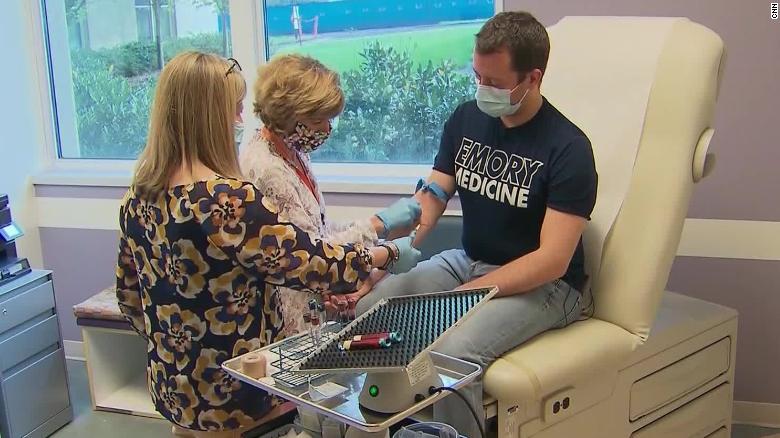








- Collaborative study of BioNTech with TRON and the University Medical Center and Research Center for Immunotherapy at Johannes Gutenberg University of Mainz introduces novel non-inflammatory mRNA vaccine encoding disease-related autoantigens that suppressed disease activity in several complex mouse models of multiple sclerosis
- Approach addresses key pitfalls in the treatment of autoimmune diseases such as the induction of systemic immune suppression
- Approach can easily be tailored to individual disease-causing antigens of patients and confers bystander tolerance to address highly complex, polyclonal and rare autoimmune disease types
- Represents the first application of BioNTech’s mRNA technology for the purpose of antigen-specific immune-modulation of autoimmune diseases, which further expands BioNTech’s diversified immunology pipeline into another category of disease relevant targets
MAINZ, GERMANY, January 7, 2021 (GLOBE NEWSWIRE) — BioNTech SE (Nasdaq: BNTX, “BioNTech” or “the Company”) announced today the publication of preclinical data on its novel mRNA vaccine approach against autoimmune diseases in the peer-reviewed journal Science. The publication titled “A non-inflammatory mRNA vaccine for treatment of experimental autoimmune encephalomyelitis” summarizes the findings on the disease-suppressing effects of a non-inflammatory, nucleoside-modified mRNA vaccine in several clinically relevant mouse models of multiple sclerosis (MS).
Autoimmune diseases like MS represent conditions in which the immune system malfunctions and attacks healthy tissue or cells of the body. In MS, the inflammation causes the destruction of the protective myelin sheath that covers the nerve fibers. This damage disrupts the ability to transmit signals between nerve cells and the target tissue resulting in a range of neurological, sensory and motor symptoms that may differ greatly between individuals.
This first application of BioNTech’s mRNA technology in MSs represents a new modality in this indication and underlines BioNTech’s potential to leverage its proprietary mRNA platform.
In the study, a non-inflammatory nanoparticulate mRNA vaccine candidate encoding a MS-associated antigen was systemically applied to mice with experimental autoimmune encephalomyelitis (EAE), which represent clinically relevant mouse models of human MS. The mRNA vaccine candidate was designed to deliver the encoded autoimmune disease target antigen into antigen-presenting cells in the lymph nodes body-wide in a non-inflammatory context to enable systemic, immune tolerance-inducing antigen presentation in lymphoid tissues.
In all investigated EAE mouse models, the vaccine was able to prevent symptomatic disease or, in mice with early-stage disease, reduced further disease progression and restored motor functions. Pro-inflammatory effector T (Teff) cell infiltration in the brain and spinal cord and demyelination of the spinal cord was considerably reduced. These effects were achieved via development of disease-suppressing regulatory T (Treg) cells directed exquisitely against the antigen encoded by the mRNA vaccine. The Treg cells also executed a strong immunosuppressive bystander effect in the different MS mouse models, demonstrating that the Treg cells, once activated by their target antigen, can also suppress Teff cells against other antigens in the inflamed tissue in a complex disease setting. This is a crucial factor to also address polyclonal diseases based on multiple, partly unknown antigens, as well as inter-individual heterogeneity between patients.
Importantly, the preclinical vaccine candidate did not suppress functional immune responses against other, non-myelin antigens (e.g. influenza vaccine antigens), therefore addressing one of the key challenges in autoimmune treatment in the preclinical studies, the induction of an unspecific, systemic immune suppression. In addition, the vaccine candidate, even after repetitive application, did not induce formation of autoantibodies against the targeted antigen, another potential pitfall in current autoimmune therapies that could exacerbate disease. Overall, these initial results regarding the immune response together with the flexibility of the mRNA approach to target individual patient antigens indicate the potential of mRNA therapeutics to address highly complex and rare autoimmune disease indications.
The publication represents results of a collaborative study of scientists from BioNTech, TRON – Translational Oncology at the University Medical Center of the Johannes Gutenberg University Mainz, the Institute for Molecular Medicine at the University Medical Center of the Johannes Gutenberg University Mainz and the Research Center for Immunotherapy (FZI) at the Johannes Gutenberg University Mainz.
About BioNTech
Biopharmaceutical New Technologies is a next generation immunotherapy company pioneering novel therapies for cancer and other serious diseases. The Company exploits a wide array of computational discovery and therapeutic drug platforms for the rapid development of novel biopharmaceuticals. Its broad portfolio of oncology product candidates includes individualized and off-the-shelf mRNA-based therapies, innovative chimeric antigen receptor T cells, bi-specific checkpoint immuno-modulators, targeted cancer antibodies and small molecules. Based on its deep expertise in mRNA vaccine development and in-house manufacturing capabilities, BioNTech and its collaborators are developing multiple mRNA vaccine candidates for a range of infectious diseases alongside its diverse oncology pipeline. BioNTech has established a broad set of relationships with multiple global pharmaceutical collaborators, including Genmab, Sanofi, Bayer Animal Health, Genentech, a member of the Roche Group, Regeneron, Genevant, Fosun Pharma, and Pfizer. For more information, please visit www.BioNTech.de.
BioNTech Forward-looking Statements
This press release contains “forward-looking statements” of BioNTech within the meaning of the Private Securities Litigation Reform Act of 1995, as amended, including, but not limited to: statements concerning the applicability of BioNTech’s mRNA technology in autoimmune diseases. In some cases, forward-looking statements can be identified by terminology such as “will,” “may,” “should,” “expects,” “intends,” “plans,” “aims,” “anticipates,” “believes,” “estimates,” “predicts,” “potential,” “continue,” or the negative of these terms or other comparable terminology, although not all forward-looking statements contain these words. The forward-looking statements in this press release are neither promises nor guarantees, and you should not place undue reliance on these forward-looking statements because they involve known and unknown risks, uncertainties, and other factors, many of which are beyond BioNTech’s control and which could cause actual results to differ materially from those expressed or implied by these forward-looking statements. Any forward-looking statements in this press release are based on BioNTech current expectations and beliefs of future events, and are subject to a number of risks and uncertainties that could cause actual results to differ materially and adversely from those set forth in or implied by such forward-looking statements. The forward-looking statements in this press release are neither promises nor guarantees, and you should not place undue reliance on these forward-looking statements because they involve known and unknown risks, uncertainties, and other factors, many of which are beyond BioNTech’s control and which could cause actual results to differ materially from those expressed or implied by these forward-looking statements.
For a discussion of these risks and uncertainties, see BioNTech’s Quarterly Report for the Three and Nine Months Ended September 30, 2020, filed as Exhibit 99.2 to its Current Report on Form 6-K filed with the SEC on November 10, which is available on the SEC’s website at www.sec.gov. All information in this press release is as of the date of the release, and BioNTech undertakes no duty to update this information unless required by law.
BioNTech Contacts:
Media Relations
Jasmina Alatovic
+49 89 62 81 75 46
Media@biontech.de
Investor Relations
Sylke Maas, Ph.D.
+49 (0)6131 9084 1074
Investors@biontech.de
University Medical Center Mainz Contact
Stabsstelle Unternehmenskommunikation
+49 06131 17 7427
pr@unimedizin-mainz.de
No comments:
Post a Comment
Note: Only a member of this blog may post a comment.