
It looks like the answer is YES.
If you aren't already aware of how incredibly effective Ivermectin is, just CLICK HERE
- Article
- Open Access
- Published:
Artemisinin inhibits neutrophil and macrophage chemotaxis, cytokine production and NET release
Scientific Reports 12, Article number: 11078 (2022)
Abstract
Immune cell chemotaxis to the sites of pathogen invasion is critical for fighting infection, but in life-threatening conditions such as sepsis and Covid-19, excess activation of the innate immune system is thought to cause a damaging invasion of immune cells into tissues and a consequent excessive release of cytokines, chemokines and neutrophil extracellular traps (NETs). In these circumstances, tempering excessive activation of the innate immune system may, paradoxically, promote recovery. Here we identify the antimalarial compound artemisinin as a potent and selective inhibitor of neutrophil and macrophage chemotaxis induced by a range of chemotactic agents. Artemisinin released calcium from intracellular stores in a similar way to thapsigargin, a known inhibitor of the Sarco/Endoplasmic Reticulum Calcium ATPase pump (SERCA), but unlike thapsigargin, artemisinin blocks only the SERCA3 isoform. Inhibition of SERCA3 by artemisinin was irreversible and was inhibited by iron chelation, suggesting iron-catalysed alkylation of a specific cysteine residue in SERCA3 as the mechanism by which artemisinin inhibits neutrophil motility. In murine infection models, artemisinin potently suppressed neutrophil invasion into both peritoneum and lung in vivo and inhibited the release of cytokines/chemokines and NETs. This work suggests that artemisinin may have value as a therapy in conditions such as sepsis and Covid-19 in which over-activation of the innate immune system causes tissue injury that can lead to death.
Introduction
An important cause of the deterioration leading to death in Covid-19 patients is thought to be an excessive release of pro-inflammatory cytokines from innate immune cells, which precipitates the severe lung condition named acute respiratory distress syndrome (ARDS)1. Other organs are also often affected in Covid-19, in a syndrome more akin to systemic sepsis2. ARDS in Covid-19 may therefore be attributable to overactivation of the innate immune system, leading to (i) neutrophil invasion of the lung3; (ii) excess release of pro-inflammatory cytokines1,2; and (iii) release of Neutrophil Extracellular Traps (NETs), sticky aggregations of DNA, histones and other proteins released from neutrophils4. In more localised infections, pro-inflammatory cytokines and chemokines perform a beneficial role, as chemoattractants that recruit immune cells to join the attack on invading pathogens, while NETs play a vital role in the physical snaring and immobilisation of pathogens. In system-wide sepsis, ARDS or Covid-19, however, excess cytokine, chemokine and NET release are thought to be critical in the events leading to clinical deterioration, organ dysfunction and death1,2,3,5.
In the present study we identified the natural compound artemisinin, which has achieved significant success as a front-line antimalarial, as a potent inhibitor of immune cell chemotaxis induced by a wide variety of chemoattractant molecules. We show that artemisinin suppresses neutrophil invasion in vivo and also successfully inhibits cytokine/chemokine secretion and NET release caused by a range of pro-inflammatory agents, including hydrogen peroxide (H2O2), bacterial lipopolysaccharide (LPS) and the SARS-CoV-2 spike protein. In view of the favourable clinical profile of artemisinin and its analogues, we suggest that these compounds may be useful as therapies in conditions such as systemic sepsis, ARDS and Covid-19.
Results
Artemisinin is a potent inhibitor of neutrophil and macrophage chemotaxis
Hydrogen peroxide (H2O2) is known to act as a potent immune cell chemoattractant6,7. In previous work we have shown that the TRPM2 ion channel, which is activated by H2O2, mediates the chemotactic action of H2O2 by preferentially inducing a calcium influx at the neutrophil leading edge8. We initially searched for inhibitors of neutrophil chemotaxis by screening a natural compound library, using neutrophil chemotaxis towards H2O2 as the assay. Figure 1A shows the forward migration index (FMI), the ratio of linear distance travelled in the direction of the H2O2 gradient to the total distance travelled, which gives an index of the directionality of cell movement. Interestingly, capsaicin, a TRPV1 agonist9 and eugenol, a TRPV3 agonist10 both significantly potentiated directional chemotaxis, perhaps because they have a weak agonist action at TRPM2. Of the compounds that caused an inhibition of chemotaxis, five were identified as interesting for further investigation (red boxes in Fig. 1A), based on a significant reduction in FMI together with a significant reduction in average speed of migration (Supplementary Fig. 1A). The dose–response relations of four of these compounds, beta-carotene, curcumin, ferulic acid and N-acetylcysteine, were similar, suggesting a common action, while artemisinin was more potent (Supplementary Fig. 1B). The four compounds showing a similar potency are antioxidants, so we investigated the possibility that they may act indirectly by dissipating the gradient of H2O2. To test this idea, we used a gradient of adenosine diphosphate ribose (ADPR), which like H2O2 is a potent neutrophil chemoattractant8,11. ADPR directly activates TRPM2 at an intracellular location12,13,14, while H2O2 does not directly activate TRPM2 but acts by increasing intracellular levels of ADPR13,14. When a gradient of ADPR was used to activate neutrophil chemotaxis none of the four antioxidant compounds was able to inhibit chemotaxis (Fig. 1B), showing that their action was indeed to dissipate the H2O2 gradient rather than to directly inhibit chemotaxis. Artemisinin, on the other hand, inhibited neutrophil chemotaxis towards both ADPR and H2O2 (Fig. 1B,C, respectively), demonstrating that its action in abolishing chemotaxis is independent of any effect on the gradient of H2O2. We next compared the ability of artemisinin to inhibit neutrophil chemotaxis with that of other well-established antimalarial compounds. Artemisinin was the only antimalarial that inhibited neutrophil migration (Fig. 1C) and therefore has a unique mechanism of action. We found that artemisinin has no effect on neutrophil viability, ruling out the possibility of a toxic action of artemisinin as a basis for its inhibition of neutrophil chemotaxis (Supplementary Fig. 2). The SARS-CoV-2 spike protein was also a potent chemoattractant for neutrophils in our in vitro assay, and artemisinin also strongly inhibited chemotaxis up a gradient of SARS-CoV-2 (Fig. 1D).

Artemisinin and its analogues are potent inhibitors of neutrophil chemotaxis. (A) Neutrophils migrated up a gradient of 10 nM H2O2 over 1 mm (see Methods). Forward migration index (FMI, vertical axis), the mean ratio of distance travelled in the direction of the chemoattractant gradient to total distance travelled, is a measure of chemoattraction (see details in ref8). Thirty-one compounds from a natural compound library were tested at 10 µM. Five compounds (red boxes) were selected on the basis that they caused the greatest inhibition of FMI, together with the greatest reduction in speed of movement (Supplementary Fig. 1A). Each bar shows mean ± SEM from n = 3 mice. Statistical analysis: For comparison with Control: **p < 0.01, ***p < 0.001, ****p < 0.0001 (One-way ANOVA and Tukey–Kramer post-hoc test). (B) Out of the five compounds inhibiting chemoattraction towards H2O2, only artemisinin inhibited migration up a gradient of ADPR, a direct activator of TRPM2. All neutrophils in each experiment from same batch; FMI in absence of inhibitor is consistent within batches but maximum value varies somewhat between batches. Each bar shows mean ± SEM from n = 3 mice. Statistical analysis: Comparison with ADPR alone: ***p < 0.001, ns = not significant. (One-way ANOVA and Tukey–Kramer post-hoc test). (C) Artemisinin (10 μM) completely inhibits neutrophil migration up a 10 nM gradient of H2O2 (FMI not significantly different from that in the absence of H2O2 gradient), while antimalarials pyrimethamine, hydroxychloroquine, mefluoquine and lumefantrine (all 10 μM) have no inhibitory effect. Each bar shows mean ± SEM from n = 3 mice. Comparison with H2O2: ***p < 0.001, ns = not significant. (One-way ANOVA and Tukey–Kramer post-hoc test). (D) Covid spike protein (SARS-CoV-2, 100 nM) is a potent neutrophil chemoattractant and chemoattraction is inhibited by artesunate (10 μM). Comparison with DMEM: ***p < 0.001, comparison with CoV + artesunate: ##p < 0.01 (One-way ANOVA and Tukey–Kramer post-hoc test). (E) Dose–response relation for inhibition of neutrophil FMI up a gradient of 10 nM H2O2 by artemisinin and its analogue artesunate. Dotted lines show mean value of FMI in gradient of H2O2 (upper) or in no chemoattractant (DMEM, lower). IC50 values: artemisinin, 0.36 nM; artesunate, 0.37 nM. No inhibition observed in presence of Fe2+ chelator desferrioxamine (DesF, 50 μM). Each point shows mean ± SEM from n = 3 mice. FMI in artemisinin /artesunate + DesF is significantly lower than artemisinin/artesunate values at all concentrations > 0.1 nM (p < 0.001). (F) Similar experiment to A, carried out with neutrophils in gradient of chemokine CXCL2 (10 nM). IC50 values: artesunate, 0.34 nM; artemisinin, 0.31 nM. Effect of artemisinin and artesunate completely abolished by desferrioxamine (50 μM). Each point shows mean ± SEM from n = 3 mice. FMI in artemisinin + DesF/artesunate + DesF is significantly lower than artemisinin/artesunate values at all concentrations > 0.1 nM (p < 0.001).
Figure 1E shows that artemisinin and artesunate (an artemisinin analogue) are both highly potent inhibitors of neutrophil chemotaxis driven by H2O2, with IC50 ≈ 0.3 nM. Artemisinin and artesunate also strongly inhibit chemotaxis towards a diverse range of other chemotactic signals, including the chemokine CXCL2 (Fig. 1E), the complement factor C5a and the bacterial cell wall component lipopolysaccharide (LPS) (Supplementary Fig. 3). In each case, the values of IC50 for artemisinin and artesunate were close to 0.3 nM, with none being significantly different from the value obtained for chemotaxis towards H2O2. Supplementary Fig. 4 shows that macrophage chemotaxis was also potently inhibited by artemisinin and artesunate, in a similar way to the effects of these compounds on neutrophils. This work identifies artemisinin as a potent inhibitor of neutrophil and macrophage chemotaxis driven by a wide variety of chemoattractant agents.
Mechanism of inhibition of chemotaxis by artemisinin
A number of active analogues of artemisinin have been developed for use as antimalarials, including arteether, artemether and artesunate (structures shown in Supplementary Fig. 5). All are rapidly metabolised in vivo to dihydroartemisinin (DHA), a more metabolically stable analogue with a longer in vivo half-life (~ 1.3 h) than any of its precursors15,16. All of these analogues, including the stable metabolite DHA, showed an equally high potency in inhibiting neutrophil chemotaxis towards a range of chemotactic signals (Fig. 1E,F and Supplementary Fig. 3; IC50 ≈ 0.3 nM for all analogues). These experiments show that none of the chemical modifications in these artemisinin analogues impacts on a site critical for the inhibitory action of artemisinin on chemotaxis.
An unusual feature of artemisinin is the endoperoxide 1,2,4-trioxane ring (top left in Supplementary Fig. 5). We found that deoxyartemisinin, which lacks the peroxide bridge but is otherwise identical to artemisinin, is completely inactive in inhibiting neutrophil chemotaxis (Fig. 2A), showing that the presence of the peroxide bridge is essential for the action of artemisinin on chemotaxis. The critical role of the peroxide suggests that artemisinin may inhibit its protein target by oxidation. It has been known for many years that hydrogen peroxide can oxidise the sulfhydryl group in cysteine, and that this reaction depends on free ferrous iron17. We therefore investigated whether the action of artemisinin on neutrophil chemotaxis also depends on iron. Removing ferrous iron with the specific chelator desferrioxamine completely abrogated the ability of both artemisinin and artesunate to inhibit neutrophil chemotaxis at all concentrations (Fig. 1E,F). Antagonism by desferrioxamine of the inhibition of chemotaxis by artemisinin was independent of whether H2O2, a chemokine, C5a or lipopolysaccharide (LPS) were used as the chemoattractant (Supplementary Fig. 6). These observations suggest that artemisinin and its active derivatives may inhibit their protein target not by reversible antagonist binding, as has previously been supposed18,19, but instead by covalent modification of a cysteine residue, catalysed by Fe2+. Artemisinin and its analogues have been shown to be capable of alkylating both cysteine itself20 and the central cysteine residue in a cysteine-containing tripeptide, glutathione21, by oxidising and combining with the cysteine sulfhydryl (Supplementary Fig. 5B, C).

Mechanism of inhibition of neutrophil chemotaxis by artemisinin and analogues. (A) Inhibition of neutrophil chemotaxis by artemisinin (10 μM, second column) is completely abolished by removal of the peroxide bond in artemisinin (deoxyartemisinin, 10 μM, third column). Final column shows FMI in absence of gradient of H2O2. Each bar shows mean ± SEM from n = 3 mice. Statistical analysis: For comparison with H2O2: **, p < 0.01; ns = not significant. (One-way ANOVA and Tukey–Kramer post-hoc test). (B) Artemisinin does not block TRPM2 ion channels. Patch clamp recording of membrane current from TRPM2-transfected HEK293 cell at + 80 mV (orange) and − 80 mV (blue); TRPM2 ion channels activated by the inclusion of 1 mM ADPR in the intracellular patch clamp solution. Moment of breaking through to whole-cell mode shown by arrow. Artemisinin (10 μM) has no effect on membrane current (fractional current change 0.99 ± 0.04 at − 80 mV, 0.97 ± 0.03 at + 80 mV, neither significantly different from 1.0, n = 6), while the known TRPM2 inhibitor N-(p-amylcinnamoyl) anthranilic acid (ACA, 20 μM) suppresses membrane current at both membrane voltages (fractional current change 0.02 ± 0.01 at − 80 mV, 0.06 ± 0.01 at + 80 mV, both significantly different from 1.0, p < 0.0001, n = 6). (C) Neutrophil forward migration index (FMI) in a gradient of H2O2 (10 nM, bar 2) and CXCL2 (10 nM, bar 5) is abolished by artesunate (10 μM) and by the selective SERCA inhibitor thapsigargin (50 nM). Each bar shows mean ± SEM from n = 4 experiments with neutrophils from 4 mice. Statistics: ***, p < 0.001 compared to DMEM control; ####, p < 0.0001 compared to H2O2 or CXCL2. 0.001, one-way ANOVA and Tukey–Kramer post-hoc test. (D) Application of SERCA inhibitor thapsigargin (black trace, 50 nM) to a neutrophil releases calcium from intracellular stores (ratio measurement with fura-2, see Methods). Calcium influx from external medium prevented with 0Ca2+/2 mM EGTA (application time shown by bar at top). Similar dose-dependent release of intracellular store calcium seen with artemisinin (pink, 10 μM; green, 100 nM; blue, 1 nM) showing that artemisinin is a SERCA inhibitor. Increase of calcium on readmission of external Ca2+ is due to activation of store-operated calcium entry (SOCE) following store discharge and is similar in all cases, showing that artemisinin does not affect SOCE. Calcium release by artemisinin (10 μM) is inhibited by Fe2+ chelator desferrioxamine (DesF, light pink, 50 μM), but DesF has no effect on calcium release by thapsigargin (brown, 50 nM). Grey trace: no application of thapsigargin or artemisinin. Left axis: fura2 fluorescence ratio; right axis: [Ca2+]i determined as in Methods. Example traces shown in D–I are typical of n = 7–12 cells imaged per cover slip, each condition repeated on 3 cover slips. (E) Similar experiment performed on HEK293 cell transfected with rSERCA1a. Calcium is released by thapsigargin (black, 1 M) but not by artemisinin (pink, 10 μM). Other conditions as in D. (G) Similar experiment with overexpressed hSERCA2a. (G) Similar experiment with overexpressed hSERCA3. In contrast to SERCA1 and 2, artemisinin (pink, 10 μM) gives a similar calcium release to thapsigargin (black, 1 M). Effect of artemisinin (10 μM) is completely inhibited by chelation of Fe2+ with desferrioxamine (light pink, 50 μM), while calcium release by thapsigargin is unaffected by desferrioxamine (brown, 50 μM). (H) Similar experiment on an untransfected HEK293 cell. Calcium stores are released by thapsigargin (black, 50 nM) but not by artemisinin (pink, 10 μM), showing that HEK293 cells, unlike neutrophils, do not express SERCA3. (I) Reversibility of inhibition of SERCA3 in neutrophils by thapsigargin but not artemisinin. Experimental protocol shown in D repeated twice with 21 min gap between applications. SOCE following 1 nM thapsigargin recovered and on readmission of 1 nM thapsigargin a second release of calcium from subcellular stores was observed. By contrast, SOCE following application of artemisinin (1 nM) diminished little and only a very small increase of calcium was observed on reapplication, showing that artemisinin is largely irreversible on the time scale tested here.
SERCA is the cellular target of artemisinin
Neutrophil chemotaxis depends on the ability of chemoattractants to generate leading-edge calcium “pulses” that determine the direction of cell migration8. Supplementary Video 1 shows the generation of calcium pulses in a neutrophil migrating up a gradient of H2O2 (left-hand video), and the complete suppression of calcium pulses, together with chemotaxis, in the presence of artemisinin (second-left video). In the presence of a gradient of ADPR, calcium pulses drive chemotaxis in a similar way to H2O2, and artemisinin also inhibits both calcium pulses and chemotaxis (pair of videos on right). These experiments suggest that artemisinin prevents chemotaxis by inhibiting the generation of leading-edge calcium pulses.
In previous work we showed that chemotaxis driven by H2O2 depends on activation of the TRPM2 ion channel8. The importance of a calcium influx via TRPM2 for chemotaxis driven by H2O2 is shown in Supplementary Fig. 7. In this experiment, neutrophils were loaded with the calcium chelator BAPTA, which completely suppressed the intracellular calcium increase caused by activation of TRPM2 by H2O2 (Supplementary Fig. 7A). In the absence of this TRPM2-mediated calcium increase, neutrophil chemotaxis towards H2O2 was abolished (Supplementary Fig. 7B).
We next carried out patch-clamp experiments on TRPM2 heterologously expressed in HEK293 cells in order to test whether artemisinin might inhibit chemotaxis by blocking TRPM2. TRPM2 was activated by alternate positive and negative voltage pulses (Fig. 2B). Artemisinin had no significant effect on the current carried by TRPM2, in contrast to the known TRPM2 blocker ACA, which caused prompt and near-complete current inhibition.
A second reason for discarding TRPM2 as a target is that artemisinin inhibits, with equal potency, chemotaxis towards H2O2, ADPR (Fig. 1B,C) and a range of other chemoattractants (Fig. 1E,F and Supplementary Fig. 3). Chemotaxis activated by H2O2 and ADPR depends on activation of TRPM28, but chemotaxis activated by cyto/chemokines depends on a separate pathway not involving TRPM28. The schematic diagram in Supplementary Fig. 12 (steps 1–4) shows how H2O2 activates calcium influx through TRPM2, which in turn generates leading-edge calcium “pulses” that steer chemotaxis8. Leading-edge calcium pulses generated by cyto/chemokines and chemoattractants such as LPS8, on the other hand, depend on a separate pathway independent of TRPM2 (Supplementary Fig. 12, steps 6, 7). The ability of artemisinin to inhibit chemotaxis activated by each of these two distinct pathways implies that the action of artemisinin must be at a point common to both pathways, such as the sarcoplasmic and endoplasmic reticulum calcium ATPase (SERCA) that is responsible for refilling subcellular calcium stores, or the store-operated calcium entry mechanism (SOCE), that mediates calcium entry and store refilling following discharge of subcellular stores22. Both SERCA and SOCE have been shown to be functional in neutrophils23.
Thapsigargin, a potent and selective SERCA blocker24, completely inhibited neutrophil chemotaxis towards both H2O2 and the chemokine CXCL2, in a similar way to the inhibition caused by artemisinin (Fig. 2C), consistent with the idea that both thapsigargin and artemisinin exhaust the internal calcium stores that are necessary to drive chemotaxis8. Thapsigargin evoked an increase in internal calcium concentration in neutrophils in the complete absence of external calcium (Fig. 2D, black trace), that must be due to release from internal stores because no calcium influx across the surface membrane is possible. The calcium release was followed by a return to baseline levels as cytoplasmic calcium was extruded by surface membrane calcium pumps. When intracellular calcium stores had been exhausted, readmission of external calcium caused a sustained calcium increase, attributable to store-operated calcium entry (SOCE) carried via activation of calcium-selective Orai channels in the surface membrane25. The protocol shown in Fig. 2D therefore shows a way of separating a potential inhibitory action of artemisinin on SERCA and on SOCE.
Artemisinin evoked a dose-dependent increase in neutrophil intracellular calcium similar to that seen with thapsigargin (Fig. 2D), showing that artemisinin, like thapsigargin, acts to release calcium from intracellular stores of neutrophils and therefore may be a SERCA inhibitor. Artemisinin was effective down to a concentration of 1 nM in releasing calcium from intracellular stores, consistent with the high potency of artemisinin in inhibiting neutrophil chemotaxis (IC50 ≈ 0.3 nM, Fig. 1E,F). The intracellular calcium release evoked by thapsigargin was unaffected by the Fe2+ chelator desferrioxamine, but calcium release by artemisinin was completely suppressed (Fig. 2D), results that echo the effect of Fe2+ chelation on chemotaxis (Fig. 1E,F). At all concentrations of artemisinin, the profile of SOCE following readmission of calcium was similar to that caused by thapsigargin, showing that artemisinin does not interact with SOCE. These experiments are consistent with SERCA being the downstream target of artemisinin in neutrophils.
Artemisinin irreversibly inhibits SERCA3
Thapsigargin is toxic to mammals26, while artemisinin has an excellent clinical safety record as an antimalarial, a difference that could arise from selective inhibition by artemisinin of a non-critical mammalian SERCA isoform, in contrast to the known ability of thapsigargin to inhibit all three SERCA isoforms equally24. SERCA1 is critical for muscle contraction, while SERCA2 is widely expressed in many essential organs27. Inhibition of either isoform would therefore be likely to cause significant toxicity. SERCA3, on the other hand, has a more limited expression pattern, which includes expression in immune cells27. These considerations suggest that SERCA3 may be the target of artemisinin in neutrophils.
In Fig. 2E–G we overexpressed mammalian SERCA1, 2 or 3 in HEK293 cells and then used the protocol shown in Fig. 2D to test for SERCA inhibition by thapsigargin or artemisinin. Thapsigargin released calcium from intracellular stores with a similar time course when applied to all SERCA isoforms, consistent with its ability to inhibit all isoforms equally24. Artemisinin, on the other hand, was inactive on cells transfected with SERCA1 and 2 (Fig. 2E,F) but released calcium with a similar time course to thapsigargin in cells transfected with SERCA3 (Fig. 2G). Removal of Fe2+ with desferrioxamine did not affect the ability of thapsigargin to inhibit SERCA3, but completely prevented inhibition of SERCA3 by artemisinin (Fig. 2G). Thapsigargin released calcium from intracellular stores of naïve HEK293 cells but artemisinin did not (Fig. 2H), consistent with expression of SERCA2 in HEK293 cells, that are derived from the kidney, where SERCA2 is the principal isoform27. As shown in Fig. 2D, artemisinin releases calcium from intracellular stores of neutrophils, consistent with the known expression of SERCA3 in cells of the immune system27. These experiments show that SERCA3 is the mammalian target of artemisinin.
Thapsigargin inhibits SERCA isoforms by binding reversibly to a location between membrane-spanning helices 3 and 7, deep within the motile machinery of the calcium pump28. The experiments above show that inhibition of SERCA3 by artemisinin depends, on the other hand, on its unusual peroxide bond, not present in thapsigargin, together with the presence of Fe2+ as a probable catalyst, suggesting a different mechanism involving irreversible covalent binding, likely to a cysteine residue. In Fig. 2I we used the protocol shown in Fig. 2D to compare the reversibility of SERCA inhibition by thapsigargin and artemisinin. Following exhaustion of calcium stores by thapsigargin, and consequent calcium influx via SOCE on readmission of external calcium, the intracellular calcium level returned slowly to its normal level over the 20 min following removal of thapsigargin, showing that SERCA had reactivated and intracellular stores had refilled, thus switching off SOCE. Readmission of thapsigargin again released calcium from intracellular stores, followed by reactivation of SOCE when extracellular calcium was readmitted, confirming the reversibility of thapsigargin binding to SERCA. However, when the same experiment was repeated using artemisinin, elevated calcium levels due to activation of SOCE persisted after store discharge, showing that stores had not refilled and that SERCA3 inhibition had therefore been maintained. On reapplying artemisinin in zero calcium, very little calcium release was observed, consistent with the lack of store refilling (Fig. 2I). This experiment confirms that inhibition of SERCA3 by artemisinin is essentially irreversible on the time scale used, in contrast to the reversible inhibition by thapsigargin.
Artemisinin and analogues suppress in vivo neutrophil invasion in response to H2O2
The potent action of artemisinin and its analogues in suppressing neutrophil chemotaxis in vitro suggests that these compounds may have a similar action in vivo, and therefore may potentially be useful as therapeutics in conditions such as ARDS and Covid-19 where excess immune cell invasion is an important driver of the pathology. We measured neutrophil invasion into mouse peritoneum following intraperitoneal injection of 10 μM H2O2, a concentration that we have found in previous work to have a maximal effect in activating neutrophil chemotaxis in vitro8. The time course of neutrophil invasion in response to i.p. H2O2 is shown in Fig. 3A. In this experiment total cell counts are shown; the background level of c. 2 × 10⁶ cells (lower dotted line) is attributable to the presence of tissue-resident macrophages8. Following injection of H2O2, neutrophil invasion causes the cell count to rise rapidly, reaching a peak of 6.5 × 10⁶ cells at 60 min, a level that is maintained until 120 min, followed by a return to baseline over the a further 90 min. Injection of artesunate s.c. 30 min prior to injection of H2O2 largely suppressed the neutrophil invasion up to 120 min, at which time the effect diminishes owing to the short in vivo lifetime of artesunate and its active metabolite dihydroartemisinin15,16. In agreement, Supplementary Fig. 8 shows that 10 μM H2O2 i.p. strongly activated an influx of neutrophils, and that neutrophil invasion was largely suppressed by injections of either artemisinin or artesunate at 28 mg/kg s.c., 30 min prior to injection of H2O2, with a slightly lesser effect at 6 mg/kg, a dose close to a typical clinically-used dose for artesunate of 2.4 mg/kg i.v. The similar in vivo inhibition by artemisinin and artesunate mirrors the similar actions of these two analogues in inhibiting neutrophil chemotaxis in vitro (Fig. 1E,F).

Artesunate suppresses neutrophil invasion and release of cytokines and NETs in response to intraperitoneal infusion of H2O2. (A) Number of cells recovered from intraperitoneal lavage following i.p. injection of H2O2 (10 µM in PBS, 10 μl/gm body weight). H2O2 causes a large influx of neutrophils that peaks at 60 min and reverses by 210 min (black points). Cells present in peritoneum before injection of H2O2 are tissue-resident macrophages, while cells entering the peritoneum following injection of H2O2 are neutrophils (see Methods for cell identification). Artesunate (6 mg/kg s.c., delivered 30 min before injection of H2O2) suppresses neutrophil influx for > 120 min (red points). Each point shows mean ± SEM from n = 4 mice. (B) Concentration of IL-1β in peritoneal lavage measured by ELISA, using i.p. lavage samples obtained as in A. Black bars show increase as function of time (mins) in vehicle-injected mice; open bars are corresponding data for mice injected with artesunate 6 mg/kg s.c., 30 min before injection of H2O2 as in A. Each bar shows mean ± SEM from n = 3 mice. (C,D,E) Similar data for IL-6, CXCL1 and CXCL2, obtained from same samples. (F) Similar data for release of NETs, quantified using Pico-Green kit. Statistical analysis: BLQ, below limit of quantitation; *, p < 0.05; **, P < 0.01; ***, p < 0.001, ****, p < 0.0001 compared with negative control (no H2O2); #, p < 0.05; ##, p < 0.01; ###, p < 0.001; ####p < 0.0001 artesunate group compared with no-artesunate group at same time point. ANOVA with Bonferroni post-hoc correction.
Excess release of cytokines/chemokines is thought to be critical in the pathology of conditions such as Covid-19 in which immune cell invasion plays an important role1,2. In Fig. 3B–E we used ELISA to measure the concentration of two pro-inflammatory cytokines, IL-1β and IL-6, and two chemokines, CXCL1 and CXCL2. In each case, the profile of increase following injection of H2O2 is similar to the profile of neutrophil invasion, rising from a low level to a broad peak at 60–120 min, followed by a return to undetectable levels by 210 min, a time at which the level of invading neutrophils had declined back to baseline. The suppression caused by prior injection of artesunate is striking, with the cytokine/chemokine increase near-completely abolished in all cases up to 120 min. An increase is seen at 150 min, in line with the recovery of neutrophil chemotaxis as the effect of artesunate wears off (Fig. 3A).
The release of neutrophil extracellular traps (NETs) from neutrophils may also augment the damaging effect of cytokines3,5,29. In Fig. 3F we examined the release of NETs by assaying cell-free DNA release. The profile is broadly similar to the release of cytokines; NET release shows a broad peak at 30–150 min, followed by a decline to low levels by 210 min as neutrophil invasion reverses. Artesunate completely inhibits NET release at times earlier than 150 min. An alternative assay of NET release using fluorescence microscopy showed a similar increase in NETs in response to in vitro application of LPS, also abolished by artemisinin (Supplementary Fig. 9).
A similar experiment carried out with infusion of H2O2 into the lung shows that neutrophil invasion, cytokine and chemokine release and NET release are all strongly suppressed by artesunate, as was found in the peritoneum (Supplementary Fig. 10). In summary, the release of cyto/chemokines and NETs in response to H2O2 parallels neutrophil invasion in both peritoneum and lung, and the ability of artesunate to inhibit neutrophil invasion has a striking effect in preventing the release of proinflammatory cytokines/chemokines and NETs.
Artemisinin and analogues suppress in vivo neutrophil invasion in response to LPS and SARS-CoV-2 spike protein
Lipopolysaccharide (LPS), a constituent of the cell wall of gram-negative bacteria, plays a critical role in the interactions of many bacterial pathogens with the innate immune system30. LPS is a potent neutrophil chemoattractant in vitro, inducing chemotaxis that is steered by leading-edge calcium pulses8. The pathway by which LPS induces neutrophil chemotaxis is different from that activated by H2O2, however, because genetic deletion or pharmacological block of TRPM2 does not suppress chemotaxis towards LPS8 (see pathway diagram in Supplementary Fig. 12). In the experiment shown in Supplementary Fig. 11, we tested the ability of LPS to induce invasion of neutrophils into the peritoneum and the effect of artemisinin on this invasion. Neutrophil invasion into the peritoneum in response to LPS was activated more slowly than that induced by H2O2, so we sampled invasion at 5 h, and gave three doses of artemisinin s.c. at intervals of 2 h to maintain systemic levels of artemisinin throughout this time. LPS activated a neutrophil invasion that was similar in magnitude to that induced by H2O2, and the invasion was also largely suppressed by artemisinin (Supplementary Fig. 11A). The production of cytokines IL1-β and IL-6 and chemokines CXCL1 and CXCL2 was also strongly suppressed by artemisinin (Supplementary Fig. 11B-E), as was NET release (Supplementary Fig. 11F).
The invasion of neutrophils into the lung has been proposed to be critical for the pathogenesis of Covid-191,2,3,5. We therefore tested whether artemisinin and its analogues are effective in suppressing neutrophil invasion into the lung, and what effect these treatments have on cytokine/chemokine and NET release. Figure 4A shows that lung neutrophil invasion in response to LPS was strongly suppressed by artesunate at both 28 mg/kg and 6 mg/kg, the latter dose being close to the clinically used dose of 2.4 mg/kg. Production of the pro-inflammatory cytokines IL1-β and IL-6, and chemokines CXCL1 and CXCL2, was strongly suppressed (Fig. 4B–E). In addition the release of NETs, as assayed from DNA release, was also inhibited (Fig. 4F).

Artesunate suppresses neutrophil invasion, release of cytokines and release of NETs in response to infusion of LPS into lung. (A) Number of neutrophils recovered from broncho-alveolar lavage fluid (BALF), 5 h after infusion of LPS (300 ng each lung). LPS causes an influx of neutrophils that is suppressed by artesunate, delivered s.c. at 30 min before and at 90 and 210 min after injection of LPS. Each bar shows mean ± SEM from n = 4 mice. (B–F) Concentrations of IL-1β, IL-6, CXCL1, CXCL2 and NETs in BALF measured by ELISA and Pico-Green quantification as in Fig. 3, using same experimental protocol as in A. Increase in cytokine concentration and NET release induced by LPS was suppressed by artesunate. Each bar shows mean ± SEM from n = 3 mice. Statistical analysis: BLQ, below limit of quantitation; *p < 0.05; **P < 0.01; ***p < 0.001, ****p < 0.0001, LPS group compared with control group; #p < 0.05; ##p < 0.01; ###p < 0.001; ####p < 0.0001, LPS group compared with LPS plus artesunate group at same time point. ANOVA with Bonferroni post-hoc correction.
A similar experiment was conducted using the SARS-CoV-2 spike protein as chemoattractant (Fig. 5). We found that the peak of neutrophil invasion in response to the SARS-CoV-2 spike protein was delayed compared to LPS, so we assayed neutrophil invasion and the release of cyto/chemokines and NETs at 24 h and maintained levels of artesunate throughout this period by regular injections (see legend to Fig. 5). As was seen with LPS injection, artesunate reduced the invasion of neutrophils into the lungs and also almost totally abolished the release of pro-inflammatory cyto/chemokines and NETs. The dose of 6 mg/kg, close to the dose of 2.4 mg/kg used clinically for malaria, gave approximately the same level of suppression as a higher dose of 28 mg/kg, suggesting that the clinical dose regime used for malaria would also be adequate for treating conditions such as ARDS and Covid-19.

Artesunate suppresses neutrophil invasion, release of cytokines and release of NETs in response to infusion of SARS-CoV-2 spike protein into lung. Number of neutrophils (A), levels of cytokines (B- E) and NETs (F) recovered from broncho-alveolar lavage fluid (BALF), 24 h after infusion of CoV (SARS-CoV-2 spike protein, 25 μg each lung). CoV causes an influx of neutrophils that is suppressed by artesunate, delivered s.c. 30 min before and then at 90 and 210, 330, 450, 570, 690, 810, 1200 and 1320 min after injection of LPS. Each bar shows mean ± SEM from n = 3 mice. BLQ, below limit of quantitation. Statistical analysis: ** p < 0.01, *** p < 0.001, **** p < 0.0001 compared to control; ## p < 0.01, #### p < 0.0001 compared to CoV. (One-way ANOVA and Bonferroni post-hoc test).
In summary, these experiments show that artemisinin and its analogues potently suppress neutrophil invasion into both peritoneum and lung in response to a wide range of pathological stimuli, and also almost totally inhibit release of cytokines, chemokines and NETs, suggesting that artemisinin may be useful therapeutically in treating conditions such as ARDS and Covid-19 in which cyto/chemokine and NET release are important contributors to morbidity.
Artemisinin directly suppresses release of cytokines, chemokines and NETs
A notable feature of the data presented in Figs. 3–5 and Supplementary Figs. 10 and 11 is that the inhibition by artemisinin of cyto/chemokine and NET release is in every case greater than the inhibition of neutrophil entry, suggesting that artemisinin may have a dual action: to suppress neutrophil chemotaxis, and in addition to directly suppress release of cyto/chemokines and NETs. In the experiment shown in Fig. 6 we examined the action of the chemoattractants H2O2 and LPS on isolated neutrophils in order to investigate the possibility of a direct action of artemisinin, independent of inhibition of neutrophil chemotaxis.

Artesunate directly suppresses cytokine and NET release in response to H2O2 or LPS in vitro. Graphs show release of cyto/chemokines and NETs from isolated neutrophils after 4 h in culture. Black bar in all graphs represent the release from neutrophils in culture incubated with DMEM only. All other additions were made before the start of the incubation period, as follows: (A–E), bars show (from left): 10 μM H2O2; 10 μM H2O2 plus 10 μM artemisinin; 10 μM H2O2 plus 10 μM artemisinin plus 50 μM desferioxamine; (F–J) LPS (10 ng/l); LPS plus artemisinin (10 μM); LPS plus 10 μM artemisinin plus 50 μM desferioxamine. BLQ, below limit of quantitation. Statistical analysis: *p < 0.05; **p < 0.01. ***p < 0.001, ****p < 0.0001 for comparison of H2O2 or LPS with DMEM control; #p < 0.05; ##p < 0.01. ###p < 0.001, ####p < 0.0001 for comparison of H2O2 or LPS with H2O2 /LPS + Artemisinin; and $p < 0.05;
A concentration of 10 μM H2O2, which maximally activates chemotaxis8, caused a small but significant enhancement of release of IL-1β, IL-6, CXCL1 and NETs (Fig. 6A–E). The enhancement caused by LPS (10 ng/ml), however, was in each case 1–2 orders of magnitude greater (Fig. 6F–J). In each case the enhanced release caused by both H2O2 and LPS was completely suppressed by artemisinin, and the action of artemisinin was in turn completely antagonised by the ferrous iron chelator desferrioxamine. These experiments highlight a second action of artemisinin, distinct from its action of inhibiting neutrophil chemotaxis, in directly suppressing release of cyto/chemokines and NETs. The mechanism of this action is currently unknown but appears to be distinct from the action on chemotaxis, suggesting the existence of a second target of artemisinin that controls the release of inflammatory mediators from neutrophils. A second target for artemisinin would not be surprising, as previous studies have also shown artemisinin to have broad effects on a number of systems in malarial parasites, including glycolytic pathways, haemoglobin degradation, antioxidant defence and protein synthesis31,32.
Discussion
The work described here shows that artemisinin and its active analogues are potent inhibitors of mammalian neutrophil and macrophage chemotaxis. We find that artemisinin inhibits chemotaxis by blocking the generation of leading-edge calcium signals that are required for innate immune cell chemotaxis. The target of artemisinin in inhibiting chemotaxis is the SERCA3 calcium pump isoform that is responsible for filling neutrophil intracellular stores with calcium, with the effect that intracellular stores are emptied and leading-edge calcium signals can therefore no longer be generated. Artemisinin inhibits only one isoform, SERCA3, out of the three mammalian SERCA isoforms, a selectivity that explains the lack of toxicity of artemisinin when used clinically as an antimalarial. We also find that artemisinin and its analogues are highly effective at reducing neutrophil chemotaxis and inhibiting cytokine/chemokine and NET release both in vivo and in vitro, and in both peritoneum and lung.
There are several reasons for thinking that the mechanism of action of artemisinin is the same for inhibition of neutrophil chemotaxis and killing of malaria parasites. In both cases the potency is high (IC50 ≈ 5 nM for malarial killing33 vs. IC50 ≈ 0.3 nM for inhibition of neutrophil chemotaxis, see Fig. 1); efficacy is completely abolished in both cases by replacing the unusual peroxide bridge with a single oxygen (ref.18 and Fig. 2A); and the action depends in both cases on low micromolar concentrations of free ferrous iron as a catalyst (refs18,34 and Fig. 1E,F). Thus, discovering the mechanism of action of artemisinin in inhibiting neutrophil chemotaxis is likely to give clues to the mechanism of action in killing malaria parasites. Understanding the molecular basis of the anti-malarial action of artemisinin will open up the possibility of designing novel antimalarials based on the artemisinin scaffold, which may become essential in the face of growing malarial resistance to artemisinin and its analogues. Previous work has identified multiple targets of artemisinin in the malaria parasite that are covalently modified by artemisinin31,32, but in these studies the artemisinin-derived probes were used at a concentration three orders of magnitude or more above the IC50 value of 0.3 nM for mammalian SERCA3 found in the present study, so the possibility of a more selective effect at lower concentrations of artemisinin cannot be excluded.
The lack of toxicity of artemisinin in mammals, which express three SERCA isoforms, is explained because the critical isoforms SERCA1 and 2 are insensitive to artemisinin (Fig. 2E–G). Malaria parasites, on the other hand, express a single SERCA isoform (also known as PfATP6)18. Malarial SERCA was proposed some years ago to be the target of artemisinin18, but subsequent studies did not confirm this work35,36 and the idea has remained controversial in the field. The work in the present paper suggests that malarial SERCA is indeed likely to be the target of artemisinin, as was originally proposed18.
How can artemisinin achieve selective inhibition of SERCA3 but not the closely-related isoforms SERCA1 and SERCA2? Alkylation of a specific cysteine residue in SERCA3 could be achieved if a high-affinity binding pocket for artemisinin was located adjacent to the target cysteine residue in SERCA3 but not in other isoforms. The SERCA pump undergoes large structural rearrangements during its active cycle37, and it is therefore plausible that the addition of a bulky residue such as artemisinin, coupled irreversibly to a cysteine residue in a critical location, could be responsible for inhibiting the calcium transporter function.
Here we also show that artemisinin and its analogues are potent inhibitors of neutrophil invasion into peritoneum and lung in vivo in response to chemoattractants such as H2O2, LPS and the SARS-CoV-2 spike protein from the virus that causes Covid-19. The knowledge that an important target of artemisinin is SERCA3 gives a molecular basis for past empirical studies using artemisinin in rodent models of lung inflammation and sepsis in vivo38,39,40,41,42,43,44,45. These studies have shown that artemisinin and its analogues inhibit cytokine release, reduce lung pathology and significantly enhance survival in response to insults such as lung infusion of lipopolysaccharide or bleomycin, exposure to cigarette smoke, and inflammation caused by systemic sepsis, and moreover that artemisinin appeared to have no adverse effects, even at large doses38,39,40,41,42,43,44,45. Our results complement these studies by showing that artemisinin and its analogues inhibit cytokine/chemokine release following injection of both LPS, a bacterial cell wall component, and the SARS-CoV-2 spike protein. Moreover, they also show a striking effect in inhibiting NET release.
Is inhibition of neutrophil chemotaxis the only mechanism by which artemisinin blocks the release of cyto/chemokines and NETs? While simply preventing the entry of neutrophils into organs such as lung or peritoneum undoubtedly makes an important contribution to inhibiting the release of pro-inflammatory factors such as cytokines and NETs in vivo, the work shown here suggests that a more direct inhibition also makes an important contribution, for two reasons: the inhibition of neutrophil chemotaxis in vivo is less complete than the inhibition of release of pro-inflammatory factors; and artemisinin has a potent effect on release of pro-inflammatory factors in vitro. An important second target of artemisinin, whose inhibition blocks synthesis or release of proinflammatory factors, therefore remains to be discovered.
Together with previous work, the results presented here suggest that artemisinin may have value in enhancing survival in conditions such as sepsis, ARDS and Covid-19. Much of the work presented in this paper formed the basis of a proposal to the World Health Organisation (WHO) for the use of artesunate as a therapy for patients seriously ill with Covid-19. This idea is now in clinical trials as part of the ‘SOLIDARITY’ initiative46,47,48.
Materials and methods
Animals
Black C57BL/6 WT mice (6–8 weeks old) were purchased from Charles River Inc. All animal work was conducted under UK Home Office personal and project licences, approved by the Animal Welfare Ethical Review Board (AWERB) of King’s College London and carried out in accordance with the Animals (Scientific Procedures) Act 1986 and in compliance with the ARRIVE guidelines.
Chemicals and reagents
Hydrogen peroxide (H2O2, 31642), adenosine 5′diphosphoribose (ADPR, A0752), thioglycolate (70157), lipopolysaccharide (LPS, LPS25), CXCL2 (SRP4251), complement component 5a (C5a, SRP4895A), N-(p-Amylcinnamoyl)anthranilic acid (ACA, A8486), lumefantrine (PHR2186), mefluoquine (PHR1705), hydroxychloroquine (PHR1782) and arteether (SML2592) were purchased from Sigma-Aldrich (Sigma-Aldrich Company Ltd., Dorset, UK). Pluronic F-127 (P3000MP), Fura-2AM (F1221) and Sytox Green (S7020) were purchased from Thermo Fisher Scientific (Thermo Fisher Scientific Life Technologies, Waltham, Massachusetts, U.S). BAPTA-AM was purchased from Stratech Scientific Ltd. The RAL DIFF-QUIK kit (a modified version of the May-Grünwald-Giemsa stain) (RAL555) was purchased from RAL Diagnostics, (RAL Diagnostics, Martillac, France). Deoxyartemisinin (2-deoxy-artemisinin) (20428) and artemether (11815) were purchased from Cambridge Bioscience (Cambridge UK). Artesunate (A3731), thapsigargin (T9033), desferrioxamine (BP987), L-cysteine (168149), pyrimethamine (BP1227) and dihydroartemsinin (1200520) were purchased from Merck Life Sciences (Feltham, UK). The XTT cell viability kit (9095S) was purchased from New England Biolabs (Ipswich, Massachusetts, U.S.) SARS-CoV-2 spike protein was sourced from R&D Systems, Bio-Techne. The natural products: artemisinin, beta-carotene, bisabolol, capsaicin, carvacrol, citral, citronellal, curcumin, D-biotin, ergosterol, eugenol, farnesene, farnesol, ferulic acid, gallic acid, geraniol, hesperidin, isoeugenol, lanosterol, lawsone, limonene, myrcene, N-acetylcysteine, neomenthol, (+)—pulegone, (−)—pulegone, quercetin, rutin-hydrate, thymol, vanillin and veratrylamine were kindly donated by Dr Suaib Luqman from CSIR-Central Institute of Medicinal and Aromatic Plants, Lucknow-226015, Uttar Pradesh, India. Artemisinin and analogues, some of which have limited solubility, were dissolved at 10 mM in 90% DMSO/10% TWEEN to make stock solutions that were then diluted as appropriate on the day of the experiment.
Isolation of mouse peritoneal neutrophils and macrophages
In vitro chemotaxis experiments
Mice were injected i.p. with 3% thioglycolate solution (10 μl/g) and, after 4 h (for neutrophils) or 4 d (for macrophages), were euthanised by cervical dislocation. The peritoneal-covering skin was removed, 5 ml PBS injected into the peritoneal cavity which was massaged gently for 60 s to dislodge cells. The peritoneal fluid was gently extracted by syringe and centrifuged for 10 min at 200 RCF. The supernatant was discarded and cells resuspended in DMEM + 10% FBS. These methods generated cell suspensions containing > 90% of either neutrophils or macrophages, identified through a fast-acting modified version of the May-Grünwald-Giemsa staining and subsequent cell type identification as shown in8 (neutrophils) and Supplementary Fig. 3 (macrophages).
In vivo peritoneal chemotaxis experiments
Mice were injected i.p. with H2O2 or LPS and at experimental time points (see methods below), mice were euthanised by cervical dislocation. The peritoneal lavage was recovered as above, and samples of the suspensions were immediately spun down onto glass slides using a cytocentrifuge (Sigma 2–7 Cyto, Shandon, Germany as described below) and leukocytes (neutrophils, macrophages) identified through a fast-acting modified version of the May-Grünwald-Giemsa staining and subsequent cell type identification as shown in8. The remaining cell suspension was then centrifuged for 10 min at 200 RCF and supernatants were collected and frozen at − 20 °C for cyto/chemokine analysis by ELISA and cf-DNA(NET) quantification using Quant-iT PicoGreen kit (Thermo Fisher).
Isolation of mouse BALF neutrophils
The nostrils of mice briefly anaesthetized were infused with H2O2, LPS or SARS-CoV-2 spike protein and at experimental time points (see lung methods below), mice were euthanised by destruction of the brain. The mice were placed in the supine position, limbs were secured and the skin around the neck was removed. Salivary glands were separated to reveal the sternal hyoid muscle and forceps used to incise the muscle around the trachea. A cotton suture was then threaded under the tracheal tissue. A needle was then used to puncture the middle of the trachea between two cartilage rings and a pre-made plastic catheter was inserted ~ 0.5 cm into the tracheal lumen and stabilised with the suture. A syringe, loaded with 1 ml PBS was then attached to the catheter and PBS slowly injected. The thorax was massaged gently for 60 s, before BAL fluid was aspirated. This was repeated 3 times to maximise the BAL fluid recovery.
Samples of the BAL fluid were immediately spun down onto glass slides using a cytocentrifuge (as described below) and neutrophils identified through a fast-acting modified version of the May-Grünwald-Giemsa staining and subsequent cell type identification as shown in 8. The remaining cell suspensions was then centrifuged for 10 min at 200 RCF and supernatants were collected and frozen at − 20 °C for cyto/chemokine analysis by ELISA and cf-DNA (NET) quantification using Quant-iT PicoGreen kit (Thermo Fisher).
Cell identification in peritoneal and BALF extracts
Cell suspension was isolated from peritonea/lungs of WT mice as above, spun down onto glass slides using a cytocentrifuge at 400 RPM for 5 min and left to air-dry overnight. A modified version of the May-Grünwald-Giemsa staining was used to identify cell types (RAL DIFF-QUIK kit, RAL diagnostics). Slides were suspended in RAL Diff-Quick fixative solution (methanol based solution to stabilize cellular components) for 1 min, in RAL Diff-Quik solution I (Xanthene solution; a buffered solution of Eosin Y) for 1 min and in RAL Diff-Quik solution II (a buffered solution of thiazine dyes, consisting of methylene blue and Azure A) for 1 min. Nuclei were meta-chromatically stained red/purple and cytoplasm pink/yellow (see ref8 and Supplementary Fig. 4).
Neutrophil and macrophage chemotaxis assays
Ibidi µ-slide chemotaxis assay chambers, precoated with collagen IV along the central migration strip, were purchased from Thistle Scientific Ltd (Uddingston, Glasgow, UK). Neutrophils or macrophages, isolated as above from peritonea of WT mice, were re-suspended within 30 min of collection in DMEM + 10% FBS at a concentration of 5 × 10⁵ cells per ml and 6 µl was seeded along the central migration strip of an Ibidi µ-slide chamber as per the manufacturer’s instructions. Slides were incubated for 1 h at 37 °C in humidified 95% air/5% CO2, to allow neutrophil/macrophage adherence to the central migration strip. DMEM (without added FBS) with and without added chemoattractant was then added to the wells on opposite sides of the central migration strip. DMEM was from Thermo Fisher Scientific Cat. No. 41966-029. For experiments in which effects of compounds were to be tested, equal concentrations were added to both DMEM + chemoattractant and DMEM wells. Slides were pre-incubated at 37 °C in 95% air/5% CO2 for 20 min to allow the generation of a gradient of chemoattractant across the 1 mm wide × 70 μm deep central cell migration strip. Live-cell time-lapse microscopy was then conducted using a 10 × lens and dark-field illumination on a Nikon Eclipse Ti-E inverted microscope equipped with the Nikon Perfect Focus System (PFS). The microscope was housed in a temperature-controlled Perspex box (Solent Scientific) at 37 °C, with slides housed in a stage-mounted block in humidified 95% air/5% CO2. A maximum of 12 individual chambers (4 individual slides, 3 chambers per slide) could be imaged per experiment by using a motorized stage. Stage movement, lens focus and image acquisition were controlled by Nikon NIS Elements software. Experiments were conducted over 2 h for neutrophils and 1 h for macrophages, with images of each assay compartment taken every 2 min. The ImageJ Fiji TrackMate plug-in was employed to track individual neutrophils/macrophages. A chemotaxis and migration plug-in, provided by Ibidi, was used to calculate speed and forwardl migration index (FMI) data from the neutrophil/macrophage tracks. For further details see ref 8.
Calcium imaging of neutrophils
Neutrophils isolated as above from the peritonea of WT mice, were re-suspended in DMEM + 10% FBS at a concentration of 5 × 10⁵ per ml. Neutrophils were plated onto a collagen-coated 13 mm round glass coverslip and incubated at 37 °C in 95%air/5% CO2 for 1 h to allow neutrophils to adhere. Fura2-AM (5 µM in DMEM) was then added to the cells on the coverslip for 30 min at 37 °C in 95% air/5% CO2. Solutions were changed as shown in the figures and fluorescence was measured during alternating illumination at 340 nm and 380 nm (OptoScan; Cairn Research Inc, Kent, UK) every 2 s using a Nikon Eclipse Ti inverted microscope with a 40 × lens and iXon 897 EM-CCD camera controlled by WinFluor 3.2 software. F340/380 ratios were obtained using FIJI (ImageJ) and converted to calcium concentrations using the equation given by Grynkiewicz et al. with values Rmax = 2.501, Rmin = 0.103, both determined experimentally.
For experiments when calcium signals during chemotaxis up a gradient of chemoattractant were to be recorded (as in Supplementary Video 1), 1 µl of Fura-2 AM solution (50 µg Fura-2 AM + 10 µl pluronic F-127 + 10 µl DMSO) was added to 500 µl of peritoneal neutrophil suspension and incubated for 1 h at 37 °C in 95%air/5% CO2. Fura-2 loaded cells in suspension were seeded into Ibidi chambers as described above and imaged in a Nikon Ti-E microscope with a 40 × phase contrast lens. Fast-moving neutrophils located in the middle of the central cell migration strip were selected, with typically only one cell imaged per field. Calcium ratio images were obtained with alternating 340 nm and 380 nm epi-illumination supplied by stable LED light sources (Fura-LED, Cairn Research), at 500 ms intervals. All images were filtered by a broad-band 510 nm filter and captured with a Photometrics Prime 95B sCMOS camera. Stage movement, focus and image acquisition were controlled by Nikon NIS Elements software. The ImageJ Fiji RatioPlus plug-in was used to generate F340/380 ratio images and a rainbow look-up table (LUT) was applied to the ratio images to indicate the level of calcium. For further details see ref8.
Loading cells with BAPTA-AM
To determine the effect of intracellular calcium chelation on intracellular calcium levels and chemotaxis induced by H2O2, extracted mouse peritoneal neutrophils were re-suspended in DMEM + 10% FBS at a concentration of 5 × 10⁵ per ml and for chemotaxis experiments were incubated with or without BAPTA-AM (50 µmol/l, Stratech Scientific Ltd) for 30 min. To measure the effect of BAPTA on intracellular calcium levels neutrophils were also incubated with Fura2-AM as described above.
Transfection of HEK293 cells
Human embryonic kidney HEK293 cells were split at a confluency of 80%, resuspended in media to a concentration of 7 × 10⁴ cells per ml and 0.5 ml was plated into a four-well plate containing 13 mm glass coverslips pre-coated with poly-d-lysine (1 mg/ml), ready for transfection the following day. Cells were transfected with 0.5 µg of a plasmid containing cDNA for SERCA1, 2 or 3 using a modified calcium-phosphate protocol, as previously described49. Cells were used for calcium imaging 2d post-transfection.
Rat SERCA1a (pMT2) was a gift from Jonathan Lytton (Addgene plasmid # 75182; http://n2t.net/addgene:75182; RRID: Addgene_75182)50. Human SERCA2a (pcDNA3.1+) was a gift from Jonathan Lytton & David MacLennan (Addgene plasmid # 75187; http://n2t.net/addgene:75187; RRID: Addgene_75187)51. Human SERCA3 (pMT2) was a gift from Jonathan Lytton & David MacLennan (Addgene plasmid # 75189; http://n2t.net/addgene:75189; RRID: Addgene_75189)52.
Patch clamp
Transfection of HEK293 cells with TRPM2, a kind gift from Prof Y. Mori, University of Kyoto, Japan, was carried out as described above. Manual whole-cell patch clamp recording was carried out as previously described53. TRPM2 ion channels were activated by the inclusion of 1 mM ADPR in the intracellular patch clamp solution.
XTT cell viability assay
Peritoneal neutrophils, isolated as above, were seeded into four individual 96 well plates (2 × 10⁵/well) and incubated for 1 h at 37 °C in 95% air/5% CO2 to allow adherence. Artemisinin was then added to half of the wells on all plates at a 10 µM concentration. Following incubation for: 0 h, 12 h, 24 h and 48 h, respectively, 50 µL of XTT/PMS solution was added to all wells, and plates were incubated for a further 2 h, before absorbance was analysed on a FLUOstar Omega microplate reader (BMG LABTECH, Buckinghamshire, UK) at 450 nm.
In vivo peritoneal H2O2 chemotaxis experiments
WT mice were injected s.c. with either sham or artemisinin/artesunate (either 28 mg/kg or 6 mg/kg for both) 30 min prior to being injected i.p. with H2O2 (10 µM in PBS, 10 µl/g body weight) or PBS alone for the control baseline group. Mice were then euthanised over 10–210 min and peritoneal lavage was extracted and cell types identified as described above, before supernatants were analysed for cytokines/chemokines by ELISA and for NETs by cf-DNA quantification.
In vivo peritoneal LPS chemotaxis experiments
WT mice were injected s.c. with either sham or artemisinin (28 mg/kg) 30 min prior to being injected i.p. with LPS (30 ng/cavity) or PBS alone for control group. Further sham/artemisinin s.c. injections were administered at 90 and 210 min, before mice were euthanised at 300 min and peritoneal lavage was extracted and cell types identified as described above, before supernatants were analysed for cytokines/chemokines by ELISA and for NETs by cf-DNA quantification.
Lung BALF H2O2 chemotaxis experiments
WT mice were injected s.c. with either sham or artesunate (28 mg/kg or 6 mg/kg) 30 min prior to having H2O2 (10 µM in PBS) or PBS alone for control group infused into both nostrils. Mice were euthanised after 60 min and bronchio-alveolar lavage fluid (BALF) was extracted and cell types identified as described above, before supernatants were analysed were analysed for cytokines/chemokines by ELISA and for NETs by cf-DNA quantification (see below).
Lung BALF LPS and SARS-CoV-2 spike protein chemotaxis experiments
WT mice were injected s.c. with either sham or artesunate (28 mg/kg or 6 mg/kg) 30 min prior to having LPS (300 ng in PBS each lung), SARS-CoV-2 spike protein (25 μg in PBS each lung) or PBS alone for the control group infused into both nostrils. Further sham/artesunate s.c. injections were administered at 90 and 210 min, before mice were euthanised at 300 min and BALF lavage was extracted and cell types identified as described above, before supernatants were analysed were analysed for cytokines/chemokines by ELISA and for NETs by cf-DNA quantification.
Analysis of cytokines and chemokines in peritoneal and lung fluid
At the indicated times after injection of the stimuli (H2O2, LPS or SARS-CoV-2 spike protein), animals were terminally anesthetized and the peritoneal lavage or BALF was collected in PBS. IL-6, IL-1β, CXCL1 and CXCL2 concentrations were measured by enzyme-linked immunosorbent assay (ELISA) using commercial kits (DuoSet; R&D Systems) as previously described54. The results are expressed as pg/mL of each cytokine/chemokine. As a control, concentrations of these cytokines/chemokines were measured in mice injected with vehicle (PBS).
Quantification of cell free DNA (NETs) in peritoneal and lung fluid
Peritoneal lavage or BALF were collected at different time points after injection of stimuli (H2O2, LPS or SARS-CoV-2 spike protein) and the amount of cell free DNA (cf-DNA) was quantified using the Quant-IT™ PicoGreen® kit (Thermo Fisher) according to the manufacturer’s instructions. The fluorescence intensity (excitation at 488 nm and emission at 525 nm wavelength), a measure of the amount of dye bound to DNA, was quantified by a fluorescence reader (FlexStation 3 Microplate Reader, Molecular Devices, CA, USA) as previously described55. The results are expressed as ng/mL of cf-DNA.
Imaging of NETs
Extracted mouse peritoneal neutrophils were re-suspended in DMEM + 10% FBS at a concentration of 5 × 10⁵ per ml and incubated with or without 10 ng/l LPS (4 h). To examine the effect of artemisinin neutrophils were pre-treated for with 10 µM artemisinin (30 min before LPS incubation). Samples were then incubated for 1 h with Sytox green nucleic acid stain (5 µM) (Thermo Fisher Scientific). Cells were plated onto coverslips and illuminated using 488 nm wavelength light at 10 × or 60 × magnification to visualise release of DNA from the neutrophils as NETs. Cells were classed as having released NETs if the diameter of the fluorescent area was > 2 × that of average for untreated cells.
References
Thwaites, R. S. et al. Inflammatory profiles across the spectrum of disease reveal a distinct role for GM-CSF in severe COVID-19. Sci. Immunol. https://doi.org/10.1126/sciimmunol.abg9873 (2021).
Jin, Y. et al. Virology, epidemiology, pathogenesis, and control of COVID-19. Viruses https://doi.org/10.3390/v12040372 (2020).
Barnes, B. J. et al. Targeting potential drivers of COVID-19: Neutrophil extracellular traps. J. Exp. Med. https://doi.org/10.1084/jem.20200652 (2020).
Papayannopoulos, V. Neutrophil extracellular traps in immunity and disease. Nat. Rev. Immunol. 18, 134–147. https://doi.org/10.1038/nri.2017.105 (2018).
Colón, D. F. et al. Neutrophil extracellular traps (NETs) exacerbate severity of infant sepsis. Crit Care 23, 113. https://doi.org/10.1186/s13054-019-2407-8 (2019).
Niethammer, P., Grabher, C., Look, A. T. & Mitchison, T. J. A tissue-scale gradient of hydrogen peroxide mediates rapid wound detection in zebrafish. Nature 459, 996–999. https://doi.org/10.1038/nature08119 (2009).
Klyubin, I. V., Kirpichnikova, K. M. & Gamaley, I. A. Hydrogen peroxide-induced chemotaxis of mouse peritoneal neutrophils. Eur. J. Cell Biol. 70, 347–351 (1996).
Morad, H., Luqman, S., Tan, C. H., Swann, V. & McNaughton, P. A. TRPM2 ion channels steer neutrophils towards a source of hydrogen peroxide. Sci. Rep. 11, 9339. https://doi.org/10.1038/s41598-021-88224-5 (2021).
Caterina, M. J. et al. The capsaicin receptor: A heat-activated ion channel in the pain pathway. Nature 389, 816–824 (1997).
Xu, H., Delling, M., Jun, J. C. & Clapham, D. E. Oregano, thyme and clove-derived flavors and skin sensitizers activate specific TRP channels. Nat. Neurosci. 9, 628–635 (2006).
Partida-Sanchez, S. et al. Chemotaxis of mouse bone marrow neutrophils and dendritic cells is controlled by adp-ribose, the major product generated by the CD38 enzyme reaction. J. Immunol. 179, 7827–7839. https://doi.org/10.4049/jimmunol.179.11.7827 (2007).
Wang, L. et al. Structures and gating mechanism of human TRPM2. Science https://doi.org/10.1126/science.aav4809 (2018).
Yu, P. et al. Identification of the ADPR binding pocket in the NUDT9 homology domain of TRPM2. J. Gen. Physiol. 149, 219–235. https://doi.org/10.1085/jgp.201611675 (2017).
Tóth, B. & Csanády, L. Identification of direct and indirect effectors of the transient receptor potential melastatin 2 (TRPM2) cation channel. J. Biol. Chem. 285, 30091–30102. https://doi.org/10.1074/jbc.M109.066464 (2010).
Morris, C. A. et al. Review of the clinical pharmacokinetics of artesunate and its active metabolite dihydroartemisinin following intravenous, intramuscular, oral or rectal administration. Malar. J. 10, 263. https://doi.org/10.1186/1475-2875-10-263 (2011).
Kouakou, Y. I. et al. Systematic review of artesunate pharmacokinetics: Implication for treatment of resistant malaria. Int. J. Infect. Dis. 89, 30–44. https://doi.org/10.1016/j.ijid.2019.08.030 (2019).
Pirie, N. W. The oxidation of sulphydryl compounds by hydrogen peroxide: Catalysis of oxidation of cysteine and glutathione by iron and copper. Biochem. J. 25, 1565–1579. https://doi.org/10.1042/bj0251565 (1931).
Eckstein-Ludwig, U. et al. Artemisinins target the SERCA of Plasmodium falciparum. Nature 424, 957–961. https://doi.org/10.1038/nature01813 (2003).
Uhlemann, A. C. et al. A single amino acid residue can determine the sensitivity of SERCAs to artemisinins. Nat. Struct. Mol. Biol. 12, 628–629. https://doi.org/10.1038/nsmb947 (2005).
Wu, W. M., Chen, Y. L., Zhai, Z., Xiao, S. H. & Wu, Y. L. Study on the mechanism of action of artemether against schistosomes: The identification of cysteine adducts of both carbon-centred free radicals derived from artemether. Bioorg. Med. Chem. Lett. 13, 1645–1647. https://doi.org/10.1016/s0960-894x(03)00293-2 (2003).
Wang, D.-Y. & Wu, Y.-L. A possible antimalarial action mode of qinghaosu (artemisinin) series compounds Alkylation of reduced glutathione by -centered primary radicals produced from antimalarial compound qinghaosu and 12-(2,4-dimethoxyphenyl)-12-deoxoqinghaosu. Chem. Commun. https://doi.org/10.1039/B006906J (2000).
Vaca, L. SOCIC: The store-operated calcium influx complex. Cell Calcium 47, 199–209. https://doi.org/10.1016/j.ceca.2010.01.002 (2010).
Geiszt, M., Káldi, K., Szeberényi, J. B. & Ligeti, E. Thapsigargin inhibits Ca2+ entry into human neutrophil granulocytes. Biochem. J. 305(Pt 2), 525–528. https://doi.org/10.1042/bj3050525 (1995).
Lytton, J., Westlin, M. & Hanley, M. R. Thapsigargin inhibits the sarcoplasmic or endoplasmic reticulum Ca-ATPase family of calcium pumps. J. Biol. Chem. 266, 17067–17071 (1991).
Lewis, R. S. Store-operated calcium channels: from function to structure and back again. Cold Spring Harb. Perspect. Biol. https://doi.org/10.1101/cshperspect.a035055 (2020).
Ollivier, A. et al. Large scale purification of the SERCA inhibitor thapsigargin from Thapsia garganica L. roots using centrifugal partition chromatography. J. Chromatogr. B Analyt. Technol. Biomed. Life Sci. 926, 16–20. https://doi.org/10.1016/j.jchromb.2013.02.015 (2013).
Martin, V. et al. Three novel sarco/endoplasmic reticulum Ca2+-ATPase (SERCA) 3 isoforms. Expression, regulation, and function of the membranes of the SERCA3 family. J. Biol. Chem. 277, 24442–24452. https://doi.org/10.1074/jbc.M202011200 (2002).
Winther, A. M. et al. Critical roles of hydrophobicity and orientation of side chains for inactivation of sarcoplasmic reticulum Ca2+-ATPase with thapsigargin and thapsigargin analogs. J. Biol. Chem. 285, 28883–28892. https://doi.org/10.1074/jbc.M110.136242 (2010).
Veras, F. P. et al. SARS-CoV-2-triggered neutrophil extracellular traps mediate COVID-19 pathology. J. Exp. Med. https://doi.org/10.1084/jem.20201129 (2020).
Calvano, S. E. et al. A network-based analysis of systemic inflammation in humans. Nature 437, 1032–1037. https://doi.org/10.1038/nature03985 (2005).
Ismail, H. M. et al. Artemisinin activity-based probes identify multiple molecular targets within the asexual stage of the malaria parasites Plasmodium falciparum 3D7. Proc. Natl. Acad. Sci. USA 113, 2080–2085. https://doi.org/10.1073/pnas.1600459113 (2016).
Wang, J. et al. Haem-activated promiscuous targeting of artemisinin in Plasmodium falciparum. Nat. Commun. 6, 10111. https://doi.org/10.1038/ncomms10111 (2015).
Price, R. N. et al. The pfmdr1 gene is associated with a multidrug-resistant phenotype in Plasmodium falciparum from the western border of Thailand. Antimicrob. Agents Chemother. 43, 2943–2949. https://doi.org/10.1128/aac.43.12.2943 (1999).
Meshnick, S. R. et al. Iron-dependent free radical generation from the antimalarial agent artemisinin (qinghaosu). Antimicrob. Agents Chemother. 37, 1108–1114. https://doi.org/10.1128/aac.37.5.1108 (1993).
Cardi, D. et al. Purified E255L mutant SERCA1a and purified PfATP6 are sensitive to SERCA-type inhibitors but insensitive to artemisinins. J. Biol. Chem. 285, 26406–26416. https://doi.org/10.1074/jbc.M109.090340 (2010).
Krishna, S., Pulcini, S., Moore, C. M., Teo, B. H. & Staines, H. M. Pumped up: Reflections on PfATP6 as the target for artemisinins. Trends Pharmacol. Sci. 35, 4–11. https://doi.org/10.1016/j.tips.2013.10.007 (2014).
Dyla, M., Basse Hansen, S., Nissen, P. & Kjaergaard, M. Structural dynamics of P-type ATPase ion pumps. Biochem. Soc. Trans. 47, 1247–1257. https://doi.org/10.1042/bst20190124 (2019).
Cao, H. et al. Effect of artemisinin on lung injury in septic rats. 951–954.
Cao, T. H. et al. Artesunate protects against sepsis-induced lung injury via heme oxygenase-1 modulation. Inflammation 39, 651–662. https://doi.org/10.1007/s10753-015-0290-2 (2016).
Ho, W. E. et al. Anti-malarial drug artesunate ameliorates oxidative lung damage in experimental allergic asthma. Free Radical Biol. Med. 53, 498–507. https://doi.org/10.1016/j.freeradbiomed.2012.05.021 (2012).
Huang, X. T. et al. Dihydroartemisinin attenuates lipopolysaccharide-induced acute lung injury in mice by suppressing NF-κB signaling in an Nrf2-dependent manner. Int. J. Mol. Med. 44, 2213–2222. https://doi.org/10.3892/ijmm.2019.4387 (2019).
Jiang, L., Zhang, Y., Sun, Y., Hu, L. & Gao, D. Artesunate attenuates lung injury in paraquat-intoxicated rats via downregulation of inflammatory cytokines. Clin. Lab. 61, 1601–1607. https://doi.org/10.7754/clin.lab.2015.141244 (2015).
Liu, Z., Qu, M., Yu, L., Song, P. & Chang, Y. Artesunate inhibits renal ischemia-reperfusion-mediated remote lung inflammation through attenuating ROS-induced activation of NLRP3 inflammasome. Inflammation 41, 1546–1556. https://doi.org/10.1007/s10753-018-0801-z (2018).
Liu, Z., Zhang, J., Li, S. & Jiang, J. Artesunate inhibits renal ischemia reperfusion-stimulated lung inflammation in rats by activating HO-1 pathway. Inflammation 41, 114–121. https://doi.org/10.1007/s10753-017-0669-3 (2018).
Ng, D. S. et al. Anti-malarial drug artesunate protects against cigarette smoke-induced lung injury in mice. Phytomedicine 21, 1638–1644. https://doi.org/10.1016/j.phymed.2014.07.018 (2014).
Ledford, H. International COVID-19 trial to restart with focus on immune responses. Nature https://doi.org/10.1038/d41586-021-01090-z (2021).
WHO’s Solidarity clinical trial enters a new phase with three new candidate drugs, <https://www.who.int/news/item/11-08-2021-who-s-solidarity-clinical-trial-enters-a-new-phase-with-three-new-candidate-drugs> (2021).
Solidarity Trial Plus: An international randomized trial of additional treatments for COVID-19 in hospitalized patients who are all receiving the local standard of care, <https://www.isrctn.com/ISRCTN18066414> (2021).
Cunningham, K. P. et al. Characterization and regulation of wild-type and mutant TASK-1 two pore domain potassium channels indicated in pulmonary arterial hypertension. J. Physiol. 597, 1087–1101. https://doi.org/10.1113/jp277275 (2019).
Wu, K. D. & Lytton, J. Molecular cloning and quantification of sarcoplasmic reticulum Ca(2+)-ATPase isoforms in rat muscles. Am. J. Physiol. 264, C333-341. https://doi.org/10.1152/ajpcell.1993.264.2.C333 (1993).
Lytton, J. & MacLennan, D. H. Molecular cloning of cDNAs from human kidney coding for two alternatively spliced products of the cardiac Ca2+-ATPase gene. J. Biol. Chem. 263, 15024–15031 (1988).
Poch, E. et al. Functional characterization of alternatively spliced human SERCA3 transcripts. Am. J. Physiol. 275, C1449-1458. https://doi.org/10.1152/ajpcell.1998.275.6.C1449 (1998).
Lainez, S., Tsantoulas, C., Biel, M. & McNaughton, P. A. HCN3 ion channels: Roles in sensory neuronal excitability and pain. J. Physiol. 597, 4661–4675. https://doi.org/10.1113/jp278211 (2019).
Pinto, L. G. et al. Joint production of IL-22 participates in the initial phase of antigen-induced arthritis through IL-1β production. Arthritis Res. Ther. 17, 235. https://doi.org/10.1186/s13075-015-0759-2 (2015).
Czaikoski, P. G. et al. Neutrophil extracellular traps induce organ damage during experimental and clinical sepsis. PLoS ONE 11, e0148142. https://doi.org/10.1371/journal.pone.0148142 (2016).
Acknowledgements
Supported by Grant 205006/Z/16/Z from the Wellcome Trust to PMcN, by a KCL Biomedical Research Centre Ph.D. studentship to HM and by an MRC studentship to GC. SL thanks the Department of Health Research (DHR), Ministry of Health & Family Welfare, Government of India for awarding a Long-Term Fellowship at KCL, UK. The datasets used and/or analysed during the current study are available from the corresponding author on reasonable request.
Author information
Authors and Affiliations
Contributions
H.O.J.M., S.L., L.G.P., K.P.C., B.V., and G.C. contributed ideas and carried out experiments. M.S.H. contributed ideas. H.O.J.M., L.G.P and P.A.M. wrote the manuscript. P.A.M. directed the project. All authors reviewed the manuscript.
Corresponding author
Ethics declarations
Competing interests
The authors declare no competing interests.
Additional information
Publisher's note
Springer Nature remains neutral with regard to jurisdictional claims in published maps and institutional affiliations.
Supplementary Information
Rights and permissions
Open Access This article is licensed under a Creative Commons Attribution 4.0 International License, which permits use, sharing, adaptation, distribution and reproduction in any medium or format, as long as you give appropriate credit to the original author(s) and the source, provide a link to the Creative Commons licence, and indicate if changes were made. The images or other third party material in this article are included in the article's Creative Commons licence, unless indicated otherwise in a credit line to the material. If material is not included in the article's Creative Commons licence and your intended use is not permitted by statutory regulation or exceeds the permitted use, you will need to obtain permission directly from the copyright holder. To view a copy of this licence, visit http://creativecommons.org/licenses/by/4.0/.
About this article

Cite this article
Morad, H.O.J., Luqman, S., Pinto, L.G. et al. Artemisinin inhibits neutrophil and macrophage chemotaxis, cytokine production and NET release. Sci Rep 12, 11078 (2022). https://doi.org/10.1038/s41598-022-15214-6
Received
Accepted
Published
DOIhttps://doi.org/10.1038/s41598-022-15214-6
Comments
By submitting a comment you agree to abide by our Terms and Community Guidelines. If you find something abusive or that does not comply with our terms or guidelines please flag it as inappropriate.
Artemisinin inhibits the replication of flaviviruses by promoting the type I interferon production
- PMID: 32360948
- DOI: 10.1016/j.antiviral.2020.104810
- doi: 10.1016/j.antiviral.2020.104810. Epub 2020 Apr 29.. 2020 Jul;179:104810.
Abstract
Flaviviruses are considered to be major emerging human pathogens globally. Currently available anti-flavivirus approaches are ineffective, thus there is a desperate need for broad-spectrum drugs that can be active against existing and emerging flaviviruses. Artemisinin has been found to cause an antiviral effect against several viruses; however, its antiviral effect against flaviviruses remains unexplored. Here the antiviral activity of artemisinin against flaviviruses such as JEV, DENV, and ZIKV was evaluated by measuring the hallmark features of virus replication both in vitro and in vivo. Mechanistically, the artemisinin-induced antiviral effect was associated with enhanced host type I interferon response. The blocking of interferon signaling inhibited the artemisinin-induced interferon-stimulated genes expression and rescued the artemisinin-suppressed virus replication. This study demonstrated for the first time the antiviral activity of artemisinin against flaviviruses with a novel antiviral mechanism. The therapeutic application of artemisinin may constitute a broad-spectrum approach to cure infections caused by flaviviruses.
Keywords: Antiviral; Artemisinin; Flavivirus; Therapy; Type I interferon.
Copyright © 2020 Elsevier B.V. All rights reserved.
Similar articles
- Antiviral Activity of Benzavir-2 against Emerging Flaviviruses.Viruses. 2020 Mar 22;12(3):351. doi: 10.3390/v12030351.PMID: 32235763 Free PMC article.
- Decanoyl-Arg-Val-Lys-Arg-Chloromethylketone: An Antiviral Compound That Acts against Flaviviruses through the Inhibition of Furin-Mediated prM Cleavage.Viruses. 2019 Oct 31;11(11):1011. doi: 10.3390/v11111011.PMID: 31683742 Free PMC article.
- Cell-type- and region-specific restriction of neurotropic flavivirus infection by viperin.J Neuroinflammation. 2018 Mar 15;15(1):80. doi: 10.1186/s12974-018-1119-3.PMID: 29544502 Free PMC article.
- The Restrictome of Flaviviruses.Virol Sin. 2020 Aug;35(4):363-377. doi: 10.1007/s12250-020-00208-3. Epub 2020 Mar 9.PMID: 32152893 Free PMC article. Review.
- Cellular Targets for the Treatment of Flavivirus Infections.Front Cell Infect Microbiol. 2018 Nov 12;8:398. doi: 10.3389/fcimb.2018.00398. eCollection 2018.PMID: 30483483 Free PMC article. Review.
Cited by 9 articles
- Worldwide Research Trends on Artemisinin: A Bibliometric Analysis From 2000 to 2021.Front Med (Lausanne). 2022 May 6;9:868087. doi: 10.3389/fmed.2022.868087. eCollection 2022.PMID: 35602470 Free PMC article.
- Network pharmacology-based predictions of active components and pharmacological mechanisms of Artemisia annua L. for the treatment of the novel Corona virus disease 2019 (COVID-19).BMC Complement Med Ther. 2022 Mar 3;22(1):56. doi: 10.1186/s12906-022-03523-2.PMID: 35241045 Free PMC article.
- The Fellowship of Privileged Scaffolds-One Structure to Inhibit Them All.Pharmaceuticals (Basel). 2021 Nov 16;14(11):1164. doi: 10.3390/ph14111164.PMID: 34832946 Free PMC article. Review.
- Artemether, Artesunate, Arteannuin B, Echinatin, Licochalcone B and Andrographolide Effectively Inhibit SARS-CoV-2 and Related Viruses In Vitro.Front Cell Infect Microbiol. 2021 Aug 30;11:680127. doi: 10.3389/fcimb.2021.680127. eCollection 2021.PMID: 34527599 Free PMC article.
- Camphor, Artemisinin and Sumac Phytochemicals as inhibitors against COVID-19: Computational approach.Comput Biol Med. 2021 Sep;136:104758. doi: 10.1016/j.compbiomed.2021.104758. Epub 2021 Aug 11.PMID: 34411900 Free PMC article.
Publication types
MeSH terms
Substances
LinkOut - more resources
Full Text Sources
Immunoregulation by Artemisinin and Its Derivatives: A New Role for Old Antimalarial Drugs
 Feifei Qiu1,2†,
  Junfeng Liu1,2†, Xiumei Mo1,2,
  Huazhen Liu1,2,
  Yuchao Chen1,2,3 and
  Zhenhua Dai1,2,3,4*
- 1Section of Immunology & Joint Immunology Program, Guangdong Provincial Academy of Chinese Medical Sciences & Guangdong Provincial Hospital of Chinese Medicine, Guangzhou, China
- 2State Key Laboratory of Dampness Syndrome of Chinese Medicine, the Second Affiliated Hospital of Guangzhou University of Chinese Medicine, Guangzhou, China
- 3Guangdong Provincial Key Laboratory of Clinical Research on Traditional Chinese Medicine Syndrome, The Second Affiliated Hospital of Guangzhou University of Chinese Medicine, Guangzhou, China
- 4Guangdong-Hong Kong-Macau Joint Lab on Chinese Medicine and Immune Diseases, Guangzhou University of Chinese Medicine, Guangzhou, China
Artemisinin and its derivatives (ARTs) are known as conventional antimalarial drugs with clinical safety and efficacy. Youyou Tu was awarded a Nobel Prize in Physiology and Medicine due to her discovery of artemisinin and its therapeutic effects on malaria. Apart from antimalarial effects, mounting evidence has demonstrated that ARTs exert therapeutic effects on inflammation and autoimmune disorders because of their anti-inflammatory and immunoregulatory properties. In this aspect, tremendous progress has been made during the past five to seven years. Therefore, the present review summarizes recent studies that have explored the anti-inflammatory and immunomodulatory effects of ARTs on autoimmune diseases and transplant rejection. In this review, we also discuss the cellular and molecular mechanisms underlying the immunomodulatory effects of ARTs. Recent preclinical studies will help lay the groundwork for clinical trials using ARTs to treat various immune-based disorders, especially autoimmune diseases.
If you read the book "Immune" you will see that our immune system releases interferons which are one of our first defenses against viruses.
Well, guess what. Artemisinin actually stimulates the production of these interferons!
Introduction
Artemisinin, originally extracted from Artemisia annua or Qinghao, is a safe and effective drug for the treatment of malaria (1). In 1972, artemisinin was first discovered by Youyou Tu (2) who was then awarded a Nobel Prize in Physiology and Medicine in 2015. The chemical structure of artemisinin was determined to be a sesquiterpene endoperoxide, which is totally different from that of other conventional antimalarial drugs (3). Subsequently, some artemisinin-based derivatives with better bioactivity or solubility have been synthesized, including dihydroartemisinin, artesunate, artemether, SM934, DC32 and ADART (9,10-Anhydrodehydroartemisin) (4). Later, artemisinin-based combination therapies were recommended by the World Health Organization (WHO) for the treatment of malaria, although artemisinin and its derivatives (ARTs) were considered as the first-line antimalarial drugs. Long known for their antimalarial effects, ARTs have recently exhibited other pharmacological properties, such as antitumor (5), antiviral (6), anti-fibrotic (7, 8) and anti-inflammatory effects (9, 10). The mechanisms underlying the effects of ARTs on inflammation have been briefly described in a review by An et al., with a focus on the impacts of quinoline- and acridine-based antimalarial drugs on innate immunity and autoimmune diseases (9). Meanwhile, immunosuppressive features of ARTs have also been briefly reviewed based on earlier studies on autoimmune diseases (10). However, in terms of immunomodulatory effects of ARTs, tremendous progress has been made during the last five to six years. We have recently demonstrated that dihydroartemisinin, an artemisinin derivative, ameliorates psoriatic skin inflammation and its relapse by selectively diminishing memory CD8+ T cells (11). In this review, we summarize recent studies that have explored the anti-inflammatory and immunomodulatory effects of ARTs on autoimmune diseases and allograft rejection as well as the mechanisms underlying their actions. We also briefly discuss the potential application of ARTs in clinic.
Artemisinin Family Drugs Exert Immunoregulatory Effects on Immune-Mediated Inflammation or Autoimmune Diseases as Well as Allograft Rejection
Immune-mediated inflammatory diseases (IMIDs) are a group of common and chronic disorders characterized by dysregulation of the immune system, resulting in inflammation and damage to target organs (12, 13). Examples of IMIDs include inflammatory bowel disease (IBD), psoriasis, rheumatoid arthritis, multiple sclerosis and systemic lupus erythematosus (SLE) (14), most of which are known to be autoimmune diseases. On the other hand, allograft rejection mediated by innate and adaptive immunity remains a main cause of graft failure after transplantation, posing a challenge to transplant patients’ quality of life and survival. Artemisinin and its derivatives (ARTs) have been shown to impact immune cells (Figure 1) and exert therapeutic effects on allograft rejection and IMIDs, including rheumatoid arthritis, psoriatic skin inflammation, IBD, multiple sclerosis, SLE and IgA nephropathy.
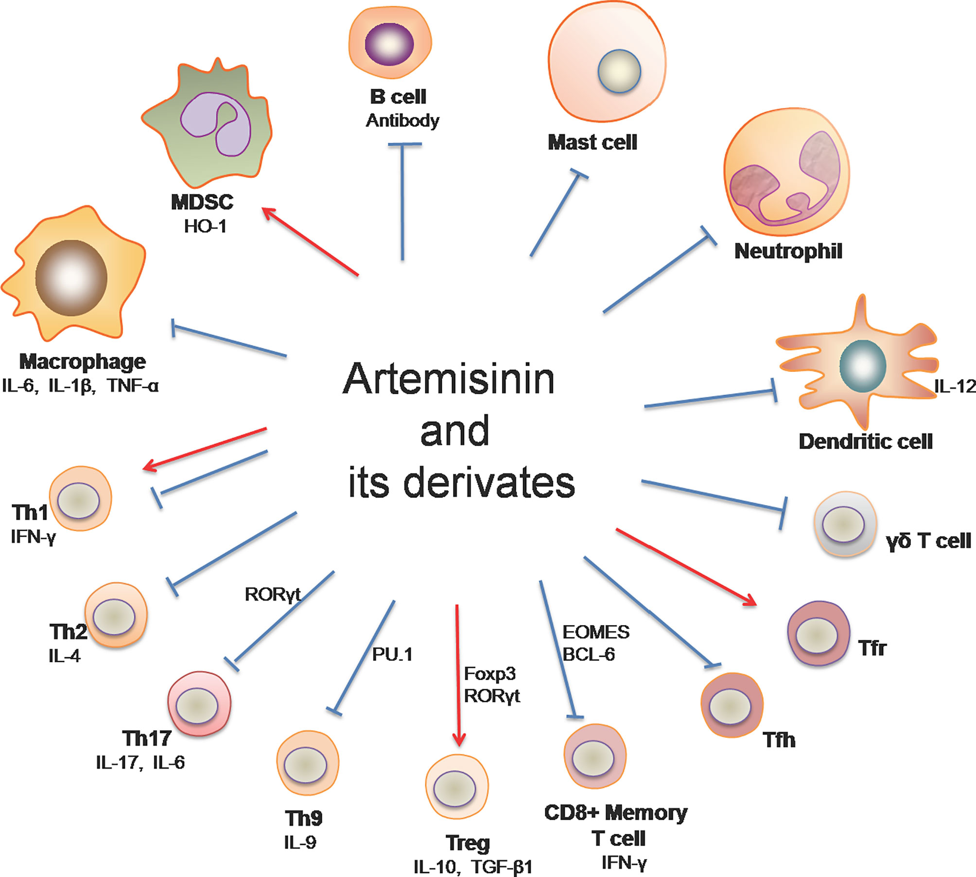
Figure 1 Artemisinin and its derivatives on both adaptive and innate immune cells. Artemisinin and its derivatives have the capacity to regulate expressions of proinflammatory and anti-inflammatory cytokines, the frequency and activation of T helper and B cells, and the responsiveness of macrophages, DCs, neutrophils, mast cells and MDSCs. “↓“ denotes “enhancing” while “⊥“ indicates “suppressing”. (Th1, T helper 1 cell; Th2, T helper 2 cell; Th9, T helper 9 cell; Th17, T helper 17 cell; Treg, regulatory T cells; Tfh, follicular helper T cells; Tfr, follicular regulatory T cells; MDSC, myeloid-derived suppressor cells).
Rheumatoid Arthritis
RA is a chronic inflammatory disease with the feature of Treg/Th17 imbalance, resulting in cartilage degradation and bone erosion within both small and large joints (15). The exact cause of RA remains unknown, posing a challenge to the diagnosis and treatment for RA (16). Accumulating evidence based on animal models has pointed to the efficacy of ARTs in RA treatment. In type II collagen-induced arthritis (CIA) in rats, artesunate treatment not only alleviated inflammation, decreased the frequency of Th17 cells and increased Treg cells in the synovium and spleen (17), but also stimulated apoptosis or autophagy in cartilage tissue and inhibited chondrocyte proliferation through the PI3K/AKT/mTOR signaling pathway (18). Moreover, artesunate exerted a suppressive effect on osteoclastogenesis and improved arthritic bone erosion in CIA rats via inhibiting the production of ROS and activating antioxidant enzyme as well as p62/Nrf2 signaling (19). In addition, artesunate significantly inhibited the migration and invasion of fibroblast-like synoviocytes (FLS) in patients with RA by suppressing PDK1-induced activation of AKT and RSK2 phosphorylation as well as MMP-2 and MMP-9 production (20). Similarly, DC32 [(9α,12α-dihydroartemisinyl)bis(2’-chlorocinnmate)], a dihydroartemisinin derivative, remarkably dampened footpad inflammation, reduced cartilage degradation through the Nrf2-p62-Keap1 feedback loop in DBA/1 mice with CIA (21), and impeded cellular infiltration and inflammation via restoration of Treg/Th17 balance and downregulation of the expression of IL-6, resulting in ultimate attenuation of RA (22). These data have suggested that ATRs can effectively alleviate RA by regulating Th17/Treg balance, FLS mobility and oxidative stress via suppressing PI3K/AKT/mTOR and AKT/RSK2 signaling pathways while activating p62/Nrf2 signaling.
Immune-Mediated Skin Diseases: Psoriasis and Atopic Dermatitis
Psoriasis, which affects over 60 million people worldwide, is an inflammatory or autoimmune skin disease (23). An important role for T cells in the pathogenesis of psoriasis was confirmed by several clinical trials (24, 25), while tissue-resident memory T cells (TRM) reportedly led to the recurrence of psoriasis at sites of previously involved skin (26). We recently found that dihydroartemisinin is more effective than methotrexate in suppressing psoriasis relapse. Dihydroartemisinin not only reduced acute skin lesions and recurrence of psoriasis in imiquimod (IMQ)-induced psoriasis-like mice, but also ameliorated psoriatic human skin lesions in humanized NSG mice receiving lesional skin from patients with psoriasis (11), mainly by diminishing CD8+ central memory T (TCM) and resident memory T (TRM) cells (11). Furthermore, artesunate was also reported to inhibit epidermal thickening and systemic inflammation in IMQ-induced psoriatic mice by reducing γδ T cells in the draining lymph nodes (27).
Atopic dermatitis (AD) is a common and chronic inflammatory skin disease with severe pruritus, cutaneous and systemic immune dysfunction, and skin lesions. Although the exact pathogenesis of AD remains unclear, it likely results from interactions of genetic and environmental factors (28, 29). It was reported that dihydroartemisinin could ameliorate AD symptoms and skin lesions in DNCB-induced AD mouse models. At high doses, dihydroartemisinin significantly alleviated mast cell infiltration into the skin lesions (30), indicating that it exerts therapeutic effects on AD by targeting mast cells in the skin. Similarly, artesunate relieved AD symptoms and mast cell infiltration, mainly by decreasing the expression of proinflammatory cytokines, including IL-6, IL-17 and IL-23, and suppressing RORγt and STAT3 phosphorylation (31). Taken together, these studies have indicated that ARTs attenuate both psoriasis and AD through inhibition of CD8+ memory T cells, Th17/γδ T cells and mast cells.
Inflammatory Bowel Disease
IBD is a chronic inflammatory bowel disorder mediated by autoimmunity, leading to the injury of gastrointestinal tracts (32, 33). IBD mainly includes ulcerative colitis (UC) and Crohn’s disease (34). In a dextran sulfate sodium (DSS)-induced mouse colitis model, artemisinin was shown to downregulate LYVE-1+ lymphatic vessel density and ameliorate the intestinal inflammation by inhibiting VEGF-C/VEGFR-3-mediated lymphangiogenesis and infiltration of macrophages and neutrophils in colon tissue (35), promoting macrophage polarization toward an M2 phenotype and/or suppressing the process of epithelial-mesenchymal transition (36). Besides, dihydroartemisinin and artesunate have been reported to effectively alleviate colitis symptoms in mice via different cellular and molecular mechanisms. For example, treatment with dihydroartemisinin suppressed activation of PI3K/AKT and NF-κB signaling pathways (37), promoted CD4+ T cell apoptosis and restored Th1/Treg cell balance through enhancing heme oxygenase-1 (HO-1) production (38). Dihydroartemisinin also regulated the expression of proinflammatory genes and cell junction-associated genes and normalized the abundance of the gut bacteria that was altered in colitis mice (39). Furthermore, artesunate reportedly reduced expression of IFN-γ, IL-17, and TNF-α in experimental colitis (40), inhibited TLR4-NF-κB signaling pathway (41), promoted apoptosis of macrophages and DCs, and reduced TNF-α and IL-12 production in vivo and in vitro (42) while suppressing excessive ER stress (43), cell apoptosis and inflammatory responses via the NF-κB pathway (44, 45). Thus, ARTs exert immunoregulatory effects on various immune cells, including T helper cells, Tregs, macrophages, neutrophils and DCs, by modulating NF-κB and PI3K/AKT signaling pathways, resulting in an improvement of colitis symptoms.
Multiple Sclerosis
MS is a chronic immune-mediated disease of the central nervous system, resulting in the destruction of oligodendrocytes and myelin sheaths and impairment of mobility and cognitive processing (46). Although MS etiology is complex and not completely elucidated, it seems to result from a combination of environmental, genetic and epigenetic factors (47). The therapeutic efficacy of artemisinin family drugs on MS was investigated using a mouse model of experimental autoimmune encephalomyelitis (EAE). It was found that artemisinin ameliorated EAE and reduced plaque formation in the brain with a decrease in IFN-γ expression and an increase in IL-4 production (48). Administration of artesunate attenuated the clinical signs and symptoms of EAE via preventing migration of pathogenic T cells to the central nervous system (49). Furthermore, Lv found that 9,10-Anhydrodehydroartemisin (ADART), a compound derived from artemisinin, effectively reduced inflammation in the central nervous system by inhibiting Th1 and Th17 cells (50). Therefore, the therapeutic effects of ARTs on MS may be attributed to their suppression of Th1 and Th17 cells.
Systemic Lupus Erythematosus
As a chronic and systemic autoimmune disease, SLE is characterized by the dysfunction of immune cells, the production of a wide range of autoantibodies and the formation of immune complexes (51, 52). It was reported that dihydroartemisinin suppressed LPS-induced activation and proliferation of spleen cells from lupus-prone MRL/lpr mice possibly through inhibiting TLR4 expression and IRF3 phosphorylation (53). Li et al. showed that senescence of myeloid-derived suppressor cells (MDSCs) promoted the pathogenesis of SLE, while dihydroartemisinin alleviated the manifestation of SLE by attenuating MDSC senescence via regulating Nrf2/HO-1 pathway (54). Subsequently, it was demonstrated that dihydroartemisinin alone, or in combination with prednisone treatment, significantly ameliorated the signs and symptoms of murine SLE through restoring the Treg/Th17 balance by reducing transcription of RORγt and increasing expression of Foxp3 in T cells (55). Serum levels of Macrophage migration inhibitory factor (MIF) in SLE patients were positively associated with the disease activity. Artesunate was shown to decrease MIF level in HUVEC culture with IFNα stimulation and in SLE patient-derived PBMC culture, partly through attenuating STAT1 phosphorylation, indicating a potential therapeutic effect of artesunate on SLE-associated atherosclerosis (56). Another study in vivo revealed that artesunate ameliorated the symptoms of lupus nephritis, decreased renal deposition of anti-dsDNA antibodies and suppressed the production of pathogenic cytokines through a reduction of follicular T helper cells (Tfh) and enhancement of follicular regulatory T cells (Tfr) as well as suppression of Jak2-Stat3 signaling pathway (57). Besides, SM934, an artemisinin derivative, extended the lifespan of MRL/lpr mice, relieved the lymphadenopathy symptoms, and suppressed B cell activation and plasma cell formation in vivo (58). Thus, ARTs appear to exert therapeutic effects on SLE, and the mechanisms underlying their effects were likely dependent on their regulation of MDSC senescence, Treg/Th17 balance and/or Tfh/Tfr ratio through Nfr2/HO-1 and Jak/STAT signaling pathways.
IgA Nephropathy
IgA nephropathy (IgAN) is a common glomerular disease and one of the leading causes of end-stage renal diseases (59). IgAN is an autoimmune disease characterized by IgA immunodeposits within the mesangium of the glomeruli, usually resulting in hematuria and renal functional insufficiency (60). An animal study performed by Bai and his colleagues demonstrated that in combination with hydroxychloroquine (AH), artemisinin significantly improved renal function, decreased IgA and IgG depositions, and reduced the expression of nuclear NF-κB and NLRP3 inflammasome-related proteins while elevating the secretion of exosomes in the kidney. They revealed that increased exosomes from HK-2 cells could inhibit the NF-κB signal pathway and NLRP3 inflammasome activation (61). On the other hand, the authors also found that artemisinin in combination with hydroxychloroquine ameliorated IgAN by reducing Th2 and Th17 cells while increasing Treg cells (62). Subsequently, it was also shown that dihydroartemisinin downregulated the mTOR/S6K1 signaling pathway in human mesangial cells (HMCs), promoted cell autophagy and suppressed cell proliferation in IgA1-stimulated HMCs, suggesting that dihydroartemisinin is a novel mTOR inhibitor and can exert an anti-proliferative effect on HMCs in IgAN (63).
Transplant Rejection
Allograft rejection represents an immune process in which the donor alloantigens evoke a vigorous immune response of a recipient against a transplant, contributing to graft loss (64). Therefore, more effective therapies need to be explored to improve transplant outcomes as current immunosuppressive agents may cause various side effects. An important study by Yang et al. revealed that artemisinin alleviated not only T cell-mediated but also antibody-mediated rejection in a cardiac transplant rat model by regulating the balance of T effector and Treg (Teff/Treg), impeding B cell activation and antibody production, and decreasing macrophage infiltration in an allograft, resulting in prolongation of graft survival. Moreover, they reported that artemisinin inhibited the activation or function of T cells, B cells and macrophages in vitro (65). Another study indicated that artemisinin remarkably extended survival time of murine skin allografts without significant changes of CD4+CD44hiCD62Lhi T cells in vivo. However, artemisinin reduced OX40+ T cell frequency and IL-6 secretion in vitro (66). Thus, artemisinin can exert immunosuppressive effects on alloimmunity or allograft rejection and may be implicated in clinical transplantation.
The Effects of Artemisinin and Its Derivatives on Cellular Signaling Pathways
NF-κB
The nuclear factor-kappa B (NF-κB) signaling plays a vital role in both immunity and inflammation (67). Upon stimulation, NF-κB translocates into the nucleus and promotes the transcription of numerous genes critical for dendritic cell function, T cell activation and sustainment of secondary lymphoid organs. In a study performed by Wang et al., TNF-α induced phosphorylation of IκBα and production of P65 in Hep3 B cells. Artemisinin suppressed the activation of NF-κB pathway induced by TNF-α via attenuating IκBα phosphorylation and activation of P65, resulting in a decrease in expression of NF-κB target genes and two adaptor proteins, RAF2 and RIP1, which in turn exerted their effects on upstream of IKK signaling (68). Dihydroartemisinin and artesunate also inhibited NF-κB signaling, leading to alleviation of colitis. Dihydroartemisinin significantly inhibited phosphorylation of IKKα, IκBα, and NF-κB (p65) in DSS-induced murine colitis and IEC-6 cells treated with LPS (37) while artesunate remarkably suppressed the NF-κB activation with a reduction in mRNA expression of IL-1β, IL-6 and TNF-α and an increase in IL-10 gene expression (41, 44, 45). Taken together, ARTs can hinder activation of NF-κB pathway and therefore suppress inflammatory responses in vivo and in vitro (Figure 2).
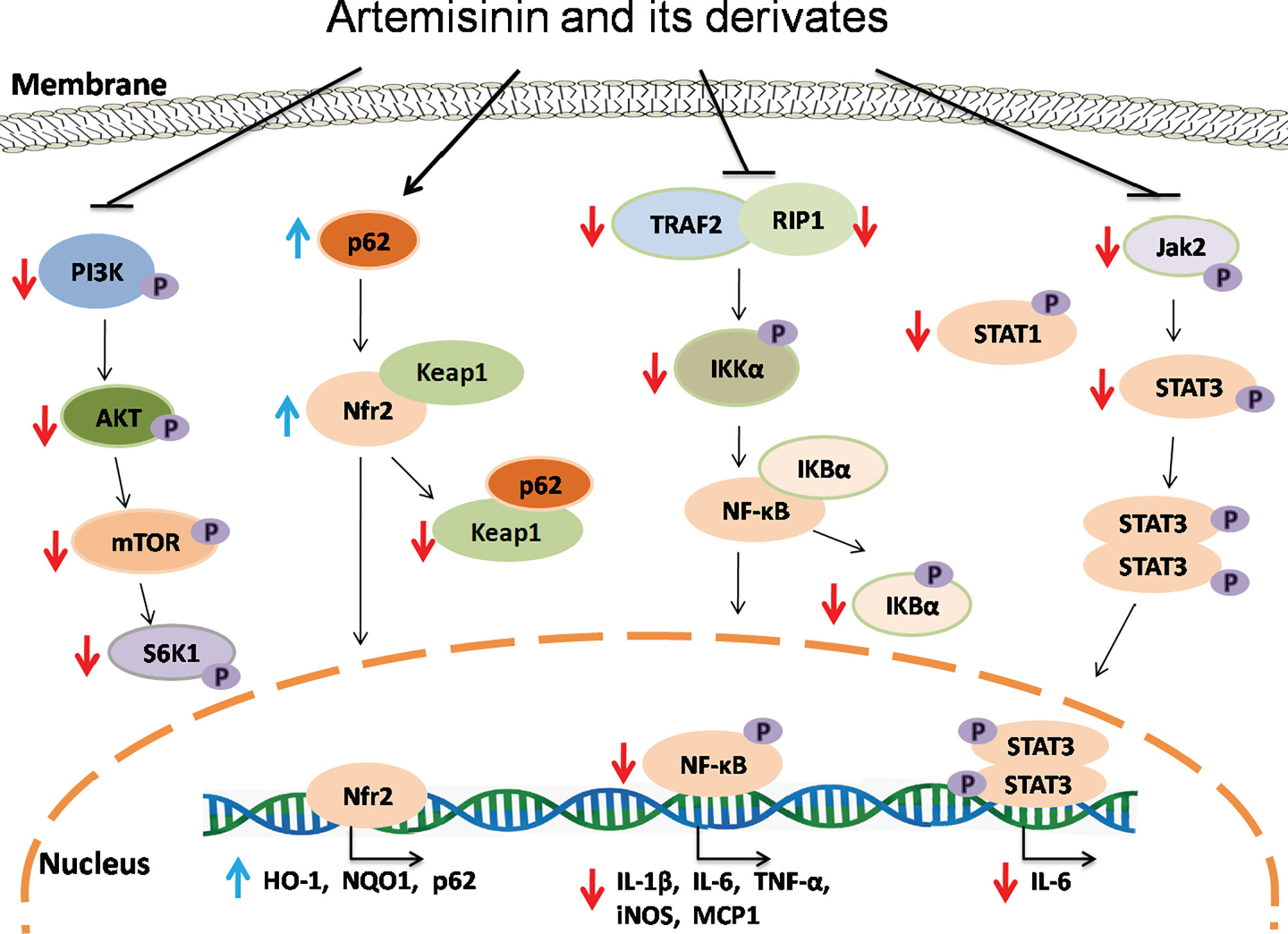
Figure 2 The signaling mechanisms underlying effects of artemisinin and its derivatives. Artemisinin and its derivatives exert immunoregulatory and anti-inflammatory effects via interfering with NF-kB, Nrf2, Jak/STAT and mTOR signaling pathways, resulting in downregulation of proinflammatory genes and upregulation of anti-inflammatory and antioxidant genes. Red downward arrows indicate suppressive effects, while blue upward arrows denote stimulating effects.
Nrf2
Transcription factor Nrf2, which is negatively regulated by Keap1, plays an important role in controlling the expression of antioxidant enzymes and suppressing inflammation via regulating transcription of anti-inflammatory genes (69, 70) while P62-mediated Keap1 degradation contributes to nuclear translocation of Nrf2 and related gene transcriptions. Dihydroartemisinin treatment elevated the expression of Nrf2 and its target gene HO-1 in MDSCs from SLE mice and attenuated the senescence of MDSCs (54). It was also found that artesunate activated Nrf2 by augmenting p62 expression in murine bone marrow macrophages, resulting in an increase in the expression of HO-1 or NQO1 (19). Similarly, DC32 strengthened Nrf2/HO-1 signaling and promoted p62 transcription or Keap1 degradation in DBA/1 mice as well as NIH-3T3 cells (21). Collectively, ARTs promote the activation of Nrf2 signaling, resulting in elevated expression of anti-inflammatory genes and a reduction in inflammation (Figure 2).
JAK/STAT
Janus kinase (JAK) and signal transducer and activator of transcription (STAT) proteins control signal transduction of many cytokines and growth factors associated with cellular growth, survival and differentiation (71). Artesunate reportedly attenuated STAT1 phosphorylation in cultured HUVECs stimulated with IFNα (56) and suppressed phosphorylation of JAK2 and STAT3 in the kidney of MRL/lpr mice, resulting in an amelioration of the symptoms of lupus nephritis (57).
mTOR
Mammalian or mechanistic target of rapamycin (mTOR) is a protein kinase that regulates eukaryotic signaling networks and diverse cellular processes upon environmental changes (72). Previous studies demonstrated that artesunate not only downregulated the mRNA and protein expressions of PI3K, AKT and mTOR, but also inhibited phosphorylation of these proteins in the cartilage tissue of RA mice and chondrocytes in vitro (18), while dihydroartemisinin suppressed phosphorylation of mTOR and S6K in human mesangial cells in the presence of IgA1 stimulation (63). Thus, ARTs may serve as a novel inhibitor of mTOR.
Conclusions and Perspectives
The number of patients treated with immunosuppressive drugs for immune-mediated inflammatory diseases (IMIDs) and transplant rejection has gradually increased over the last decades (73). IMIDs or autoimmune diseases affect 5-7% of the population in western countries (12), resulting in substantial personal and societal costs due to disease chronicity. Lifelong administration of conventional immunosuppressive agents poses a huge economic burden to patients with autoimmune diseases or organ transplantation, accompanied by some complications, such as infections, tumors and lymphoproliferative diseases. In spite of advances in remedies, IMIDs and allograft rejection remain linked to a high risk of morbidity and mortality (74). Thus, finding a cost-effective and efficacious treatment with few side effects is warranted.
ARTs have been widely used in humans for treating malaria with only mild side effects (75). Other clinical trials showed that artesunate was safe and well-tolerated in patients with breast cancer (76, 77), while artemisinin treatment in pregnant women did not elevate the risk of miscarriage, stillbirth or congenital malformation (78). Collectively, ARTs are generally considered to be safe and effective in clinical practice.
In this review, we summarize the latest studies demonstrating the efficacy of ARTs in the treatment of IMIDs or autoimmune diseases and allograft rejection. ARTs mainly regulate adaptive and innate immune cells, including subsets of CD4+ T cells (Th1/Th2/Th9/Th17/Tfh/Tfr/Treg), CD8+ memory T cells, γδ T cells, B cells, dendritic cells, neutrophils, mast cells, macrophages, and MDSCs, through altering cellular apoptosis and differentiation, pro-inflammatory and anti-inflammatory cytokine secretion, and signal transduction. So far, systematic researches into ARTs for their efficacy in IMIDs and allograft rejection have been largely confined to animal models due to the lack of large randomized and controlled clinical trials, although previous clinical evidence has indicated that ARTs may have been immunosuppressive in lupus patients (79–81). Therefore, more clinical studies are warranted to evaluate their doses, efficacy and side effects in the treatment of IMIDs and allograft rejection. ARTs may present a promising therapeutic alternative for treating IMIDs and allograft rejection in the near future. Alternatively, they could be used in combination with an immunosuppressant to either enhance therapeutic efficacy or reduce side effects.
Author Contributions
FQ and JL wrote the original manuscript together. XM, HL, and YC searched some of the literature. ZD provided the general idea and edited the manuscript. All authors contributed to the article and approved the submitted version.
Funding
This work was supported by National Natural Science Foundation of China (82071800), the State Key Laboratory of Dampness Syndrome of Chinese Medicine (SZ2020ZZ16 and SZ2020ZZ18), the 2020 Guangdong Provincial Science and Technology Innovation Strategy Special Fund (Guangdong-Hong Kong-Macau Joint Lab 2020B1212030006), the Specific Research Fund for TCM Science and Technology of Guangdong Provincial Hospital of Chinese Medicine (YN2019MJ03, YN2019QJ07 and YN2019QJ02), and the Key-Area Research and Development Program of Guangdong Province (NO. 2020B1111100010).
Conflict of Interest
The authors declare that the research was conducted in the absence of any commercial or financial relationships that could be construed as a potential conflict of interest.
Publisher’s Note
All claims expressed in this article are solely those of the authors and do not necessarily represent those of their affiliated organizations, or those of the publisher, the editors and the reviewers. Any product that may be evaluated in this article, or claim that may be made by its manufacturer, is not guaranteed or endorsed by the publisher.
Acknowledgments
The authors thank Prof. Daniel Kreisel (Washington University in St Luis, USA) for his minor editing of the manuscript.
References
1. Tu YY. The Discovery of Artemisinin (Qinghaosu) and Gifts From Chinese Medicine. Nat Med (2011) 17(10):1217–20. doi: 10.1038/nm.2471
2. Ma N, Zhang ZY, Liao FL, Jiang TL, Tu YY. The Birth of Artemisinin. Pharmacol Therapeut (2020) 216:107658. doi: 10.1016/j.pharmthera.2020.107658
3. Liu JM, Ni MY, Fan JF, Tu YY, Zhou WS. Structure and Reaction of Arteannuin. Acta Chim Sin (1979) 37(2):129–43.
4. Xu CC, Deng T, Fan ML, Lv WB, Liu JH, Yu BY. Synthesis and In Vitro Antitumor Evaluation of Dihydroartemisinin-Cinnamic Acid Ester Derivatives. Eur J Med Chem (2016) 107:192–203. doi: 10.1016/j.ejmech.2015.11.003
5. Khanal P. Antimalarial and Anticancer Properties of Artesunate and Other Artemisinins: Current Development. Monatsh Chem (2021) 152:387–400. doi: 10.1007/s00706-021-02759-x
6. Efferth T. Beyond Malaria: The Inhibition of Viruses by Artemisinin-Type Compounds. Biotechnol Adv (2018) 36(6):1730–7. doi: 10.1016/j.biotechadv.2018.01.001
7. Dolivo D, Weathers P, Dominko T. Artemisinin and Artemisinin Derivatives as Anti- Fibrotic Therapeutics. Acta Pharmacol Sin B (2021) 11(2):322–39. doi: 10.1016/j.apsb.2020.09.001
8. Wang YK, Wang YT, You FM, Xue JX. Novel Use for Old Drugs: The Emerging Role of Artemisinin and Its Derivatives in Fibrosis. Pharmacol Res (2020) 157:104829. doi: 10.1016/J.Phrs.2020.104829
9. An J, Minie M, Sasaki T, Woodward JJ, Elkon KB. Antimalarial Drugs as Immune Modulators: New Mechanisms for Old Drugs. Annu Rev Med (2017) 68:317–30. doi: 10.1146/annurev-med-043015-123453
10. Hou L, Huang H. Immune Suppressive Properties of Artemisinin Family Drugs. Pharmacol Therapeut (2016) 166:123–7. doi: 10.1016/j.pharmthera.2016.07.002
11. Chen YC, Yan YH, Liu HZ, Qiu FF, Liang CL, Zhang QF, et al. Dihydroartemisinin Ameliorates Psoriatic Skin Inflammation and Its Relapse by Diminishing CD8(+) T-Cell Memory in Wild-Type and Humanized Mice. Theranostics (2020) 10(23):10466–82. doi: 10.7150/thno.45211
12. El-Gabalawy H, Guenther LC, Bernstein CN. Epidemiology of Immune-Mediated Inflammatory Diseases: Incidence, Prevalence, Natural History, and Comorbidities. J Rheumatol Suppl (2010) 85:2–10. doi: 10.3899/jrheum.091461
13. Gonzalez-Serna D, Villanueva-Martin G, Acosta-Herrera M, Marquez A, Martin J. Approaching Shared Pathophysiology in Immune-Mediated Diseases Through Functional Genomics. Genes (2020) 11(12):1482. doi: 10.3390/genes11121482
14. Bunte K, Beikler T. Th17 Cells and the IL-23/IL-17 Axis in the Pathogenesis of Periodontitis and Immune-Mediated Inflammatory Diseases. Int J Mol Sci (2019) 20(14):3394. doi: 10.3390/ijms20143394
15. Myngbay A, Manarbek L, Ludbrook S, Kunz J. The Role of Collagen Triple Helix Repeat-Containing 1 Protein (CTHRC1) in Rheumatoid Arthritis. Int J Mol Sci (2021) 22(5):2426. doi: 10.3390/Ijms22052426
16. Littlejohn EA, Monrad SU. Early Diagnosis and Treatment of Rheumatoid Arthritis. Primary Care (2018) 45(2):237–55. doi: 10.1016/j.pop.2018.02.010
17. Liu J, Hong X, Lin D, Luo X, Zhu M, Mo H. Artesunate Influences Th17/Treg Lymphocyte Balance by Modulating Treg Apoptosis and Th17 Proliferation in a Murine Model of Rheumatoid Arthritis. Exp Therapeut Med (2017) 13(5):2267–73. doi: 10.3892/etm.2017.4232
18. Feng FB, Qiu HY. Effects of Artesunate on Chondrocyte Proliferation, Apoptosis and Autophagy Through the PI3K/AKT/mTOR Signaling Pathway in Rat Models With Rheumatoid Arthritis. BioMed Pharmacother (2018) 102:1209–20. doi: 10.1016/j.biopha.2018.03.142
19. Su X, Guo W, Yuan B, Wang D, Liu L, Wu X, et al. Artesunate Attenuates Bone Erosion in Rheumatoid Arthritis by Suppressing Reactive Oxygen Species via Activating P62/Nrf2 Signaling. BioMed Pharmacother (2021) 137:111382. doi: 10.1016/j.biopha.2021.111382
20. Ma JD, Jing J, Wang JW, Yan T, Li QH, Mo YQ, et al. A Novel Function of Artesunate on Inhibiting Migration and Invasion of Fibroblast-Like Synoviocytes From Rheumatoid Arthritis Patients. Arthritis Res Ther (2019) 21:153. doi: 10.1186/S13075-019-1935-6
21. Fan ML, Li YN, Yao CH, Liu XF, Liu JH, Yu BY. DC32, a Dihydroartemisinin Derivative, Ameliorates Collagen-Induced Arthritis Through an Nrf2-P62-Keap1 Feedback Loop. Front Immunol (2018) 9:2762. doi: 10.3389/Fimmu.2018.02762
22. Fan ML, Li YN, Yao CH, Liu XF, Liu XM, Liu JH. Dihydroartemisinin Derivative DC32 Attenuates Collagen-Induced Arthritis in Mice by Restoring the Treg/Th17 Balance and Inhibiting Synovitis Through Down-Regulation of IL-6. Int Immunopharmacol (2018) 65:233–43. doi: 10.1016/j.intimp.2018.10.015
23. Griffiths CEM, Armstrong AW, Gudjonsson JE, Barker JNWN. Psoriasis. Lancet (2021) 397(10281):1301–15. doi: 10.1016/S0140-6736(20)32549-6
24. Brookes DB. Clearance of Psoriasis With Low Dose Cyclosporin. Brit Med J (Clin Res Ed) (1986) 293(6554):1098–9. doi: 10.1136/bmj.293.6554.1098-c
25. Ellis CN, Gorsulowsky DC, Hamilton TA, Billings JK, Brown MD, Headington JT, et al. Cyclosporine Improves Psoriasis in a Double-Blind Study. JAMA (1986) 256(22):3110–6. doi: 10.1001/jama.1986.03380220076026
26. Cheuk S, Wiken M, Blomqvist L, Nylen S, Talme T, Stahle M, et al. Epidermal Th22 and Tc17 Cells Form a Localized Disease Memory in Clinically Healed Psoriasis. J Immunol (2014) 192(7):3111–20. doi: 10.4049/jimmunol.1302313
27. Huang ZZ, Xu Y, Xu M, Shi ZR, Mai SZ, Guo ZX, et al. Artesunate Alleviates Imiquimod-Induced Psoriasis-Like Dermatitis in BALB/c Mice. Int Immunopharmacol (2019) 75:105817. doi: 10.1016/J.Intimp.2019.105817
28. Yang G, Seok JK, Kang HC, Cho YY, Lee HS, Lee JY. Skin Barrier Abnormalities and Immune Dysfunction in Atopic Dermatitis. Int J Mol Sci (2020) 21(8):2867. doi: 10.3390/Ijms21082867
29. Torres T, Ferreira EO, Goncalo M, Mendes-Bastos P, Selores M, Filipe P. Update on Atopic Dermatitis. Acta Med Portug (2019) 32(9):606–13. doi: 10.20344/amp.11963
30. Xue X, Dong Z, Deng Y, Yin S, Wang P, Liao Y, et al. Dihydroartemisinin Alleviates Atopic Dermatitis in Mice by Inhibiting Mast Cell Infiltration. Nan Fang Yi Ke Da Xue Xue Bao (2020) 40(10):1480–7. doi: 10.12122/j.issn.1673-4254.2020.10.14
31. Bai XY, Liu P, Chai YW, Wang Y, Ren SH, Li YY, et al. Artesunate Attenuates 2, 4-Dinitrochlorobenzene-Induced Atopic Dermatitis by Down-Regulating Th17 Cell Responses in BALB/c Mice. Eur J Pharmacol (2020) 874:173020. doi: 10.1016/J.Ejphar.2020.173020
32. Kelsen JR, Russo P, Sullivan KE. Early-Onset Inflammatory Bowel Disease. Immunol Allergy Clin (2019) 39(1):63–79. doi: 10.1016/j.iac.2018.08.008
33. Koliani-Pace JL, Siegel CA. Prognosticating the Course of Inflammatory Bowel Disease. Gastrointest Endosc Clin North Am (2019) 29(3):395–404. doi: 10.1016/j.giec.2019.02.003
34. Mentella MC, Scaldaferri F, Pizzoferrato M, Gasbarrini A, Miggiano GAD. Nutrition, IBD and Gut Microbiota: A Review. Nutrients (2020) 12(4):944. doi: 10.3390/Nu12040944
35. Lee AS, Hur HJ, Sung MJ. The Effect of Artemisinin on Inflammation-Associated Lymphangiogenesis in Experimental Acute Colitis. Int J Mol Sci (2020) 21(21):8086. doi: 10.3390/ijms21218068
36. Huai MX, Zeng JX, Ge WS. Artemisinin Ameliorates Intestinal Inflammation by Skewing Macrophages to the M2 Phenotype and Inhibiting Epithelial-Mesenchymal Transition. Int Immunopharmacol (2021) 91:107284. doi: 10.1016/J.Intimp.2020.107284
37. Li N, Sun W, Zhou X, Gong H, Chen Y, Chen D, et al. Dihydroartemisinin Protects Against Dextran Sulfate Sodium-Induced Colitis in Mice Through Inhibiting the PI3K/AKT and NF-κb Signaling Pathways. BioMed Res Int (2019) 2019:1415809. doi: 10.1155/2019/1415809
38. Yan SC, Wang YJ, Li YJ, Cai WY, Weng XG, Li Q, et al. Dihydroartemisinin Regulates the Th/Treg Balance by Inducing Activated Cd4+T Cell Apoptosis via Heme Oxygenase-1 Induction in Mouse Models of Inflammatory Bowel Disease. Molecules (2019) 24(13):2475. doi: 10.3390/Molecules24132475
39. Lei ZL, Yang YH, Liu SM, Lei YT, Yang LX, Zhang XY, et al. Dihydroartemisinin Ameliorates Dextran Sulfate Sodium Induced Inflammatory Bowel Diseases in Mice. Bioorg Chem (2020) 100:103915. doi: 10.1016/J.Bioorg.2020.103915
40. Yang Z, Ding J, Yang C, Gao Y, Li X, Chen X, et al. Immunomodulatory and Anti-Inflammatory Properties of Artesunate in Experimental Colitis. Curr Med Chem (2012) 19(26):4541–51. doi: 10.2174/092986712803251575
41. Chen YX, Zhang XQ, Yu CG, Huang SL, Xie Y, Dou XT, et al. Artesunate Exerts Protective Effects Against Ulcerative Colitis via Suppressing Toll-Like Receptor 4 and Its Downstream Nuclear Factor-Kappa B Signaling Pathways. Mol Med Rep (2019) 20(2):1321–32. doi: 10.3892/mmr.2019.10345
42. Sun WL, Han X, Wu SY, Wu JH, Yang CH, Li XH. Unexpected Mechanism of Colitis Amelioration by Artesunate, a Natural Product Fromartemisia annuaL. Inflammopharmacology (2020) 28(4):851–68. doi: 10.1007/s10787-019-00678-2
43. Yin SJ, Li LH, Tao Y, Yu J, Wei SM, Liu MJ, et al. The Inhibitory Effect of Artesunate on Excessive Endoplasmic Reticulum Stress Alleviates Experimental Colitis in Mice. Front Pharmacol (2021) 12:629798. doi: 10.3389/Fphar.2021.629798
44. Yin S, Yang H, Tao Y, Wei S, Li L, Liu M, et al. Artesunate Ameliorates DSS-Induced Ulcerative Colitis by Protecting Intestinal Barrier and Inhibiting Inflammatory Response. Inflammation (2020) 43(2):765–76. doi: 10.1007/s10753-019-01164-1
45. Yang ZB, Qiu LZ, Chen Q, Lin JD. Artesunate Alleviates the Inflammatory Response of Ulcerative Colitis by Regulating the Expression of miR-155. Pharm Biol (2021) 59(1):97–105. doi: 10.1080/13880209.2020.1867196
46. Diaz C, Zarco LA, Rivera DM. Highly Active Multiple Sclerosis: An Update. Mult Scler Relat Dis (2019) 30:215–24. doi: 10.1016/j.msard.2019.01.039
47. Olsson T, Barcellos LF, Alfredsson L. Interactions Between Genetic, Lifestyle and Environmental Risk Factors for Multiple Sclerosis. Nat Rev Neurol (2017) 13(1):25–36. doi: 10.1038/nrneurol.2016.187
48. Khakzad MR, Ganji A, Ariabod V, Farahani I. Artemisinin Therapeutic Efficacy in the Experimental Model of Multiple Sclerosis. Immunopharmacol Immunotoxicol (2017) 39(6):348–53. doi: 10.1080/08923973.2017.1379087
49. Thome R, de Carvalho AC, da Costa TA, Ishikawa LLW, Fraga-Silva TFD, Sartori A, et al. Artesunate Ameliorates Experimental Autoimmune Encephalomyelitis by Inhibiting Leukocyte Migration to the Central Nervous System. CNS Neurosci Ther (2016) 22(8):707–14. doi: 10.1111/cns.12561
50. Lv J, Zhuang W, Zhang Y, Xie L, Xiang ZL, Zhao QJ, et al. 9,10-Anhydrodehydroartemisinin Attenuates Experimental Autoimmune Encephalomyelitis by Inhibiting Th1 and Th17 Cell Differentiation. Inflammation (2021). doi: 10.1007/s10753-021-01456-5
51. Tang WY, Liu JH, Peng CJ, Liao Y, Luo JS, Sun X, et al. Functional Characteristics and Application of Mesenchymal Stem Cells in Systemic Lupus Erythematosus. Arch Immunol Ther Ex (2021) 69(1):7. doi: 10.1007/s00005-021-00603-y
52. Di Bartolomeo S, Alunno A, Carubbi F. Respiratory Manifestations in Systemic Lupus Erythematosus. Pharmaceuticals (2021) 14(3):276. doi: 10.3390/ph14030276
53. Huang X, Xie Z, Liu F, Han C, Zhang D, Wang D, et al. Dihydroartemisinin Inhibits Activation of the Toll-Like Receptor 4 Signaling Pathway and Production of Type I Interferon in Spleen Cells From Lupus-Prone MRL/lpr Mice. Int Immunopharmacol (2014) 22(1):266–72. doi: 10.1016/j.intimp.2014.07.001
54. Li D, Qi J, Wang J, Pan Y, Li J, Xia X, et al. Protective Effect of Dihydroartemisinin in Inhibiting Senescence of Myeloid-Derived Suppressor Cells From Lupus Mice via Nrf2/HO-1 Pathway. Free Rad Biol Med (2019) 143:260–74. doi: 10.1016/j.freeradbiomed.2019.08.013
55. Chen Y, Tao TJ, Wang WL, Yang BT, Cha XS. Dihydroartemisinin Attenuated the Symptoms of Mice Model of Systemic Lupus Erythematosus by Restoring the Treg/Th17 Balance. Clin Exp Pharmacol P (2021) 48(4):626–33. doi: 10.1111/1440-1681.13461
56. Feng X, Chen W, Xiao L, Gu F, Huang J, Tsao BP, et al. Artesunate Inhibits Type I Interferon-Induced Production of Macrophage Migration Inhibitory Factor in Patients With Systemic Lupus Erythematosus. Lupus (2017) 26(1):62–72. doi: 10.1177/0961203316651738
57. Dang WZ, Li H, Jiang B, Nandakumar KS, Liu KF, Liu LX, et al. Therapeutic Effects of Artesunate on Lupus-Prone MRL/lpr Mice Are Dependent on T Follicular Helper Cell Differentiation and Activation of JAK2-STAT3 Signaling Pathway. Phytomedicine (2019) 62:152965. doi: 10.1016/J.Phymed.2019.152965
58. Wu Y, He S, Bai B, Zhang L, Xue L, Lin Z, et al. Therapeutic Effects of the Artemisinin Analog SM934 on Lupus-Prone MRL/lpr Mice via Inhibition of TLR-Triggered B-Cell Activation and Plasma Cell Formation. Cell Mol Immunol (2016) 13(3):379–90. doi: 10.1038/cmi.2015.13
59. Rajasekaran A, Julian BA, Rizk DV. IgA Nephropathy: An Interesting Autoimmune Kidney Disease. Am J Med Sci (2021) 361(2):176–94. doi: 10.1016/j.amjms.2020.10.003
60. Hassler JR. IgA Nephropathy: A Brief Review. Semin Diagn Pathol (2020) 37(3):143–7. doi: 10.1053/j.semdp.2020.03.001
61. Bai LX, Li JC, Li HL, Song JP, Zhou Y, Lu RR, et al. Renoprotective Effects of Artemisinin and Hydroxychloroquine Combination Therapy on IgA Nephropathy via Suppressing NF-Kappa B Signaling and NLRP3 Inflammasome Activation by Exosomes in Rats. Biochem Pharmacol (2019) 169:113619. doi: 10.1016/J.Bcp.2019.08.021
62. Bai LX, Li HL, Li JC, Song JP, Zhou Y, Liu BH, et al. Immunosuppressive Effect of Artemisinin and Hydroxychloroquine Combination Therapy on IgA Nephropathy via Regulating the Differentiation of CD4+T Cell Subsets in Rats. Int Immunopharmacol (2019) 70:313–23. doi: 10.1016/j.intimp.2019.02.056
63. Xia M, Liu D, Tang XF, Liu YX, Liu HY, Liu Y, et al. Dihydroartemisinin Inhibits the Proliferation of IgAN Mesangial Cells Through the mTOR Signaling Pathway. Int Immunopharmacol (2020) 80:106125. doi: 10.1016/J.Intimp.2019.106125
64. Stolp J, Zaitsu M, Wood KJ. Immune Tolerance and Rejection in Organ Transplantation. Meth Mol Biol (Clifton NJ) (2019) 1899:159–80. doi: 10.1007/978-1-4939-8938-6_12
65. Yang Z, Han F, Liao T, Zheng H, Luo Z, Ma M, et al. Artemisinin Attenuates Transplant Rejection by Inhibiting Multiple Lymphocytes and Prolongs Cardiac Allograft Survival. Front Immunol (2021) 12:634368. doi: 10.3389/fimmu.2021.634368
66. Liu L, Zhao J, Li A, Yang X, Sprangers B, Li S. Prolongation of Allograft Survival by Artemisinin Treatment Is Associated With Blockade of OX40-OX40L. Immunopharmacol Immunotoxicol (2021) 43(4):291–8. doi: 10.1080/08923973.2021.1902347
67. Taniguchi K, Karin M. NF-κb, Inflammation, Immunity and Cancer: Coming of Age. Nat Rev Immunol (2018) 18(5):309–24. doi: 10.1038/nri.2017.142
68. Wang KS, Li J, Wang Z, Mi C, Ma J, Piao LX, et al. Artemisinin Inhibits Inflammatory Response via Regulating NF-kappaB and MAPK Signaling Pathways. Immunopharmacol Immunotoxicol (2017) 39(1):28–36. doi: 10.1080/08923973.2016.1267744
69. Ahmed SM, Luo L, Namani A, Wang XJ, Tang X. Nrf2 Signaling Pathway: Pivotal Roles in Inflammation. Biochim Biophys Acta Mol Basis Dis (2017) 1863(2):585–97. doi: 10.1016/j.bbadis.2016.11.005
70. Saha S, Buttari B, Panieri E, Profumo E, Saso L. An Overview of Nrf2 Signaling Pathway and Its Role in Inflammation. Molecules (2020) 25(22):5474. doi: 10.3390/molecules25225474
71. Goropevsek A, Holcar M, Pahor A, Avcin T. STAT Signaling as a Marker of SLE Disease Severity and Implications for Clinical Therapy. Autoimmun Rev (2019) 18(2):144–54. doi: 10.1016/j.autrev.2018.08.010
72. Saxton RA, Sabatini DM. mTOR Signaling in Growth, Metabolism, and Disease. Cell (2017) 169(2):361–71. doi: 10.1016/j.cell.2017.03.035
73. Croce E, Hatz C, Jonker EF, Visser LG, Jaeger VK, Buhler S. Safety of Live Vaccinations on Immunosuppressive Therapy in Patients With Immune-Mediated Inflammatory Diseases, Solid Organ Transplantation or After Bone-Marrow Transplantation - A Systematic Review of Randomized Trials, Observational Studies and Case Reports. Vaccine (2017) 35(9):1216–26. doi: 10.1016/j.vaccine.2017.01.048
74. David T, Ling SF, Barton A. Genetics of Immune-Mediated Inflammatory Diseases. Clin Exp Immunol (2018) 193(1):3–12. doi: 10.1111/cei.13101
75. Zani B, Gathu M, Donegan S, Olliaro PL, Sinclair D. Dihydroartemisinin-Piperaquine for Treating Uncomplicated Plasmodium Falciparum Malaria. Cochr Datab Systemat Rev (2014) 2014(1):CD010927. doi: 10.1002/14651858.CD010927
76. Konig M, von Hagens C, Hoth S, Baumann I, Walter-Sack I, Edler L, et al. Investigation of Ototoxicity of Artesunate as Add-on Therapy in Patients With Metastatic or Locally Advanced Breast Cancer: New Audiological Results From a Prospective, Open, Uncontrolled, Monocentric Phase I Study. Can Chemother Pharmacol (2016) 77(2):413–27. doi: 10.1007/s00280-016-2960-7
77. von Hagens C, Walter-Sack I, Goeckenjan M, Osburg J, Storch-Hagenlocher B, Sertel S, et al. Prospective Open Uncontrolled Phase I Study to Define a Well-Tolerated Dose of Oral Artesunate as Add-on Therapy in Patients With Metastatic Breast Cancer (ARTIC M33/2). Breast Can Res Treat (2017) 164(2):359–69. doi: 10.1007/s10549-017-4261-1
78. D'Alessandro S, Menegola E, Parapini S, Taramelli D, Basilico N. Safety of Artemisinin Derivatives in the First Trimester of Pregnancy: A Controversial Story. Molecules (2020) 25(15):3505. doi: 10.3390/molecules25153505
79. Huang XX. Clinical Study of Artesunate on Immune Function in Patients With SLE. Lishizhen Med Mater Med Res (2011) 22(7):1673–4.
80. Zhang JY, Zhong JX, Peng SQ, Zhang FX. Study on the Effect of Artesunate and Lingdan Tablet on IL-2 and sIL-2R in Patients With SLE. J Henan Univ Chin Med (2003) 18(2):38–9.
Keywords: artemisinin, adaptive immunity, innate immunity, autoimmune disease, immunoregulation, cellular signaling
Citation: Qiu F, Liu J, Mo X, Liu H, Chen Y and Dai Z (2021) Immunoregulation by Artemisinin and Its Derivatives: A New Role for Old Antimalarial Drugs. Front. Immunol. 12:751772. doi: 10.3389/fimmu.2021.751772
Received: 02 August 2021; Accepted: 25 August 2021;
Published: 09 September 2021.
Edited by:
Fadi G. Lakkis, University of Pittsburgh, United StatesReviewed by:
Qiquan Sun, Third Affiliated Hospital of Sun Yat-sen University, ChinaJamil Roumanos Azzi, Harvard Medical School, United States
Copyright © 2021 Qiu, Liu, Mo, Liu, Chen and Dai. This is an open-access article distributed under the terms of the Creative Commons Attribution License (CC BY). The use, distribution or reproduction in other forums is permitted, provided the original author(s) and the copyright owner(s) are credited and that the original publication in this journal is cited, in accordance with accepted academic practice. No use, distribution or reproduction is permitted which does not comply with these terms.
*Correspondence: Zhenhua Dai, zdai2009@outlook.com
†These authors have contributed equally to this work
Disclaimer: All claims expressed in this article are solely those of the authors and do not necessarily represent those of their affiliated organizations, or those of the publisher, the editors and the reviewers. Any product that may be evaluated in this article or claim that may be made by its manufacturer is not guaranteed or endorsed by the publisher.
https://www.sciencedirect.com/science/article/abs/pii/S0014299911007515
ReviewArtemisinins and immune system
Keywords
- 1
These authors equally contributed to this work.
https://link.springer.com/article/10.1007/s11427-019-9550-4
- Research Paper
- Published:
Dihydroartemisinin regulates the immune system by promotion of CD8+ T lymphocytes and suppression of B cell responses
Science China Life Sciences 63, 737–749 (2020)
Abstract
Artemisia annua is an anti-fever herbal medicine first described in traditional Chinese medicine 1,000 years ago. Artemisinin, the extract of A. annua, and its derivatives (dihydroartemisinin (DHA), artemether, and artesunate) have been used for the treatment of malaria with substantial efficacy. Recently, DHA has also been tested for the treatment of lupus erythematosus, indicating that it may function to balance the immune response in immunocompromised individuals. In the present study, the regulatory effect of artemisinin on the murine immune system was systematically investigated in mice infected with two different protozoan parasites (Toxoplasma gondii and Plasmodium berghei). Our results revealed that the mouse spleen index significantly increased (spleen enlargement) in the healthy mice after DHA administration primarily due to the generation of an extra number of lymphocytes and CD8+ T lymphocytes in both the spleen and circulation. DHA could increase the proportion of T helper cells and CD8+ T cells, as well as decrease the number of splenic and circulatory B cells. Further, DHA could reduce the production of proinflammatory cytokines. Our study revealed that apart from their anti-parasitic activity, artemisinin and its derivatives can also actively modulate the immune system to directly benefit the host.
This is a preview of subscription content, access via your institution.
References
Abdin, A.A., Ashour, D.S., and Shoheib, Z.S. (2013). Artesunate effect on schistosome thioredoxin glutathione reductase and cytochrome c peroxidase as new molecular targets in Schistosoma mansoni-infected mice. Biomed Environ Sci, 26, 953–961.
Aderka, D., Wysenbeek, A., Engelmann, H., Cope, A.P., Brennan, F., Molad, Y., Hornik, V., Levo, Y., Maini, R.N., Feldmann, M., et al. (2010). Correlation between serum levels of soluble tumor necrosis factor receptor and disease activity in systemic lupus erythematosus. Arthrit Rheumat 36, 1111–1120.
Bhadra, R., Gigley, J.P., and Khan, I.A. (2011a). The CD8 T-cell road to immunotherapy of toxoplasmosis. Immunotherapy 3, 789–801.
Bhadra, R., Gigley, J.P., Weiss, L.M., and Khan, I.A. (2011b). Control of Toxoplasma reactivation by rescue of dysfunctional CD8+ T-cell response via PD-1-PDL-1 blockade. Proc Natl Acad Sci USA 108, 9196–9201.
Cheng, C., Ho, W.E., Goh, F.Y., Guan, S.P., Kong, L.R., Lai, W.Q., Leung, B.P., and Wong, W.S.F. (2011). Anti-malarial drug artesunate attenuates experimental allergic asthma via inhibition of the phosphoinositide 3-kinase/Akt pathway. PLoS ONE 6, e20932.
Chimanuka, B., Francois, G., Timperman, G., Heyden, Y.V., Holenz, J., Plaizier-Vercammen, J., and Bringmann, G. (2001). A comparison of the stage-specific efficacy of chloroquine, artemether and dioncophylline B against the rodent malaria parasite Plasmodium chabaudi chabaudi in vivo. Parasitol Res 87, 795–803.
Couper, K.N., Roberts, C.W., Brombacher, F., Alexander, J., and Johnson, L.L. (2005). Toxoplasma gondii-specific immunoglobulin M limits parasite dissemination by preventing host cell invasion. Infect Immun 73, 8060–8068.
Cruz-González, D.J., Gómez-Martin, D., Layseca-Espinosa, E., Baranda, L., Abud-Mendoza, C., Alcocer-Varela, J., González-Amaro, R., and Monsiváis-Urenda, A.E. (2018). Analysis of the regulatory function of natural killer cells from patients with systemic lupus erythematosus. Clin Exp Immunol 191, 288–300.
Dong, Y.J., Li, W.D., and Tu, Y.Y. (2003) Effect of dihydro-qinghaosu on auto-antibody production, TNF alpha secretion and pathologic change of lupus nephritis in BXSB mice (in Chinese). Zhongguo Zhong Xi Yi Jie He Za Zhi 23, 676–679.
Du, X.X., Li, Y.J., Wu, C.L., Zhou, J.H., Han, Y., Sui, H., Wei, X.L., Liu, L., Huang, P., Yuan, H.H., et al. (2013). Initiation of apoptosis, cell cycle arrest and autophagy of esophageal cancer cells by dihydroartemisinin. Biomed Pharmacother 67, 417–424.
Dunay, I.R., Chan, W.C., Haynes, R.K., and Sibley, L.D. (2009). Artemisone and artemiside control acute and reactivated toxoplasmosis in a murine model. Antimicrobial Agents Chemother 53, 4450–4456.
Efferth, T. (2017). From ancient herb to modern drug: Artemisia annua and artemisinin for cancer therapy. Seminars Cancer Biol 46, 65–83.
Feng, Y., Zhu, X., Wang, Q., Jiang, Y., Shang, H., Cui, L., and Cao, Y. (2012). Allicin enhances host pro-inflammatory immune responses and protects against acute murine malaria infection. Malar J 11, 268.
Gordon, C., Li, C.K., and Isenberg, D.A. (2009). Systemic lupus erythematosus. N Engl J Med 38, 73–80.
Guo, Y., Xu, P., Xuan, Y., Wu, L., and Li, S. (1997) Effect of artesunate on vltrastructure of schistosomula Schistosoma japonicum (in Chinese). Chin J Schistosom Contr 9, 34–36.
He, Y., Fan, J., Lin, H., Yang, X., Ye, Y., Liang, L., Zhan, Z., Dong, X., Sun, L., and Xu, H. (2011). The anti-malaria agent artesunate inhibits expression of vascular endothelial growth factor and hypoxia-inducible factor-1α in human rheumatoid arthritis fibroblast-like synoviocyte. Rheumatol Int 31, 53–60.
Hou, L.F., He, S.J., Li, X., Yang, Y., He, P.L., Zhou, Y., Zhu, F.H., Yang, Y. F., Li, Y., Tang, W., et al. (2011). Oral administration of artemisinin analog SM934 ameliorates lupus syndromes in MRL/lpr mice by inhibiting Th1 and Th17 cell responses. Arthrit Rheumat 63, 2445–2455.
Hou, L., Block, K.E., and Huang, H. (2014). Artesunate abolishes germinal center B cells and inhibits autoimmune arthritis. PLoS ONE 9, e104762.
Huang, X., Xie, Z., Liu, F., Han, C., Zhang, D., Wang, D., Bao, X., Sun, J., Wen, C., and Fan, Y. (2014). Dihydroartemisinin inhibits activation of the Toll-like receptor 4 signaling pathway and production of type I interferon in spleen cells from lupus-prone MRL/lpr mice. Int Immunopharmacol 22, 266–272.
Klonis, N., Crespo-Ortiz, M.P., Bottova, I., Abu-Bakar, N., Kenny, S., Rosenthal, P.J., and Tilley, L. (2011). Artemisinin activity against Plasmodium falciparum requires hemoglobin uptake and digestion. Proc Natl Acad Sci USA 108, 11405–11410.
Langermans, J.A., Van der Hulst, M.E., Nibbering, P.H., Hiemstra, P.S., Fransen, L., and Van Furth, R. (1992). IFN-gamma-induced L-arginine-dependent toxoplasmastatic activity in murine peritoneal macrophages is mediated by endogenous tumor necrosis factor-alpha. J Immunol 148, 568–574.
Lewis, J.E., Fu, S.M., and Gaskin, F. (2013). Autoimmunity, end organ damage, and the origin of autoantibodies and autoreactive T cells in systemic lupus erythematosus. Discov Med 15, 85–92.
Li, G.Q., Guo, X.B., Fu, L.C., Jian, H.X., and Wang, X.H. (1994). Clinical trials of artemisinin and its derivatives in the treatment of malaria in China. Trans R Soc Trop Med Hygiene 88, 5–6.
Li, T., Chen, H., Wei, N., Mei, X., Zhang, S., Liu, D., Gao, Y., Bai, S., Liu, X., and Zhou, Y. (2012). Anti-inflammatory and immunomodulatory mechanisms of artemisinin on contact hypersensitivity. Int Immunopharmacol 12, 144–150.
Li, T., Chen, H., Yang, Z., Liu, X.G., Zhang, L.M., and Wang, H. (2013a). Evaluation of the immunosuppressive activity of artesunate in vitro and in vivo. Int Immunopharmacol 16, 306–312.
Li, W., Dong, Y., Tu, Y., and Lin, Z. (2006). Dihydroarteannuin ameliorates lupus symptom of BXSB mice by inhibiting production of TNF-alpha and blocking the signaling pathway NF-kappa B translocation. Int Immunopharmacol 6, 1243–1250.
Li, X., Li, T.T., Zhang, X.H., Hou, L.F., Yang, X.Q., Zhu, F.H., Tang, W., and Zuo, J.P. (2013b). Artemisinin analogue SM934 ameliorates murine experimental autoimmune encephalomyelitis through enhancing the expansion and functions of regulatory T cell. PLoS ONE 8, e74108.
Li, Y. (2012). Qinghaosu (artemisinin): Chemistry and pharmacology. Acta Pharmacol Sin 33, 1141–1146.
Listed, N. (1979). Antimalaria studies on Qinghaosu. Chin Med J (Engl) 92, 811–816.
Lourenco, E.V., Procaccini, C., Ferrera, F., Iikuni, N., Singh, R.P., Filaci, G., Matarese, G., Shi, F.D., Brahn, E., Hahn, B.H., et al. (2009). Modulation of p38 MAPK activity in regulatory T cells after tolerance with anti-DNA Ig peptide in (NZB × NZW)F1 lupus mice. J Immunol 182, 7415–7421.
Mack, D.G., and Mcleod, R. (1992). Human Toxoplasma gondii-specific secretory immunoglobulin A reduces T. gondii infection of enterocytes in vitro. J Clin Invest 90, 2585–2592.
Mahmoudvand, H., Ziaali, N., Ghazvini, H., Shojaee, S., Keshavarz, H., Esmaeilpour, K., and Sheibani, V. (2016). Toxoplasma gondii infection promotes neuroinflammation through cytokine networks and induced hyperalgesia in BALB/c mice. Inflammation 39, 405–412.
Matowicka-Karna, J., Dymicka-Piekarska, V., and Kemona, H. (2009). Does Toxoplasma gondii Infection Affect the Levels of IgE and Cytokines (IL-5, IL-6, IL-10, IL-12, and TNF-alpha)? Clin Dev Immunol 2009(1), 1–4.
Meira, C.S., Pereira-Chioccola, V.L., Vidal, J.E., de Mattos, C.C.B., Motoie, G., Costa-Silva, T.A., Gava, R., Frederico, F.B., and de Mattos, L.C. (2014). Cerebral and ocular toxoplasmosis related with IFN-γ, TNF-α, and IL-10 levels. Front Microbiol 5, 492.
Meshnick, S.R. (2002). Artemisinin: mechanisms of action, resistance and toxicity. Int J Parasitol 32, 1655–1660.
Munoz, M., Liesenfeld, O., and Heimesaat, M.M. (2015). Immunology of Toxoplasma gondii.Immunol Rev 240, 269–285.
Nagamune, K., Beatty, W.L., and Sibley, L.D. (2007). Artemisinin induces calcium-dependent protein secretion in the protozoan parasite Toxoplasma gondii. Eukaryot Cell 6, 2147–2156.
Qinghaosu Research Group, Institute of Biophysics Academia Sinica. (1980). Crystal structure and absolute configuration of Qinghaosu. Sci China Ser A, 380–396.
Shakoor, N., Michalska, M., Harris, C.A., and Block, J.A. (2002). Drug-induced systemic lupus erythematosus associated with etanercept therapy. Lancet 359, 579–580.
Schofield, L., Villaquiran, J., Ferreira, A., Schellekens, H., Nussenzweig, R., and Nussenzweig, V. (1987). γ Interferon, CD8+ T cells and antibodies required for immunity to malaria sporozoites. Nature 330, 664–666.
Shi, X., Wang, L., Li, X., Bai, J., Li, J., Li, S., Wang, Z., and Zhou, M. (2017). Dihydroartemisinin induces autophagy-dependent death in human tongue squamous cell carcinoma cells through DNA doublestrand break-mediated oxidative stress. Oncotarget 8, 45981–45993.
Shlomchik, M.J., Craft, J.E., and Mamula, M.J. (2001). From T to B and back again: positive feedback in systemic autoimmune disease. Nat Rev Immunol 1, 147–153.
Sibley, L.D., Adams, L.B., Fukutomi, Y., and Krahenbuhl, J.L. (1991). Tumor necrosis factor-alpha triggers antitoxoplasmal activity of IFN-gamma primed macrophages. J Immunol 147, 2340–2345.
Skinner, T.S., Manning, L.S., Johnston, W.A., and Davis, T.M.E. (1996). In vitro stage-specific sensitivity of Plasmodium falciparum to quinine and artemisinin drugs. Int J Parasitol 26, 519–525.
Studnicka-Benke, A., Steiner, G., Petera, P., and Smolen, J.S. (1996). Tumour necrosis factor alpha and its soluble receptors parallel clinical disease and autoimmune activity in systemic lupus erythematosus. Rheumatology 35, 1067–1074.
Stumhofer, J.S., Laurence, A., Wilson, E.H., Huang, E., Tato, C.M., Johnson, L.M., Villarino, A.V., Huang, Q., Yoshimura, A., Sehy, D., et al. (2006). Interleukin 27 negatively regulates the development of interleukin 17-producing T helper cells during chronic inflammation of the central nervous system. Nat Immunol 7, 937–945.
Sun, H., Meng, X., Han, J., Zhang, Z., Wang, B., Bai, X., and Zhang, X. (2013). Anti-cancer activity of DHA on gastric cancer—an in vitro and in vivo study. Tumor Biol 34, 3791–3800.
Tajima, M., Wakita, D., Noguchi, D., Chamoto, K., Yue, Z., Fugo, K., Ishigame, H., Iwakura, Y., Kitamura, H., and Nishimura, T. (2008). IL-6-dependent spontaneous proliferation is required for the induction of colitogenic IL-17-producing CD8+ T cells. J Exp Med 205, 1019–1027.
Tsokos, G.C. (2011). Systemic lupus erythematosus. N Engl J Med 365, 2110–2121.
Utzinger, J., Chollet, J., You, J., Mei, J., Tanner, M., and Xiao, S. (2001). Effect of combined treatment with praziquantel and artemether on Schistosoma japonicum and Schistosoma mansoni in experimentally infected animals. Acta Trop 80, 9–18.
Villegas-Mendez, A., de Souza, J.B., Murungi, L., Hafalla, J.C.R., Shaw, T. N., Greig, R., Riley, E.M., and Couper, K.N. (2011). Heterogeneous and tissue-specific regulation of effector T cell responses by IFN-gamma during Plasmodium berghei ANKA infection. J Immunol 187, 2885–2897.
Wilson, M.S., Feng, C.G., Barber, D.L., Yarovinsky, F., Cheever, A.W., Sher, A., Grigg, M., Collins, M., Fouser, L., and Wynn, T.A. (2010). Redundant and pathogenic roles for IL-22 in mycobacterial, protozoan, and helminth infections. J Immunol 184, 4378–4390.
Wu, L., Xu, Y., Guo, Y., Xu, P., and Li, S. (1996). Studies on the effect of artesunate to the energy-metabolic enzymes (in Chinese). Chin J Schistosom Contr 5, 267–269.
Wen, X., Zhang, D., Kikuchi, Y., Jiang, Y., Nakamura, K., Xiu, Y., Tsurui, H., Takahashi, K., Abe, M., Ohtsuji, M., et al. (2004). Transgene-mediated hyper-expression of IL-5 inhibits autoimmune disease but increases the risk of B cell chronic lymphocytic leukemia in a model of murine lupus. Eur J Immunol 34, 2740–2749.
Xiao, S.H., Booth, M., and Tanner, M. (2000). The prophylactic effects of artemether against Schistosoma japonicum infections. Parasitol Today 16, 122–126.
Xing, C., Zhu, G., Xiao, H., Fang, Y., Liu, X., Han, G., Chen, G., Hou, C., Shen, B., Li, Y., et al. (2017). B cells regulate thymic CD8+T cell differentiation in lupus-prone mice. Oncotarget 8, 89486–89499.
Xu, C.H., Liu, Y., Xiao, L.M., Guo, C.G., Zheng, S.Y., Zeng, E.M., and Li, D.H. (2017). Dihydroartemisinin treatment exhibits antitumor effects in glioma cells through induction of apoptosis. Mol Med Rep 16, 9528–9532.
Xu, L.M., Chen, X.R., and Tu, Y.Y. (2002). Effect of hydroartemisinin on lupus BXSB mice (in Chinese). Chin J Dermatovenerol Integr Trad West Med 1, 19–20.
Xu, H., He, Y., Yang, X., Liang, L., Zhan, Z., Ye, Y., Yang, X., Lian, F., and Sun, L. (2007). Anti-malarial agent artesunate inhibits TNF-alpha-induced production of proinflammatory cytokines via inhibition of NF-kappaB and PI3 kinase/Akt signal pathway in human rheumatoid arthritis fibroblast-like synoviocytes. Rheumatology 46, 920–926.
Zhang, S., Shi, L., Ma, H., Li, H., Li, Y., Lu, Y., Wang, Q., and Li, W. (2017). Dihydroartemisinin induces apoptosis in human gastric cancer cell line BGC-823 through activation of JNK1/2 and p38 MAPK signaling pathways. J Recept Signal Transduct 37, 174–180.
Zhao, X., Zhong, H., Wang, R., Liu, D., Waxman, S., Zhao, L., and Jing, Y. (2015) Dihydroartemisinin and its derivative induce apoptosis in acute myeloid leukemia through Noxa-mediated pathway requiring iron and endoperoxide moiety. Oncotarget 6, 5582–5596.
Zhao, Y.G., Wang, Y., Guo, Z., Gu, A., Dan, H.C., Baldwin, A.S., Hao, W., and Wan, Y.Y. (2012). Dihydroartemisinin ameliorates inflammatory disease by its reciprocal effects on Th and regulatory T cell function via modulating the mammalian target of rapamycin pathway. J Immunol 189, 4417–4425.
Zhou, W., Wu, J., Wu, Q., Wang, J., Zhou, Y., Zhou, R., He, P., Li, X., Yang, Y., Zhang, Y., et al. (2010). A novel artemisinin derivative, 3-(12-beta-artemisininoxy) phenoxyl succinic acid (SM735), mediates immunosuppressive effects in vitro and in vivo. Acta Pharmacol Sin 26, 1352–1358.
Acknowledgements
This work was supported by the National Key Research and Development Program of China (2017YFD0500400), the National Natural Science Foundation of China (81420108023, 81772219) and distinguished scientist grant from Shenyang Agricultural University.
Author information
Authors and Affiliations
Corresponding author
https://www.bitchute.com/video/NiQHes8OS4Iy/
Another great article
https://vitalityscience.com/could-fenbendazole-treat-cancer-in-dogs-and-cats/
Check out this one too.
No comments:
Post a Comment
Note: Only a member of this blog may post a comment.