
THE TREE THAT COULD HELP STOP THE PANDEMIC
The rare Chilean soapbark tree produces compounds that can boost the body’s reaction to vaccines.
in early april, Paul Hiley was kicking back in the executive suite at Desert King International LLC, gazing out the window at the San Diego sunshine and daydreaming about his golf game. California had issued its initial stay-at-home order for COVID-19, but apart from the hand sanitizer around the office, life was more or less normal. Retirement was on the horizon for Hiley. Maybe he’d sell the business. Maybe his son, Damian, would take over.
For more than 42 years, Hiley has been a leading purveyor of certain plant-based food additives such as saponins, foaming agents used in root beer and Slurpees. Most of us never think about these compounds, and Hiley has always liked it that way. “My theory of business is the only two people who need to know my name are my wife and my banker,” he told me recently.
Then, one day—April 14th, to be exact—his son told him that they had a call with Stanley Erck. Erck is the CEO of Novavax, a Maryland-based maker of vaccines. Not a seller of vaccines, mind you: The company had yet to bring one of its candidates to market. But like other companies around the world, Novavax had thrown its hat into the coronavirus-vaccine race. And its success, Erck believed, depended on that odd ingredient in Slurpees.
The inner bark of the Chilean soapbark tree, Quillaja saponaria, is the source material for some of these saponins. Pulverized and soaked in water at the Desert King factory in Chile, the bark is transformed into a brown, bitter, bubbly fluid. This precious goo does many things well, and it happens to be the raw material for one of the world’s most coveted vaccine adjuvants: QS-21. Adjuvants are compounds that boost the body’s immune reaction to a vaccine. Owing to their potential risks to human health, however, only a handful of adjuvants have been approved by the U.S. Food and Drug Administration, and QS-21 is one of the newest.
A single gram of powdered QS-21 costs more than $100,000, though only about $5 worth is needed for each shot. Nine years ago, researchers estimated that the global supply of pharmaceutical-grade Quillaja extract was sufficient for just 6 million doses of vaccine. Everyone in the business knew the story of the Pacific yew tree, whose bark was the original source of the chemotherapy drug paclitaxel, and which was threatened by large-scale harvesting in the 1980s. “If you take out all the trees in one shot and deplete the source of saponin, you are in deep shit in the future,” says Garo Armen, whose company, Agenus, helped bring QS-21 to market. Novavax has its own saponin-based adjuvant, called Matrix-M, and warned investors last year that their vaccines could be delayed if they failed to “secure sufficient supplies” of high-quality extract. And the Hileys practically had a monopoly on it.
During his call with the Hileys, Erck asked if Desert King could increase their production for Novavax a hundredfold. Paul Hiley’s jaw dropped to the table. Novavax was on the cusp of receiving $384 million in international funding to help it produce 100 million doses of its COVID-19 vaccine for the world by the end of the year, and a billion doses by the end of 2021. It would also soon be on the short list of vaccine candidates funded by the Trump administration’s Operation Warp Speed. Novavax needed guarantees of 1,500 pounds of saponin now, and up to three times as much next year.
Hiley’s immediate concern was that his Chilean operation had already missed the bark-harvesting window—typically during the trees’ spring growth, between September and December. And last year he had made the ill-timed decision to postpone expanding their pharmaceutical operations in favor of investing in Desert King’s booming animal-feed business.
In the end, Hiley knew there was only one way to answer Erck: “Of course, we can deliver it,” he said. Three months later, inside the Desert King conference room in early July, sitting across from a shelf displaying Slurpee cups and bottles of Stewart’s Root Beer, Hiley let out a chuckle through his surgical mask. “I had no idea if we could.”
For all the talk about the cutting-edge vaccines that may just get us out of the COVID-19 mess, little has been written about adjuvants. Perhaps that shouldn’t be surprising: The late Yale professor Charles Janeway famously called adjuvants the “immunologist’s dirty little secret.”
These unheralded helpers can turn a half-baked vaccine into an effective one, or stretch a scarce vaccine supply during a pandemic. Not every vaccine requires an adjuvant, but many do: Of the more than 200 vaccines listed in the Milken Institute’s COVID-19 vaccine tracker, approximately 40 percent are protein-based vaccines, which rarely work without an adjuvant. Yet adjuvants have never attracted much funding from industry and government. “Adjuvants have been the weak link in vaccines for the last hundred years,” says Nikolai Petrovsky, a vaccine researcher at Flinders University in Adelaide, Australia.
The discovery of adjuvants is credited to a bearded veterinarian named Gaston Ramon, who worked at the Pasteur Institute in Paris in the 1920s. At the time, horses were routinely injected with toxins from tetanus and diphtheria so their bodies would produce antibodies that could be used in human therapies. Ramon, who was trying to develop the first human vaccines for these life-threatening diseases, observed that the horses’ circulating antibodies generally declined over time—even if the animals were re-injected with bacterial toxins every few weeks. Every so often, however, a new injection would cause waning antibody levels to rebound.
When he examined the horses whose antibody levels rebounded, Ramon discovered abscesses at their injection sites. Those pus-filled lumps, he thought, could be temporarily trapping the toxins, giving the horses’ immune systems more time to ramp up their responses. Ramon experimented with ways of artificially slowing the absorption of the injected toxins, mixing them with bread crumbs, powdered infant formula, and tapioca starch—which happens to contain high levels of saponins—to produce local swelling without a full-blown abscess. One horse in his study, injected with a mix of toxins and tapioca, produced five times the normal levels of antibodies.
Meanwhile, a British researcher found that aluminum salts, injected into guinea pigs, had similar but more predictable effects on antibody production. For the next 70 years, they would be the only adjuvants used in vaccines. (While aluminum adjuvants can produce swelling and pain that lasts for a few days, abscesses and other side effects are uncommon.)
The earliest viral vaccines actually did fine without adjuvants. The polio and measles vaccines were initially made from weakened or inactivated whole viruses, which were more than 90 percent effective after several doses. Both vaccine types generate an antibody response, and the live ones also activated another part of the immune system, the T-cell response, which is important for fighting more complex pathogens and can even kill the body’s own cells if they become infected.
The risk of live-virus vaccines is that they can possibly revert to their more dangerous selves or replicate uncontrollably in people with weakened immune systems. Inactivated-whole-virus vaccines have also raised alarm after episodes where they altered the immune system in unpredictable ways. During a clinical trial in the 1960s, 31 infants received a vaccine made with inactivated respiratory syncytial virus (RSV). Those who later caught the virus ended up with a more severe form of the disease; two of the 23 who were infected died. By the early 1980s, the quest for ever-safer vaccines against ever-trickier viruses, such as RSV, hepatitis B, and HIV, led researchers to develop vaccines that contained just a fragment of the virus, typically a protein.
But the immune system seemed blind to these new vaccines—until researchers added just the right adjuvant. Aluminum didn’t stimulate an appropriate T-cell response, which scientists noticed could be induced with other substances, such as heat-killed tuberculosis bacteria. “Why do we need to use adjuvants?” Janeway asked in 1989. “To be quite honest, the answer is not known.”
Adjuvants posed their own dangers. During a pilot study of an adjuvanted flu vaccine in the 1990s, some subjects got triple-digit fevers and egg-shaped lumps on their arms. “That scared people,” says Tyler Martin, who once worked at the Chiron Corporation, which developed that vaccine. Adjuvants became a frequent target for the anti-vaccination community, which contributed to the FDA’s conservative approach to them. “At advisory-committee meetings, people come out to rail against adjuvants,” Peter Marks, the director of the FDA’s Center for Biologics Evaluation and Research, says. “We want to make sure they are safe.”
In any event, the side effects were proof that adjuvants weren’t simply slowing down the spread of the antigen through the body. Perhaps, as Janeway himself theorized, they were flipping on some ancient danger switch to alert our immune system of an invader. In 1997, scientists located that switch: Our dendritic cells—the tentacled sentries lurking in our tissues—have at least 10 receptors attuned to pathogens. Some adjuvants act on those receptors. Martin, now the CEO of Adjuvance Technologies in Lincoln, Nebraska, told me, “Once we understand what’s the nature of the immune response we really want to create to COVID, then we can pick the right adjuvants to sculpt that response.”
Acouple of weeks after meeting the Hileys, I stood across the street from a Starbucks on the wooded edge of the University of California at Berkeley. It was 10 o’clock on a Wednesday morning in mid-July, but the place was uncannily quiet—all summer classes had moved online. During the previous week, the county had reported more than 1,000 new cases of COVID-19, the highest totals since the outbreak began.
After a few minutes, a man rolled up on his bicycle, a buff cinched around his nose and mouth. Intense dark eyes peeked out from under his fluorescent-green helmet. This was Ricardo San Martin, a scientist who had helped develop the Chilean soapbark industry. He had moved on to other projects, but he still consulted for Desert King. In April, he said, he got a WhatsApp message from Damian Hiley that said simply: “Google Novavax.”
When San Martin heard that Novavax was going to need several thousand pounds of Quillaja extract each year, he started doing the calculations in his head. Since 2000, Chile has cleared 11 percent of its native forests, and mature, accessible Quillaja saponaria trees have become rare. Under Chilean law, landowners need a special permit to cut down Quillaja trees, but they are allowed to prune up to 35 percent of their biomass every five years. Over the next few years, the industry was on course to exceed one published estimate of the maximum sustainable harvest of 27,000 tons, or about 67,500 trees. The Hileys say that number is a significant underestimate of what the forests can bear. Regardless, just one major vaccine rollout would require bark from the equivalent of 5,000 to 7,000 trees per year—or more if you’re only relying on prunings. San Martin realized that while most of the world was thinking about the pandemic’s risk to the human species, someone needed to be thinking about its antidote’s risk to Quillaja. “I feel like if I don’t do it,” he told me, “then who’s going to do it?”
Keeping a safe distance from each other, San Martin and I walked around the perimeter of a small grove of trees just within the campus boundary, mostly eucalyptus and redwoods. We came to a tree about 60 feet tall with gray, sandpapery bark and waxy, oval-shaped leaves with rippled margins. This was it: a Chilean soapbark, one of a handful planted on the Berkeley campus starting in the late 1800s. San Martin—a chemical engineer, not a botanist—doesn’t know how they all ended up here, just that he’s lucky to live near them. (The soapbark is uncommon in the United States and tends to grow well only in California, which has a climate similar to Chile’s.)
Pulling a pair of garden clippers out of a pannier on the side of his bike, he snipped off a few leafy branches to tuck inside a paper bag. Later, back in his garage laboratory a few blocks from campus, San Martin ground up the dried leaves and soaked them in warm water to produce an extract. As he shook the liquid up in a large graduated cylinder that looked like a theater prop, counting to 30, it produced a dense foam like the head of a beer. The amount of that foam, he explained, was roughly equivalent to the extract’s saponin content.

Leaves aren’t currently used by industry because they represent just 5 percent of a mature tree’s weight. In a sapling, however, they may account for 30 to 50 percent of the tree’s biomass and are the only material that can be harvested sustainably. In the face of the current public-health emergency and the potentially life-saving role of saponin adjuvants, San Martin believes we should be establishing new soapbark-tree plantations inside and outside of Chile, and preparing to harvest leaves from the young trees. He’s now testing local soapbark stock inside Berkeley’s greenhouses, hunting for the plants best suited to found plantations on American soil. “What I want now, urgently, is to provide a second source of this natural raw material,” he said.
He hopes to finish a mission he began long ago.
Flash back to the early 1990s: Picture, if you will, San Martin sitting on the toilet. He was a newly minted Ph.D., working at the Catholic University in the Chilean capital of Santiago. He was looking for a project with commercial potential, something that might contribute to the Chilean economy as the country recovered from the cruelty and corruption of the Pinochet dictatorship. He had brought a copy of a United Nations newsletter into the bathroom, and one article caught his eye. It said that a “South American tree” was being tested in a vaccine against HIV.
Only later did he realize that one of those very trees was growing in his own backyard. Chilean soapbarks were once abundant in the sun-soaked hills around the capital, climbing up the flanks of the Andes to about 6,000 feet. For hundreds, if not thousands, of years, the tree bark, ground up and mixed with water, was used as soap by the indigenous Mapuche people. In his 1782 treatise on the natural history of Chile, the Jesuit priest Juan Ignacio Molina wrote of its use by locals: “There is never to be seen on their clothes the least spot or dirt.”
Over the next century, soapbark became an international commodity. American magazines offered up recipes for hair-curling liquids and wool detergents made with the soap-like saponins. Sozodont toothpaste advertised itself as “the only dentifrice” that contained “this salubrious botanical product.” (“When rosy lips part, pearls should glitter behind them.”) In the early 1900s, makers of carbonated beverages discovered that adding soapbark extract to their drinks created a coating of bubbles on the surface that kept the carbonation from escaping. From there, the applications of saponin expanded rapidly: During World War II, it was used in surveillance efforts as both a lens cleaner and an ingredient in photographic reagents.
In 1949, a U.S. government report noted that careless bark-stripping methods were destroying “thousands of trees each year” and regulations were poorly enforced. Some harvesters simply peeled off the vital bark from around the main trunk, girdling the trees and leaving behind a slowly dying forest. Felling 25-year-old trees for about 35 pounds of bark was also wasteful: Up to 95 percent of their weight was being left to rot in the field. Landowners were paid just $30 for a tree’s worth of bark, which was then sent overseas for processing. Chile was destroying its natural heritage for a pittance, and when San Martin met with forestry experts, they asked if there was anything he could do to help.
Back in his lab, San Martin began to study the chemical makeup of the tree’s fibers. Although saponins are most abundant in, and easiest to extract from, the bark, he found he could also obtain them from the trunk and the branches. Using this method, a single tree could replace five or six destroyed in the past. He also found that through judicious pruning, one could improve the condition of the scraggly stump-sprout trees left behind from previous clearcutting.
San Martin hoped to develop a soapbark-processing industry in Chile, and he set out to find international buyers for saponin. With the help of a university loan, he started a company called Natural Response and spent several years hunting for customers—with little luck. With three kids to support, his bank account was shrinking, and interest on the loan was piling up. “I had one car from 1970-something with no brakes,” San Martin said. “I was bankrupted, honestly.”
In 1995, one of San Martin’s employees sent a fax to Paul Hiley, whose business at that time revolved around saponins from Mexican yucca. Within days, Hiley was stepping off a plane in Chile. San Martin took him to the university to show him a small barrel of highly purified, powdered white saponin. “He was proud of his little production facility,” Hiley told me.
But Hiley wasn’t interested in the high-grade stuff back then. He pointed at the cola-colored syrup that San Martin had yet to process. The crude extract was exactly what Hiley needed for his clients in the soft-drink business. “I’ll buy 10 tons,” Hiley said. He purchased that first shipment and then wired San Martin an extra $300,000 to partner with him and expand the business.
San Martin was still in a hole: He needed his extract to be a pure liquid, but it came out cloudy, filled with microscopic particles. Every attempt to refine it in the factory negated the cost-saving efficiencies he’d achieved in the field, and he wasn’t hitting the price targets he had promised Hiley. “I couldn’t say, ‘Paul, pay me 12, and you sell it at 11.’”
In the late 1990s, while visiting his children in Montpellier, France, he stopped in a bookstore. He plucked a two-volume treatise on wine making off the shelf. “Why did I pull that out? I don’t know,” San Martin said. As he started reading, he realized that the food-safe processes that vintners used to remove clumps of tannin from their fermented grape juice could be applied to soapbark extract: “I rushed to Chile with that thing.” It was his Eureka moment.
It was also his introduction to the Jevons paradox, the frustrating phenomenon by which technological increases in efficiency fuel increased demand. With San Martin’s innovations, the annual harvest of Quillaja declined from a high of 20,000 tons per year to around 5,000 tons. Then it started creeping up, exceeding 11,000 tons in 2012. The annual harvest is now approaching 20,000 tons again, according to Hiley.
To some degree, this was San Martin’s own fault. He couldn’t stop inventing new applications for the soapy substance. He discovered that it could be used as a bio-pesticide for nematodes on grapes. (Saponins likely evolved as a defense against pests.) Then he found that it reduced the toxic mist of sulphuric acid that rises out of copper-extraction tanks.
San Martin sold his remaining stake in the business to Hiley in 2005, and moved to Berkeley in 2013. Over the past 15 years, the fastest-growing part of Desert King’s business has been their saponin-based animal supplements, which can improve growth rates and reduce Salmonella infections in chickens. Desert King says its saponins, manufactured and sold by major feed companies, are now fed to more than 50 percent of antibiotic-free poultry in the U.S. The supplements are also showing promise in preventing infections of viruses and parasites in fish, including farmed salmon, a big business in Chile.
Everyone at Desert King was enough of a believer in Quillaja’s juju that they began putting a few drops of extract into their coffee or orange juice each day to ward off disease. “It tastes like soap,” Damian Hiley told me. Whether this did anything for their health was doubtful, but the profits were undeniable: By the late 2000s, the company was bringing in tens of millions of dollars per year. Then, three years ago, the Food and Drug Administration gave QS-21 the nod of approval. “Everyone was knocking on our door,” Damian said.
It is often said that vaccines are one of the most successful public-health interventions in human history. They are also bad business propositions. Two-thirds of vaccines fail in clinical trials. Once approved, they are often less profitable than drugs for cancer or rare diseases. In 2004, just five companies were manufacturing vaccines for Americans, down from 26 in 1967.
Since then, vaccine makers have lost money trying to develop vaccines for Zika and Ebola, because the outbreaks subsided and government funding dried up. When the new coronavirus landed on U.S. shores, the major vaccine makers sat on the sidelines for weeks—a situation that Anthony Fauci, the director of the National Institute of Allergy and Infectious Diseases, characterized as “very frustrating” during a February event at the Aspen Institute.
Novavax, however, was the eager kid waving a hand in the back of the room. Founded in 1987, the company had so far failed to bring a vaccine to market, and stayed afloat through private investment, research contracts, and licensing deals. Last year, its clinical trial for an RSV vaccine was a bust. Tens of millions of dollars went down the drain, employees were laid off, and two development and manufacturing facilities were sold. Soul searching ensued. “If you get bad data, everybody thinks you’re a failure,” Gregory Glenn, the company’s president of R&D, says. “I have PTSD from that.”
They got a gold star for attendance, though. Over the past decade, Glenn’s scientists have repeatedly pursued vaccines for emerging diseases including swine flu, Ebola, and Middle East respiratory syndrome, another coronavirus. Their laboratories maintained a stock of cells originally plucked from the ovaries of caterpillars in the 1970s. These cells were little factories that could be induced to pump out just about any kind of virus protein, including the coronavirus spike.
Before Glenn joined Novavax in 2010, the company wasn’t a believer in adjuvants, arguing that an unadjuvanted flu vaccine would be faster to win FDA approval. But Glenn, a pediatrician who had worked in the laboratory of an adjuvant expert at the Walter Reed Army Institute of Research, thought it was time to embrace them.
Adjuvants had undergone a renaissance, and QS-21 was its poster child. A crude saponin extract had been used in veterinary vaccines since the 1950s, but it was too toxic for humans, causing red blood cells to burst. In the 1990s, a researcher named Charlotte Kensil separated some of the 50 or so saponins in Quillaja saponaria extract, then tested them individually in mice. QS-7 was a potent adjuvant, but there wasn’t a lot of it. QS-18 proved to be the most toxic. QS-21 was relatively mild and generated both an antibody and a T-cell response.
GlaxoSmithKline licensed QS-21 from the maker. In order to tune the immune response, it combined QS-21 with a second adjuvant, a fat-like substance derived from Salmonella bacteria. Three years ago, this potent combo came onto the market in their shingles vaccine, called Shingrix. “That vaccine hit the ball out of the park,” says Janet McElhaney, an expert on aging and immunity at Health Sciences North in Ontario.
Shingrix conferred immunity on 91 percent of people over 70 years old, more than double that of a previous shingles vaccine. Last year, the same adjuvant combo was rolled out in parts of the world in GSK’s malaria vaccine, Mosquirix, and it is also a component of a late-stage-tuberculosis vaccine candidate.
Novavax, meanwhile, obtained the rights to a different saponin-based adjuvant, now called Matrix-M, which was developed by a Swedish researcher who had worked on that HIV vaccine San Martin first read about. Recently, Novavax has tested Matrix-M as part of its NanoFlu vaccine, which not only provided a stronger antibody response than existing flu vaccines but also offered cross-protection against multiple strains of influenza.
By the time COVID-19 arrived, the company was finishing up Phase 3 clinical trials of NanoFlu, which would demonstrate Matrix-M’s safety in 2,650 human subjects. In February, it began testing its COVID-19 vaccine with Matrix-M in animals, and the results coming out in the spring were promising. “We all need to be humble in front of trying to make a billion doses,” Glenn told me a couple of months ago. “But, so far, things have gone exceptionally well for us.”
No matter how effective a COVID-19 vaccine is, it won’t put a dent in the pandemic unless it can be produced on a massive scale. The downside of an adjuvant is that it adds one more link to the global supply chain, one more crucial connection that can be broken. And by the time Novavax was preparing for its first human tests, the Hileys were struggling to keep their doors open.
As COVID-19 started to circulate in the U.S., Desert King had to provide van transportation for its 220 Chilean workers to replace the public buses forced out of service due to pandemic restrictions. The company pulled strings with local officials for lockdown waivers, and solicited letters of support from Coca-Cola and GSK. By the middle of May, however, they no longer had enough employees coming to work to run their boilers, and they had to shut down. “Every company had the same sob story,” Damian Hiley said. “Maybe our messaging was falling on deaf ears.”
The impending closure triggered a red alert at Novavax’s headquarters, some 5,000 miles away. The company sent an official letter to Chilean President Sebastián Piñera, requesting his assistance to help them put a halt to the pandemic. A couple of days later, Desert King got the exemption it needed and was cranking out Quillaja extract around the clock. It also resolved to break ground on a new pharmaceutical manufacturing suite. In July, Novavax made headlines with a $1.6 billion commitment from Operation Warp Speed, the largest award at the time.
The Hileys knew that their harvesting practices in Chile were now under the global microscope. During my visit to Desert King’s headquarters, Damian showed a brand-new company video that included drone photos of vast stands of Quillaja, an upbeat soundtrack, and the soothing voice of a female narrator describing the company’s “sustainable objectives” and “responsible management.”
“We hadn’t had to do this before,” he said of the public-relations campaign. “We really want to make sure that people understand, especially in Chile, that we are doing this in a responsible, sustainable, renewable, kosher, dotting-every-i-and-crossing-every-t way.”
“I’m not an environmentalist by any stretch,” his father added. “As a capitalist, which I am, if I can make a buck, honestly, legally, and help people and not damage Mother Earth, well, check, check, check.”
Desert King doesn’t own much land in Chile. Instead, they make agreements with local landowners. Their harvesters prune trees at intervals ranging from seven years to 20, using San Martin’s low-impact methods. They peel off all the bark they need for vaccines and use the rest of the tree biomass they harvest for their other businesses. They have mapped and tested thousands of trees to track their saponin makeup, which varies greatly by location. “Any fool can go to Chile and harvest a few trees,” Damian said. “The problem with QS-21 is that out of 100 trees, maybe five of them have the right profile.”
As part of its long-term growth plan, Desert King gives out seedlings to Chilean landowners and encourages them to plant native Quillaja instead of exotic eucalyptus and pine. The country’s forestry managers have also distributed Quillaja seedlings around the country—139,000 last year, more than any other species. Five years ago, Desert King invested in a plantation specifically for their pharmaceutical contracts, using cloned trees high in QS-21. Those trees are now large enough for harvesting, but the company still has to ensure that the adjuvant produced from their extract will be equivalent in makeup and quality to what they were using before.
Desert King plans to establish additional plantations in Chile, and possibly elsewhere, to match the needs of Novavax’s adjuvant, but any seedlings planted now will take years to produce harvestable bark—which is why San Martin’s work on Quillaja leaves is so critical. “Who knows what’s going to happen in Chile?” Damian said. “What if they say it’s illegal to harvest Quillaja?” The current supply is vulnerable in other ways: In January 2017, the country had the worst fires in its history, which burned more than a million acres of central-south Chile—a region home to Quillaja trees.
Such concerns have provided an opening for Desert King’s competitors. Tyler Martin of Adjuvance Technologies told me that they can increase the adjuvant yield from a tree by a factor of a hundred, using its semisynthetic version of QS-21. Meanwhile, Agenus is now working with another company to grow QS-21 inside vats filled with cultured plant cells. Its partner, Phyton Biotech, used this method to wean the world off the Pacific yew, and is now the world’s largest supplier of paclitaxel.
The other drugmakers partnering on a protein-subunit vaccine in Operation Warp Speed aren’t taking any chances with the saponin supply chain. GSK and Sanofi are using GSK’s less-potent oil-in-water adjuvant. It, too, contains a natural product—an oily compound from shark livers—but it is unlikely to face a supply constraint and has already been stockpiled. “That’s the way I would have gone,” Carl Alving, a retired Army adjuvant expert, says. “It’s much less expensive and much less difficult to formulate and put together in a very rapid period of time.”
Damian Hiley brushes off the suggestion that the world shouldn’t rely on Quillaja for a COVID-19 vaccine. “That’s complete bullshit,” he said. “There’s plenty of material.” What his naysayers don’t realize, he said, is that San Martin recently revamped their process of extracting saponin from bark, allowing the company to double its efficiency. The company also believes Chile’s forests can sustain four times the current annual harvest. If supplies become tight and saponins are needed for multiple vaccines, they’d just shift production away from, say, chicken feed. “Maybe one day,” he said, “we’ll have to say to those customers, ‘Sorry, guys, we’re no longer supplying this.’”
On august 4th, Novavax released the first data from its initial safety trials, which had tested the vaccine on 131 human subjects. The immune response was stellar, activating both antibody and T-cell production. “This is the first time I’m looking at something and saying, ‘Yeah, I’d take that,’” the Cornell virologist John Moore told The New York Times. As with other COVID-19 vaccines, some patients had experienced headaches, fatigue, and swelling at the injection site, but there were no serious side effects.
Novavax’s protein-based vaccine will likely only arrive on the U.S. market after the faster-to-develop RNA vaccines from Moderna Therapeutics and Pfizer have received emergency approval. But unlike those gene-based vaccines, which require ultra-low-temperature freezers, protein-based vaccines can be stored in refrigerators, simplifying global distribution. Novavax has entered Phase 3 trials in the United Kingdom, while interim data from their Phase 2 trial in the U.S. is expected by the end of the year. Japan, Canada, South Korea, and the United Kingdom have now secured purchase agreements with Novavax, and the company expects to be able to produce 2 billion doses of vaccine annually in 2021.
San Martin wants a safe, effective vaccine as much as anyone on Earth. While I was in Berkeley, he told me that two of his old friends from Chile had recently died from COVID-19. He and his wife had decided to temporarily increase their social-distancing measures. Six feet wasn’t far enough; he wanted 10.
He is looking forward to being able to have a beer with friends, listen to live music, and talk about old times with Paul Hiley without wearing a mask. At times, the thought crosses his mind that he’s the only person who can save the trees in Chile. He brushes such anxious thoughts aside, though, because they prevent him from focusing on the science.
As San Martin and I stood next to Berkeley’s soapbark trees, a groundskeeper rolled up behind us in a maintenance vehicle, a weed whacker and trash can in its bed. “What are we doing?” she hollered.
San Martin spun around, a bouquet of leaves in his hand. “I’m taking a sample,” he said. “This tree has some compounds that are now being used in the best candidates for the COVID vaccine.”
“Wow! That’s amazing,” the woman replied. “We’ll have to plant a lot of those, huh?”
We took a moment to laugh, and to appreciate a brief social connection in dark times. The woman zoomed away. San Martin turned back to the tree with a serious look in his eyes. He craned his neck up toward the drooping branches overhead, then back at the promising leaves in his hand. “Okay,” he said. “So, here we go.”


Effect of synthetic surfactants and soapwort (Saponaria officinalis L.) extract on skin-mimetic model lipid monolayers

Keywords
1. Introduction
Skin is the largest organ of the human body. With thickness varying between 1.5 and 5 mm [1], its main task is to protect the body against any mechanical damage, water loss and penetration by chemical and biological irritants. It participates in reception of the stimuli from the external environment and constitutes an important route for transdermal absorption of numerous substances [2,3]. Mammalian skin consists of three major layers: subcutaneous tissue, dermis and epidermis. The latter constitutes an ultimate barrier between the organism and its external environment. Human epidermis is composed of four layers. The most intrinsic one is called stratum basale and consists of keratinocytes, melanocytes, Merkel cells and Langerhans cells, participating in the immune response of the body. Together with the neighboring stratum spinosum, they form viable epidermis. The third layer - stratum granulosum, consisting of several rows of keratinocytes, is a reservoir of ceramides, important constituents of the intercellular lipids in the outermost layer of the epidermis - stratum corneum [1,2]. The latter consists of 10–25 layers of dead mature keratinocytes – corneocytes (“bricks”) [4], which are surrounded by intercellular lipids (“mortar”), composed of ceramides (45–50%), cholesterol (25%), cholesterol sulphate (5%) and free fatty acids (10–15%) [3]. Ceramides are mainly responsible for the skin renewal process [5]. So far, at least 12 different classes of ceramides have been isolated from the human epidermis [6]. The stratum corneum is additionally covered with a protective coat of sebum, consisting essentially of triglycerides, wax esters, squalene, fatty acids and lesser amounts of cholesterol, cholesterol esters and diglycerides [7].
Synthetic surfactants present in household cleaning agents often remove the sebum layer [7,8] and can also reach deeper skin layers, causing disorganization and denaturation of proteins. Consequent increase of the fluidity and permeability of the lipid layers may lead to skin irritations [[9], [10], [11]]. The effect of surfactants and other chemicals on human skin can be investigated both in vivo [12] and in vitro, using different skin models [[13], [14], [15]], e.g. explants of the skin of other mammals: pigs [16], rabbits [17], guinea pigs [18], rat [17] or mouse [19]. To replace animal tests, alternative methods based on reconstruction of human skin in vitro have been proposed, using single- or multi-layered cultures of human keratinocytes (as a model of the epidermis), or keratinocytes and fibroblasts (as a full-skin model), on synthetic matrices [20,21]. Tests employing biological material are experimentally difficult and prone to numerous biases, thus chemical models gain increasing popularity, especially when the effect of different products under similar conditions needs to be compared. Chemical models typically consist of an appropriate lipid mixture in a form of mono- [22,23] or bilayers [2]. Usually, the lipids employed are mixtures of ceramide/cholesterol [24], ceramide/cholesterol/fatty acids [23,25,26] or ceramide/cholesterol/fatty acids/cholesterol sulphate [27].
Saponaria officinalis L., commonly known as “soapwort”, “bouncing-bet” or “soapweed”, is a perennial herb belonging to the Caryophyllaceae family that can be found all over the world [28]. In the past, its roots and rhizomes' extracts have been traditionally used as household detergents and cosmetics, mainly due to their high content of glycoside biosurfactants – saponins. For the same reason, one of the major current applications of soapwort is in production of halva, where it serves as a natural emulsifier. Besides the emulsifying, cleansing and foaming properties, the saponin-rich soapwort extracts show strong biological activity. This renders them valuable herbal medicine ingredients, employed e.g. as an expectorant in bronchitis or for skin and rheumatic disorders [29,30]. The soapwort's saponins display also acaricidal activity against Tetranychus urticae mites [31,32] and inhibit growth of tumorigenic human breast cancer and prostate cancer cells [29]. Numerous saponins show moderate to strong hemolytic or cytotoxic activity [33], which is often linked to their affinity to membrane lipids and especially to sterols (e.g. cholesterol) [34,35]. Our previous studies on the effect of saponins from Quillaja saponaria Molina [36], Hedera helix, Glycyrrhiza glabra [37] and Digitalis purpurea [38,39] on model lipid membranes clearly show that at low-to-intermediate cholesterol content, saponins act as very gentle surfactants and do not solubilize lipids from the monolayers. In contrast to typical synthetic surfactants (SDS, CTAB, Triton X-100), they can easily accommodate in between even tightly packed phospholipids, altering the lipids order and fluidity [40].
The aim of the present paper is to extend our previous studies to model lipid monolayers mimicking the outer skin layer. We compare the effect of selected synthetic surfactants used commonly in household and cosmetic industries (sodium lauryl sulphate, ammonium lauryl sulphate, sodium laureth sulphate, cocamidopropyl betaine) with that of a saponin-rich extract from rhizomes of Saponaria officinalis, L. For this purpose, two model lipid monolayers, composed of dipalmitoylphosphatidylcholine/cholesterol and ceramide [AP]/stearic acid/cholesterol were employed. The monolayers were exposed to 1% solutions of the synthetic surfactants and of the soapwort extract, using a dedicated Langmuir trough. The effect of (bio)surfactants on the model lipid monolayers was followed by the fluorescence microscopy observations supported by the surface pressure relaxation and surface dilational rheology measurements.
2. Material and methods
Dried rhizomes of Soapwort (Saponaria officinalis L.) were purchased from “Dary Podlasia,” herbal provider (Bielsk Podlaski, Poland). The aqueous extract was prepared by maceration in water at room temperature for 24 h. The extract was dried using a YC-015A lab spray dryer, Pilotech, China (chamber temperature 120 °C, outlet temperature 70 °C). The dried extract was stored at room temperature. The saponins present in the soapwort Extract were qualitatively and quantitatively analyzed using a Waters ACQUITY UPLC system (Milford, MA, USA) equipped with a binary solvent manager and coupled to a Waters ACQUITY TQD (tandem quadruple mass detector) with an electrospray ionization (ESI) source was used for quantitation. A Waters Peptide BEH C18 column (2.1 × 100 mm, 1.7 μm; Milford, MA, USA) was used to separate the analytes. The elution program used for chromatographic separation consisted of solvents A (Milli-Q water containing 0.1% (v/v) HCOOH) and B (MeCN containing 0.1% (v/v) HCOOH) delivered as follows: 0.0–5.0 min (18% B), 53.0 min (40% B), 54.0 min (95% B), 56 min (95% B), 56.1 min (18% B), and finished at 60.0 min. The flow rate was 0.3 ml/min, and the column temperature was 30 °C. The injection wash solvents were MeCN/H2O (5:95, v/v) and MeCN/MeOH/iPrOH (1:1:1, v/v) for weak (900 μL) and strong wash (300 μL), respectively. A 2.5 μL sample was injected for analysis. Identification of analytes was based on mass spectrometry data acquired using electrospray ionization in the full scan negative ion mode. Spectra were acquired over a mass range from m/z 150 to m/z 2000 with the following parameters of mass spectrometer: capillary voltage 2.8 kV; cone voltage varied from 60.0 to 63.5 V; source temperature, 150 °C; desolvation temperature 450 °C; con and desolvation gas flow was 100 and 900 l/h, respectively. The peak areas of saponins were selected, and the concentration was determined for each of them separately based on calibration curves of saponarioside I (Table 1). All data were acquired and processed using Waters MassLynx 4.1 software.
Table 1. The phytochemical profile of the examined extract of Saponaria officinalis.
| No. | m/z [M−H]− | tR [min] | Concentration [mg/g of dry mass] ± SD | Aglycone | Compound |
|---|---|---|---|---|---|
| 1 | 1443 | 15.01 | 7.01 ± 0.30 | 16α-Hydroxygypsogenic acid | Saponina [43] |
| 2 | 1281 | 17.48 | 8.57 ± 0.87 | 16α-Hydroxygypsogenic acid | saponarioside I [44] |
| 3 | 1427 | 19.27 | 1.13 ± 0.02 | Gypsogenic acid | Unknown saponin |
| 4 | 1281 | 22.43 | 3.34 ± 0.30 | 16α-Hydroxygypsogenic acid | Saponarioside F [45] |
| 5 | 1265 | 21.25 | 3.64 ± 0.79 | Gypsogenic acid | Saponarioside C [45] |
| 6 | 1119 | 22.02 | 6.71 ± 1.33 | 16α-Hydroxygypsogenic acid | Saponarioside G [45] |
| 7 | 1265 | 25.64 | 3.48 ± 0.84 | Gypsogenic acid | Saponarioside D [45] |
| 8 | 1103 | 26.31 | 2.75 ± 0.69 | Gypsogenic acid | Saponarioside L [44] |
| 9 | 1879 | 26.91 | 1.19 ± 0.19 | Unknown | Unknown saponin |
| 10 | 1715 | 27.48 | 1.06 ± 0.05 | Unknown | Unknown saponin |
| 11 | 795 | 30.02 | 1.63 ± 0.29 | 16α-Hydroxygypsogenic acid | Dianchinenoside B [46] |
| 12 | 1687 | 33.57 | 19.04 ± 1.55 | Quillaic acid | Unknown saponin |
| 13 | 1525 | 33.94 | 4.35 ± 1.38 | Unknown | Unknown saponin |
| 14 | 1657 | 34.01 | 7.11 ± 1.22 | Unknown | Unknown saponin |
| 15 | 1819 | 37.16 | 7.78 ± 0.15 | Quillaic acid | Unknown saponin |
| 16 | 1789 | 37.58 | 1.93 ± 0.31 | Quillaic acid | Unknown saponin |
| 17 | 1699 | 39.59 | 4.55 ± 0.50 | Quillaic acid | Saponinb [43] |
| 18 | 1891 | 41.78 | 1.46 ± 0.36 | Quillaic acid | Unknown saponin |
| 19 | 1861 | 42.54 | 50.59 ± 3.42 | Quillaic acid | Saponinc [43] |
| 20 | 1729 | 43.02 | 20.26 ± 1.54 | Quillaic acid | Saponind [43] |
| 21 | 1831 | 43.42 | 27.41 ± 2.69 | Quillaic acid | Saponarioside A [47] |
| 22 | 1699 | 43.55 | 11.04 ± 1.08 | Quillaic acid | Saponarioside B [47] |
| 23 | 1903 | 44.49 | 41.18 ± 2.83 | Quillaic acid | Saponine [43] |
| 24 | 1771 | 45.39 | 1.13 ± 0.29 | Quillaic acid | Saponinf [43] |
- a
3-O-β-d-xylopyranosyl-16α-hydroxygypsogenic acid-28-O-[β-d-glucopyranosyl-(1 → 3)]-[α-d-galactopyranosyl-(1 → 6)-α-d-galactopyranosyl-(1 → 6)-β-d-glucopyranosyl-(1 → 6)]-β-d-glucopyranoside;
- b
3-O-β-d-galactopyranosyl-(1 → 2)-[β-d-xylopyranosyl-(1 → 3)]-β-d-glucuronopyranosyl-quillaic acid-28-O-β-d-xylopyranosyl-(1 → 4)-α-L-rhamnopyranosyl-(1 → 2)-[β-d-xylopyranosyl-(1 → 3)-(4-O-acetyl)-β-d-quinovopyranosyl-(1 → 4)]-β-d-fucopyranoside;
- c
3-O-β-d-galactopyranosyl-(1 → 2)-[β-d-xylopyranosyl-(1 → 3)]-β-d-glucuronopyranosyl-quillaic acid-28-O-β-d-glucopyranosyl-(1 → 3)-β-d-xylopyranosyl-(1 → 4)-α-L-rhamnopyranosyl-(1 → 2)-[β-d-xylopyranosyl-(1 → 3)-(4-O-acetyl)-β-d-quinovopyranosyl-(1 → 4)]-β-d-fucopyranoside;
- d
3-O-β-d-galactopyranosyl-(1 → 2)-[β-d-xylopyranosyl-(1 → 3)]-β-d-glucuronopyranosyl-quillaic acid-28-O-β-d-glucopyranosyl-(1 → 3)-β-d-xylopyranosyl-(1 → 4)-α-L-rhamnopyranosyl-(1 → 2)-[(4-O-acetyl)-β-d-quinovopyranosyl-(1 → 4)]-β-d-fucopyranoside;
- e
3-O-β-d-galactopyranosyl-(1 → 2)-[β-d-xylopyranosyl-(1 → 3)]-β-d-glucuronopyranosyl-quillaic acid-28-O-(6-O-acetyl)-β-d-glucopyranosyl-(1 → 3)-[β-d-xylopyranosyl-(1 → 4)]-α-L-rhamnopyranosyl-(1 → 2)-[β-d-xylopyranosyl-(1 → 3)-(4-O-acetyl)-β-d-quinovopyranosyl-(1 → 4)]-β-d-fucopyranoside;
- f
3-O-β-d-galactopyranosyl-(1 → 2)-[β-d-xylopyranosyl-(1 → 3)]-β-d-glucuronopyranosyl-quillaic acid-28-O-(6-O-acetyl)-β-d-glucopyranosyl-(1 → 3)-[β-d-xylopyranosyl-(1 → 4)]-α-L-rhamnopyranosyl-(1 → 2)-[(4-O-acetyl)-β-d-quinovopyranosyl-(1 → 4)]-β-d-fucopyranoside.
Immediately before starting the measurements, the dried soapwort extract was dissolved in Milli-Q water (Merck Millipore, France) to achieve the concentration of 2,5% (w/w), which after dilution with the water from the trough during the subphase exchange, produced the final concentration of 1% (SAP). Synthetic surfactants: sodium lauryl sulphate (SLS, Rosulfan L), sodium laureth sulphate (SLES, Sulforokanol L170/1), ammonium lauryl sulphate (ALS, Rosulfan A) and cocamidopropyl betaine (CAPB, Rokamina K30) were obtained from PCC Exol (Brzeg Dolny, Poland). Their stock solutions in Milli-Q water were prepared analogously to those of SAP, in a way to produce the final concentration of 1% (w/w) after dilution with the water from the Langmuir trough. 1,2-Dipalmitoyl-sn-glycero-3-phosphocholine, DPPC (purity ≥ 99%, semisynthetic), stearic acid, SA (purity ≥ 98,5%) and cholesterol, CHOL (purity ≥ 99%) were obtained from Sigma–Aldrich, Poland. Ceramide [AP], CER (purity ≥ 95%, Evonik Industries AG, Germany) was kindly donated by Adara Ltd., Poland. 1-palmitoyl-2-(dipyrrometheneboron difluoride)undecanoyl- sn-glycero-3-phosphocholine, TopFluor PC (purity > 99%) was purchased from Avanti Polar Lipids (Alabaster, AL, USA). All lipids for monolayer deposition were dissolved in chloroform (purity ≥ 99.8%) and methanol (purity ≥ 99.9%), 9:1, both purchased from Sigma-Aldrich, Poland and used without any further purification. Milli-Q water (Merck Millipore, France) was used to prepare all solutions. Its surface purity was verified by monitoring dynamic surface tension for 1 h using a drop shape analysis tensiometer PAT-1 from Sinterface, Germany. All glassware was cleaned with Hellmanex II solution (Hellma, Worldwide) and acetone prior to rinsing with Milli-Q water.
Foaming properties were evaluated using a home-built setup consisting of three glass columns (40 cm high, 20 mm diameter) with glass frits (G4) connected to the compressed nitrogen bottle through a 3-way valve. After filling the three columns with 5 ml of the 1% (bio)surfactant solution, nitrogen gas (99,9%) was purged through each frit for 30 s at a 3 l/h flow to produce the foam. The foam height in all three columns was used to calculate the average and standard deviation for a given extract. The foam height was monitored for 10 min in each column.
Two types of lipid monolayer composition were used in Langmuir trough experiments:
- 1)
DPPC/CHOL, containing DPPC and cholesterol in a molar ratio of 7:3
- 2)
CER/SA/CHOL, containing Ceramide [AP] [41] stearic acid and cholesterol in a molar ratio of 14:14:10.
The surface pressure relaxation curves after a quick compression Π(t), for both lipid monolayers on pure water and on the (bio)surfactant solution were recorded using a home-built Langmuir trough equipped with a Wilhelmy plate made of filter paper (ashless Whatman Chr1, Sigma Aldrich, Poland) connected to a KSV NIMA electrobalance (Biolin Scientific, Sweden). The subphase temperature of 21 °C was controlled by means of a thermostat. The experimental details are given in [39]. Briefly, the trough with a total area of 194,25 cm2 equipped with two connection ports for the subphase exchange ensuring minimum distortion of the monolayer was used. The appropriate mixture of lipids dissolved in a chloroform/methanol (9:1 v/v) mixture was deposited onto a Milli-Q water subphase with a Hamilton micro syringe, and left to evaporate for 15 min. Next, the monolayer was compressed to Π0 = 30 mN/m at the rate of 7 mm/min and the subphase exchange procedure was initiated. For this purpose, a peristaltic pump with a flow rate of 9 ml/min was used. The surface pressure was monitored for 6000 s. The experiments with the respective Gibbs layers (without the lipid monolayer) were performed analogously, omitting the monolayer deposition and compression steps.
Surface dilational rheology experiments were performed at the end of each monolayer relaxation in order to probe mechanical properties of the monolayer after relaxation. Surface dilational visco-elasticity modulus | E| is defined as(1)where A is the mean molecular area at a given surface pressure, Π.
The modulus |E| has a real and an imaginary component (E′ – storage modulus and E″ – loss modulus), their values were obtained from harmonic compressions and expansions of the monolayer using the movable barriers, with a constant frequency of 0.1 Hz and relative amplitude of 2%. The Wilhelmy plate made of the filter paper was oriented parallel to the direction of barrier movements. Such measurements allow determining the storage E′ and loss E″ surface dilational parameters of the monolayer [42] with a typical reproducibility of ±5%. The surface compression modulus (Cs−1) was calculated from the compression isotherm:where: A – is the mean area per molecule at a given surface pressure (π) value.
Theoretical values of area per molecule at Π = 30 mN/m (Atheor) for DPPC/CHOL and CER/SA/CHOL were calculated as weighted average of the areas per molecule in single-component monolayers at the same surface pressure:
For DPPC/CHOL: =ADPPC · XDPPC + ACHOL · XCHOL; XDPPC + XCHOL = 1
For CER/SA/CHOL: =ACER · XCER + APA · XPA + ACHOL · XCHOL; XDPPC + XPA + XCHOL = 1
For morphological characterization of the monolayers before and after exposure to the appropriate (bio)surfactant solution, an OLYMPUS BX51WI (Olympus Corp., Japan) epifluorescence microscope with U-MWB2 mirror unit (excitation filter: 460–490 nm; dichromatic mirror: 500 nm) was employed. The images were acquired, displayed and analyzed with a cellSens software. The monolayers for the microscopic observations were spiked with a fluorescent dye, TopFluor PC (0.5% v/v). The monolayers on the subphase exchanged against the synthetic surfactants solutions could be visualized in real time. However, because of the presence of highly fluorescent components in the Soapwort extract, in order to visualize the SAP-penetrated monolayers, an additional subphase exchange step was necessary prior to the microscopic observations. To remove SAP from bulk of the subphase, the latter was re-exchanged with 5-times its volume of Milli-Q water. After removing most of the SAP, the fluorescence pattern of the layers could be observed again.
3. Results and discussion
Chemical structures of lipids constituting the two skin-mimicking monolayer models employed in this study are shown in Fig. 1. To model general bilayer structure of keratinocytes, a mixture of DPPC with cholesterol (DPPC/CHOL) in a molar ratio of 7:3 was used [48,49]. The intercellular lipids constituting the “mortar” filling the space between corneocytes (“bricks”) in stratum corneum are modelled using Ceramide [AP] (CER), stearic acid (SA) and cholesterol (CHOL) mixed in a molar ratio of 14:14:10. The compression isotherms for Langmuir monolayers of individual lipids and their respective mixtures (DPPC/CHOL and CER/SA/CHOL) are shown in Fig. 2. The two models differ significantly in the collapse pressure (45 mN/m for DPPC/CHOL vs 39 mN/m for CER/SA/CHOL) and in the slope of the isotherms. In both cases the condensing effect of cholesterol is evident from the comparison of theoretical (calculated assuming no interaction between the individual components, see Material and methods part), Atheor, and experimental, Aexp, values of area per molecule. For DPPC/CHOL and CER/SA/CHOL at Π = 30 mN/m, Atheor = 42,85 and 36,85 Å2/molecule, respectively. The experimentally determined values, Aexp = 40,15 and 36,45 Å2/molecule are in both cases smaller than the theoretical values.

Fig. 1. Chemical structures of lipids and (bio)surfactants used in the study: dipalmitoylphosphatidylcholine (DPPC), stearic acid (SA), Ceramide [AP] (CER), cholesterol (CHOL), sodium lauryl sulphate (SLS), ammonium lauryl sulphate (ALS), sodium laureth sulphate (SLES), cocamidopropyl betaine (CAPB) and a representative saponin from Saponaria officinalis (SAP).

Fig. 2. Compression isotherms for DPPC/CHOL (molar ratio 7:3) and CER/SA/CHOL (molar ratio of 14:14:10) and the corresponding pure components (DPPC, CHOL, SA, CER). The insets show the compressibility moduli, Cs−1.
In order to compare the compressibility characteristics of the two selected models, the surface elasticity modulus (Cs−1) was calculated from the compression isotherms (insets in Fig. 2). For the DPPC/CHOL mixed monolayer, the compressibility is close to that for bare DPPC and at Π = 30 mN/m - the elasticity modulus, Cs−1 = 200 mN/m. On the other hand, for the second model (CER/SA/CHOL), the two highly elastic components (SA and CHOL) dominate the mechanical properties, and the resulting Cs−1 at 30 mN/m reaches 450 mN/m, typical for highly condensed phases.
Before analyzing the effect of (bio)surfactants on model lipid monolayers, surface properties of sodium lauryl sulphate (SLS), ammonium lauryl sulphate (ALS), sodium laureth sulphate (SLES), cocamidopropyl betaine (CAPB) and a saponin-rich aqueous extract from Soapwort (Saponaria officinalis), SAP, were compared (see Fig. 1 for their chemical structures). In terms of foamability and foam stability, the foam heights for SLS, ALS, SLES, CAPB and SAP were very similar: (5,9 ± 0,1) cm; (6,0 ± 0,1) cm; (6,0 ± 0,1) cm; (6,3 ± 0,2) cm; (6,3 ± 0,1) cm, respectively. The height of all foams did not change for 10 min, although some bubble coarsening could be noticed for SAP (Fig. S1 in Supplementary material). Surface pressure of 1% (w/w) (bio)surfactants solutions was measured using Langmuir trough after 6000 s of adsorption (in the absence of lipid monolayers) and the results are shown in Fig. 3. All tested synthetic surfactants are able to increase surface pressure to about 39–41 mN/m, while the saponin-rich extract of Soapwort (SAP) is much less efficient at this concentration (Π = 21 mN/m). On the other hand, the adsorbed layers of SAP show significantly higher values of surface dilational storage (elasticity) modulus, suggesting an extensive network formation within the surface layer. Many saponins have been recently shown to form exceptionally highly elastic Gibbs layers [[50], [51], [52]], so the value E′ = 48 mN/m obtained for the present SAP extract is not surprising. In contrast, for the synthetic surfactants E′ < 20 mN/m. For SLES and ALS, E′ does not even exceed 3 mN/m (Fig. 5, Fig. S2 in Supplementary material).

Fig. 3. Surface pressure values after 6000 s subphase exchange for Milli-Q water or 1% (w/w) (bio)surfactant solutions in the absence of any lipid monolayer and for DPPC/CHOL and CER/SA/CHOL monolayers compressed initially to Π0 = 30 mN/m.
It is generally accepted that the molecular packing of lipids in real biological membranes is similar to that in lipid monolayers compressed to Π0 in the range 30–35 mN/m. Thus, to mimic the exposition of the keratinocyte bilayer lipid membrane and intercellular lipids of stratum corneum, to the selected (bio)surfactants, both mixed monolayers were first spread on Milli-Q water subphase and compressed to Π0 = 30 mN/m. The subphase was then exchanged for the respective (bio)surfactant solution and the changes of surface pressure were monitored for 6000 s, which is much longer than necessary for the complete exchange of the subphase in the present setup [40]. To decouple changes of surface pressure induced by the presence of the respective (bio)surfactant from hydrodynamic effects related to the flow in the subphase, control experiments with the subphase exchange for Milli-Q water were also performed (Fig. 3). In the case of DPPC/CHOL model, a decrease of surface pressure to ca. 27 mN/m could be noticed within the first minutes of subphase exchange, after which the pressure readings remained stable. On the other hand, for CER/SA/CHOL a very slow but constant reduction of surface pressure was noticed during the whole experiment, and consequently the final surface pressure, Π6000 = 25 mN/m. Nevertheless, for both monolayers no sudden drops of surface pressure could be noticed (see Fig. S3 in Supplementary material) and the overall changes should be considered minor in comparison with those observed when Milli-Q water was replaced with the (bio)surfactants solutions. In the case of all synthetic surfactants: SLS, ALS, SLES and CAPB, surface pressure increased to Π6000 between 39 and 42 mN/m (Fig. 3). During the subphase exchange several jumps of surface pressure could be clearly noticed, probably related to Marangoni flow in the surface layer due to temporary gradients of surface pressure. The surface pressure jumps appeared in irregular time intervals, hence the final values, Π6000, are shown as average of three measurements with standard deviation. Within experimental errors, both model monolayers responded in a similar way to all four synthetic surfactants. Interestingly, in all cases surface pressure reached values very close to those for the Gibbs adsorption layers of the surfactants in the absence of any lipid monolayer (Fig. 3). This suggests that either the lipid monolayers are penetrated by these surfactants in a way that surface pressure increases accidentally to the same values of Π6000, or that the surfactants simply replace the lipids in the surface layer. It should be noted that for CER/SA/CHOL monolayers, the surface pressure values observed after the subphase exchange exceed even the value of the collapse pressure for this monolayer on Milli-Q water.
To answer the question about the fate of lipids in the monolayers exposed to the solutions of synthetic surfactants, dilational surface rheology tests were performed at the end of subphase exchange process (after 6000 s), as described in the Material and methods part. The surface dilational elasticity modulus, E′, was first determined for the respective model monolayers on Milli-Q water (Fig. 4 and Fig. S2 in Supplementary material). The values of E′ = 181,7 ± 18,8 mN/m and 332,4 ± 7,5 mN/m, for DPPC/CHOL and CER/SA/CHOL, respectively, confirm that both monolayers are highly elastic after 6000 s in the absence of surfactants. The values obtained from surface dilational rheology tests agree well with those calculated from the compression isotherms (Cs−1, see insets in Fig. 2). Interestingly, after exchange of the Milli-Q subphase with SLS, ALS, SLES or CAPB, the surface dilational elasticity modulus dropped to almost zero. The very low E′ values after the subphase exchange are in fact close to the respective values obtained for the Gibbs layers of these surfactants in the absence of lipid monolayer. Given the much higher E′ values observed for both lipid monolayers on water, it is very likely that under the employed conditions all synthetic surfactants replaced the lipids in monolayers.

Fig. 4. Dilational storage modulus, E′, in the absence of any lipid monolayer (free solutions) and for the DPPC/CHOL and CER/SA/CHOL monolayers compressed initially to Π0 = 30 mN/m and exposed subsequently to Milli-Q water or 1% (w/w) (bio)surfactant solutions for 6000 s. The numbers on top of each bar refer to E′ values with the corresponding error values.
The extent of lipids removal can be visualized using fluorescence microscopy. For this purpose, both model monolayers were doped with 0,5% (mol/mol) of a fluorescent probe, Top Fluor PC, and the changes in monolayer morphology during surface pressure relaxation were recorded. The fluorescence microphotographs of DPPC/CHOL and CER/SA/CHOL monolayers compressed to Π0 = 30 mN/m (before the start of subphase exchange) are shown in Fig. 5. For DPPC/CHOL, a characteristic network of the remainders of the liquid expanded (LE) phase within the liquid condensed (LC) one can be noticed. The difference between the theoretical and experimental values of the area per molecule for the binary mixture (Atheor − Aexp) at Π = 30 mN/m amounts to 2,7 Å2/molecule, confirming a significant contraction of the monolayer due to miscibility of DPPC and CHOL under these conditions. As seen in Fig. 5, the LE phase reminders do not disappear even after 6000 s of monolayer relaxation. This fact facilitates interpretation of morphological changes of the DPPC/CHOL monolayer upon contact with the synthetic surfactants. In the case of ALS, SLES and CAPB, the first morphological changes can be observed even before the subphase exchange is completed (see photographs after 1000 s in Fig. 6). At the end of the subphase exchange process (6000 s) only traces of the initial lipid phase can be noticed, confirming that all the synthetic surfactants remove significant amounts of the lipids from the DPPC/CHOL monolayer. Unexpectedly, SLS, which is generally considered as the most harmful to skin among the tested synthetic surfactants, under the employed conditions proved the least destructive to the DPPC/CHOL monolayer. This observation agrees also with the surface dilational rheology results (Fig. 5 and Fig. S2 in Supplementary material), which indeed showed a slightly higher E′ value for the SLS-penetrated monolayer than for the Gibbs layer of SLS (22,9 ± 8,5 mN/m vs 10,9 ± 5,7 mN/m).

Fig. 5. Fluorescence microphotographs of DPPC/CHOL (a, b, c) and CER/SA/CHOL (d, e, f) monolayers containing 0,5% (mol/mol) TopFluor PC on Milli-Q water, compressed to Π0 = 30 mN/m at t = 0 (a, d), t = 1000 s (b, e) and 6000 s (c, f).

Fig. 6. Fluorescence microphotographs of DPPC/CHOL monolayers containing 0,5% (mol/mol) TopFluor PC compressed to Π0 = 30 mN/m during subphase exchange for: SLS (a, b), SLES (c, d), ALS (e, f), CAPB (g, h) and SAP (i, j). The brightness of the microphotograph for SAP at t = 1000 s (i) is due to fluorescence of SAP from the subphase, and (j) shows the same monolayer after t = 6000 s, followed by washing off SAP from the subphase with Milli-Q, see details in the text.
The second lipid model (CER/SA/CHOL) also shows a stable fluorescence microscopy pattern on water (Fig. 7). In this case, however, the condensing effect of cholesterol is less pronounced (Atheor − Aexp = 0,4 Å2/molecule at Π = 30 mN/m), and the more complex fluorescence pattern suggests that the components are less miscible than for the binary DPPC/CHOL mixture. In addition to parallelly aligned lighter and darker stripes, bright circular domains, corresponding to a relatively fluid phase, can be noticed in Fig. 7. In presence of the synthetic surfactants, the first changes become evident for SLES, ALS and CAPB already after 1000 s, and the pictures point to a possible increase of miscibility of the components of the synthetic surfactant-penetrated lipid monolayers. It is likely that some phase-separated components are preferentially removed by the first portions of the surfactants, except for SLS. At 6000 s, for SLES and ALS, the microscopic picture shows practically no remaining lipids, while for SLS and CAPB still some trace amounts of a fluorescent lipid phase can be seen. Also in this case, a correlation between the surface dilational rheology and fluorescence microscopy results exists (see Fig. 5 and Fig. S2 in Supplementary material). This suggests that SLS and CAPB may indeed display slightly lower tendency to remove the lipids from the monolayer mimicking the intercellular lipids of stratum corneum, than SLES and ALS.

Fig. 7. Fluorescence microphotographs of CER/SA/CHOL monolayers containing 0,5% (mol/mol) TopFluor PC compressed to Π0 = 30 mN/m during subphase exchange for: SLS (a, b), SLES (c, d), ALS (e, f), CAPB (g, h) and SAP (i, j). The brightness of the microphotograph for SAP at t = 1000 s (i) is due to fluorescence of SAP from the subphase, and (j) shows the same monolayer after t = 6000 s, followed by washing off SAP from the subphase with Milli-Q, see details in the text.
While all four synthetic surfactants showed similar effect on both model lipid monolayers (yet with some minor differences in efficiency of the lipid removal), the saponin-rich extract from Saponaria officinalis (SAP) shows in many aspects opposite trends. First, surface pressure for the extract in absence of any lipid increases only to Π = 21 mN/m, while for SAP-penetrated DPPC/CHOL and CER/SA/CHOL monolayers the values as high as 44 and 49 mN/m are achieved (Fig. 2). It should be noted that for the former mixture, the surface pressure exceeds the collapse pressure on pure water by about 10 mN/m. This suggests that the lipids from DPPC/CHOL monolayer are not simply replaced with the components of the extract, but rather a new structure, comprising both the lipids and the components of the extract, is formed. The same applies to the other model, CER/SA/CHOL.
The penetration of both model monolayers with components of the Soapwort extract is also confirmed by surface rheology results. In the present system, the surface dilational storage modulus for both monolayers practically doubled upon penetration of SAP (Figs. 5, S2). As suggested previously, this effect probably corresponds to formation of a mixed monolayer, where a laterally interconnected surface network, initially formed solely by the lipids, gets further strengthened by saponins and/or other components of the Soapwort extract. The increase of E′ above the level of both the bare saponins and the lipid monolayer on pure water is typical for saponins, and similar effects have been observed earlier [[36], [37], [38]].
The resistance of both model monolayers to removal by SAP could also be proven using fluorescence microscopy. In this case, however, an additional step of subphase exchange was necessary, because of a strong fluorescence emission of the extract itself, overlapping with the emission spectrum of the employed fluorescent dye (see Fig. S4 in in Supplementary material for the fluorescence spectrum of SAP). The strong emission of SAP from the subphase prohibited direct observation of the monolayers during the subphase exchange for SAP. However, its subsequent removal with Milli-Q water in the same setup revealed fluorescence patterns similar to the original ones, observed prior to SAP introduction (Figs. 6j and 7j). They were also similar to the fluorescence patterns in the monolayers on Milli-Q water compressed mechanically to similar values of surface pressure (not shown). Interestingly, surface pressure did not return to the starting value of Π0 = 30 mN/m, but remained at 41 and 38 mN/m, for DPPC/CHOL and CER/SA/CHOL, respectively, despite an extensive subphase exchange (SAP in the subphase was exchange with 5-times its volume of Milli-Q water). The high value of surface dilational storage modulus for CER/SA/CHOL (E′ = 512 mN/m) also confirm that after the subphase re-exchange, the monolayer still contains both the lipid and the SAP components. For the second mixture, DPPC/CHOL, the monolayer collapse prevented reliable measurements of the storage modulus, nevertheless, even for the collapsed layer, E′ as high as 200 mN/m were recorded. Thus, both model monolayers employed in the present study could be easily penetrated by SAP without the lipid removal.
4. Conclusions
Despite different chemical composition and mechanical properties, both employed models of stratum corneum responded in a similar way to the saponin-rich extract from Saponaria officinalis (SAP) and to four synthetic surfactants (SLS, SLES, ALS, CAPB). For SAP, the saponins and possibly other components of the extract, incorporated into the monolayers increasing their surface pressure and enhancing their elastic properties. These components could not be washed away from the monolayer by simple replacement of the extract from the subphase with a Milli-Q water. In contrast, the synthetic surfactants easily solubilized most of the lipids from the monolayers. Only for SLS (in the case of DPPC/CHOL and CER/SA/CHOL) and CAPB (in the case of DPPC/CHOL), some evidence of incomplete lipids removal could be noticed under applied conditions.
Basing on the present models we can speculate that all four synthetic surfactants are capable of solubilizing the lipids associated with human skin to a large extent. In contrast, saponin-rich extracts from Soapwort probably do not remove the stratum corneum lipids, but may alter their physical state, affecting their permeability. The latter issue will be subject of further studies in the future.
Transparency document
Transparency document.
Acknowledgments
This work was financially supported by The National Centre for Research and Development (Poland), project “Development of a series of natural shampoos without the addition of synthetic surfactants, particularly SLS and SLES” ( POIR.01.02.00-00-0005/16). Evonik Industries AG (Germany) and Adara (Poland) companies are acknowledged for provision of Ceramide [AP]. PCC Exol, Brzeg Dolny, Poland is acknowledged for provision of the synthetic surfactants samples. Prof. A. Stochmal is especially acknowledged for chromatographic characterization of SAP extract.
Conflicts of interest
The authors do not declare any conflict of interest
Appendix A. Supplementary data
Supplementary figures
People also ask
https://www.creative-biolabs.com/vaccine/saponin-adjuvant.htm
Saponin
Creative Biolabs is a world leader in the field of vaccine adjuvants development. With our extensive experience and advanced platform, we are therefore confident in offering the best services for vaccine adjuvants for use in vaccine development and guarantee the finest results for our customers all over the world.
Saponin Adjuvant
Saponins are natural glycosides of steroid or triterpene which are widely distributed in the plant and exhibit many different biological and pharmacological activities such as antiphlogistic, antiallergic, cytotoxic, antitumor and antitumor-promoting, antiviral and so on. They can activate and modulate the mammalian immune system, therefore attract significant interest as immunological adjuvants for use in vaccines. The most extensively used saponin-based adjuvants are saponin vaccine adjuvant and its derivatives QS-21, isolated from the bark of the South American tree Quillaja saponaria. Immunostimulatory effects of saponins from Quillaja saponaria have been known since nearly 100 years ago.
Immunostimulation by Saponin Adjuvants
Saponin adjuvants show a strong adjuvant potential which appears to be associated with their ability to induce cytokine production. Depending on their oligosaccharides, saponins may induce Th1 or Th2 immunities, thus, the mechanisms of action (MOAs) must be established only after the type of immune response has been certainly identified, and also induce strong cytotoxic CD8+ lymphocyte responses and potentiate the response to mucosal antigens. The most widely used triterpene saponin adjuvants like QS-21 may act independently or in concert on T cells and dendritic cells, via receptor-mediated and non-receptor-mediated mechanisms. It can induce either proinflammatory Th1/Th2 or sole anti-inflammatory Th2 immunities. QS-21 comprises four structural domains, with a central quillaic acid triterpene core flanked by a branched trisaccharide, a bridging linear tetrasaccharide, and a glycosylated acyl side chain. About the analysis of QS-21 structure, studies have shown that imine-forming carbonyl groups are needed for T cell activation leading to induction of Th1/Th2 immunities. Although saponins with different triterpenoid aglycons and oligosaccharide chains can activate DCs to induce Th1/Th2 immunoresponses, fucopyranosyl residues from their oligosaccharides by binding to the DC-SIGN receptor can bias DCs toward a sole Th2 immunity.
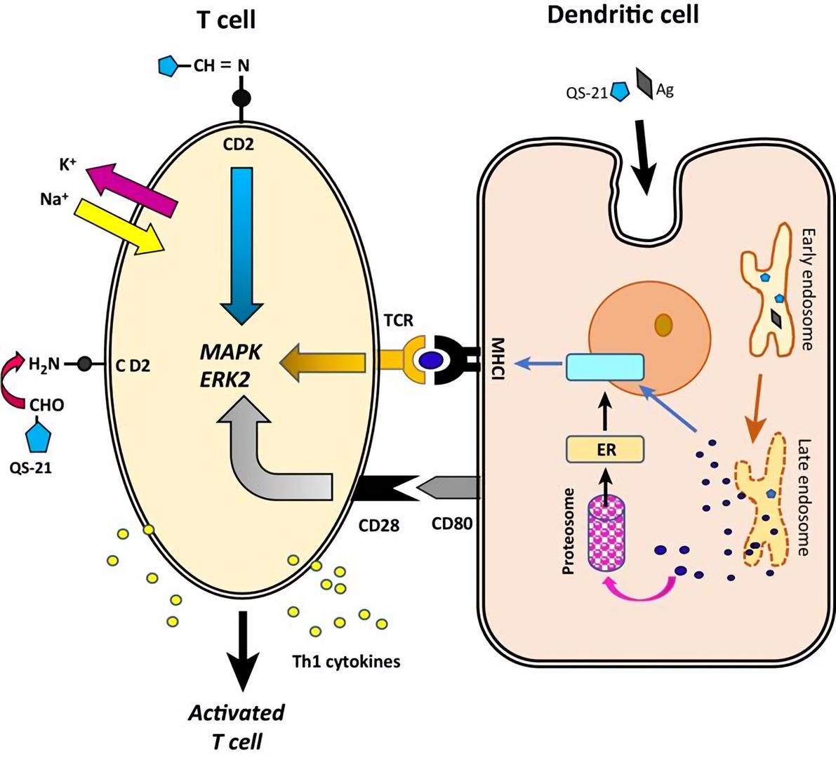
Fig. 2 Schematic diagram of QS-21.
The Potential of Saponin Adjuvants
Saponin-type adjuvants have hydrophobic domains from which their surface-active properties arise. They are ideal for use in subunit vaccines and vaccines directed against intracellular pathogens as well as for therapeutic cancer vaccines due to their unique capacity which can stimulate both the Th1 immune response and the production of cytotoxic T-lymphocytes (CTLs) against exogenous antigens. Quil A has been used successfully for veterinary applications. In a trial with HIV-1 env antigen, QS-21 was able to allow a significant dose reduction for the antigen and also enhanced proliferative T-cell responses but not CTL. QS-21 has also been claimed to perform as an adjuvant for DNA vaccines, following both systemic and mucosal administration. But saponin-based adjuvants have some drawbacks such as high toxicity, undesirable haemolytic effect and instability in aqueous phase, which limit their use as adjuvant in the human vaccine development. It has encouraged the research for saponin-based adjuvant from other kinds of natural products. QS-21 significantly outperformed the other classes of adjuvants including glucan formulations, peptidoglycans, amphophilic block copolymers, bacterial nucleosides and bacterial lipopolysaccharides to better serve the broad market.
Creative Biolabs is a leader in the field of vaccine adjuvant development and has focused on the saponin adjuvants for many years. We have experts who are able to help you with the development of the saponin adjuvants for use in vaccines. If you are interested in our services, please contact us for more details.
Related Products
Reference
- Dante J. Marciani. (2018). “Elucidating the Mechanisms of Action of Saponin-Derived Adjuvants.” TRENDS PHARMACOL SCI 39 (6), 573-585.
 Our services are for research use only. We do not provide services directly to individuals.
Online Inquiry
CONTACT US
USA
45-1 Ramsey Road, Shirley, NY 11967, USA
Tel: 1-631-466-5530
Fax: 1-631-207-8356
Email: info@creative-biolabs.com Europe
Heidenkampsweg 58, 20097 Hamburg, Germany
Tel: 44-207-097-1828 Email: info@creative-biolabs.com
People also ask
https://www.ncbi.nlm.nih.gov/books/NBK8423/

General Concepts
Viral Activation of Immunity
Immunity to viral infection is caused by a variety of specific and nonspecific mechanisms. The activation of different immune functions and the duration and magnitude of the immune response depend on how the virus interacts with host cells (on whether it is a cytolytic, steady-state, latent, and/or integrated infection) and on how the virus spreads (by local, primary hematogenous, secondary hematogenous, and/or nervous system spread). Therefore, viral antigens may be present in different parts of the body depending on the route of spread and phase of infection. Local infections at surfaces such as the mucosa can elicit local cell-mediated and humoral (IgA) immune responses, but not necessarily systemic immunity. The host has multiple immune defense functions that can eliminate virus and/or viral disease.
Humoral Immunity: Virus and/or virus-infected cells can stimulate B lymphocytes to produce antibody (specific for viral antigens) Antibody neutralization is most effective when virus is present in large fluid spaces (e.g., serum) or on moist surfaces (e.g., the gastrointestinal and respiratory tracts). IgG, IgM, and IgA have all been shown to exert antiviral activity. Antibody can neutralize virus by: 1) blocking virus-host cell interactions or 2) recognizing viral antigens on virus-infected cells which can lead to antibody-dependent cytotoxic cells (ADCC) or complement-mediated lysis. IgG antibodies are responsible for most antiviral activity in serum, while IgA is the most important antibody when viruses infect mucosal surfaces.
Cell-Mediated Immunity: The term cell-mediated immunity refers to (1) the recognition and/or killing of virus and virus-infected cells by leukocytes and (2) the production of different soluble factors (cytokines) by these cells when stimulated by virus or virus-infected cells. Cytotoxic T lymphocytes, natural killer (NK) cells and antiviral macrophages can recognize and kill virus-infected cells. Helper T cells can recognize virus-infected cells and produce a number of important cytokines. Cytokines produced by monocytes (monokines), T cells, and NK cells (lymphokines) play important roles in regulating immune functions and developing antiviral immune functions.
Virus-Induced Immunopathology
Immune-mediated disease may develop in certain virus infections in which viral antigens and uncontrolled immune hypersensitivity to them persist for a long period. Immune-mediated disease can be mediated by both humoral and cell-mediated immune functions. Immune-complex syndrome can be mediated by virus/virus antigen antibody complexes. T cells (cytotoxic and helper) can also mediate immunopathologic injuries via a number of mechanisms. Immunopathology can result from tissue/organ damage via cytotoxic T cells, inflammation induced via cytokines, antibody plus complement, antibody-antigen complexes and/or ADCC.
Roles of Immune Functions during Viral Infections
The early, nonspecific responses (nonspecific inhibition, natural killer cell activity, and interferon) limit virus multiplication during the acute phase of virus infections. The later specific immune (humoral and cell-mediated) responses function to help eliminate virus at the end of the acute phase, and subsequently to maintain specific resistance to reinfection.
Introduction
The general principles of immunology are presented in Chapter 1. The present chapter discusses viral activation of immunity, humoral and cell-mediated immunity, virus-induced immunopathology, and roles of immune functions during viral infections.
Viral Activation of Immunity
The term immunity as used in this chapter covers the mechanisms by which a host may specifically recognize and react to viruses. The nonspecific defenses are considered in Chapter 49. The host immune response may be beneficial, detrimental, or both. An immune response to a virus appears first during the primary infection of a susceptible, nonimmune host (Table 50-1) and increases during reinfection of an immune host. The specific immune responses that are effective against viruses are (1) cell-mediated immunity involving T lymphocytes and cytotoxic effector T lymphocytes, (2) antibody, with and without its interaction with complement and antibody-dependent cell-mediated cytotoxicity (ADCC), (3) natural killer (NK) cells and macrophages, and (4) lymphokines and monokines (Fig. 50-1). Some of these immune functions may interact, often synergistically, with nonimmune defense mechanisms (see Ch. 49).
Table 50-1
Host Effector Functions Important against Primary Viral Infections.

Figure 50-1
The immune system response to a virus. (1) Virus bearing an antigenic epitope. (2) Processing of antigen to fragments. (3) Presentation of antigen (Ag) to T cells (on the infected cell surface) and B cells (free antigenic pieces or viruses). (4 and 5) Regulator (more...)
Viral Antigens
The degree to which viral antigens are exposed to the host immune defenses is governed by the intracellular replication of viruses and by the several possible types of virus-host cell interaction (Fig. 50-2; see also Ch. 1).

Figure 50-2
Virus-host cell interactions. The degree to which viral antigens are exposed to the host immune defenses is governed by the obligate intracellular replication of viruses. This exposure varies according to the virus-host cell interactions shown here; i.e., (more...)
Acute Cytolytic Infection
Acute cytolytic infection, the most common form of virus-host cell interaction (Fig. 50-2a-c), results in destruction of the infected cell. There are three ways in which the immune system can encounter the virus or virus-specific antigens of cytolytic viruses. In some cases, the immune system encounters viral antigen only when cell lysis releases the virions (Fig. 50-2a). Many viruses (e.g., reoviruses and coxsackieviruses), however, also induce virus-specific antigens on the cell surface before cell death occurs and sometimes before viral multiplication is complete (Fig. 50-2b). In the third type of cytolytic infection (Fig. 50-2c), common among enveloped viruses (e.g., herpesviruses, poxviruses, paramyxoviruses), virus-specific antigens are present on the cell surface and the cells release the infectious virions by budding for a short period before cell death. These viruses (e.g., herpesviruses, poxviruses, and paramyxoviruses) sometimes are disseminated by contiguous spread from cell to cell without exposure to extracellular antibody. Cell-mediated immune responses are believed to be important in controlling the local spread of this type of infection.
Persistent Infections
Some viruses produce a chronic (steady-state) infection rather than an acute infection of the host cell: progeny virions are released continuously, with little adverse effect on cellular metabolism. These cells express virus-specific antigens on their surface and produce abundant virus progeny, but are not killed by the infectious process. In some steady-state infections the progeny virus is released by budding through the cell membrane, and virus can spread from cell to cell without being exposed to the extracellular environment. DNA viruses do not produce steady-state infections, but some RNA viruses (paramyxoviruses and retroviruses) do.
Latent Infections
Latent infections result when an infecting virus (e.g., a herpesvirus) is maintained within a cell for a long time (sometimes years) without giving rise to progeny virus or damaging the cell. Cells infected in this way may express virus-specific antigens on their cell surface. Months to years after infection, the virus in these cells can be reactivated, replicate, and cause disease. The mechanisms by which viruses are maintained intracellularly for long periods and then reactivated are only incompletely understood. Many latent infections occur in sequestered areas of the body (such as the nervous system), where recognition of infected cells by the immune system is believed to be difficult. In addition, any cell that harbors a virus but does not express viral antigens is not recognized by the immune system.
Integrated Virus Infection
There is another type of persistent virus-host cell interaction, integrated virus infection, in which all or part of the viral nucleic acid becomes integrated into the genome of the host cell. Progeny virions may never be assembled or released from the host cell. New virus-specific antigens, however, can be detected within the cell or on the cell surface. Infection with retroviruses is a classic example of this mechanism.
In most cases, the immune system is activated because the virus and its antigens appear in the extracellular fluid or on the cell membrane.
Virus Spread
Another important consideration in how viral infections trigger an immune response is the way in which a particular virus spreads in the host. In animal hosts, four types of viral spread are recognized: (1) local spread, in which the infection is confined largely to a mucosal surface or organ (as in infection of the respiratory epithelium by rhinoviruses or of the gastrointestinal epithelium by rotaviruses); (2) primary hematogenous spread, in which the virus is inoculated directly into the bloodstream (e.g., insect-transmitted viruses) and then disseminates to target organs; (3) secondary hematogenous spread, in which the initial virus infection and replication (often relatively asymptomatic) occur on a mucosal surface with subsequent dissemination to target organs via the bloodstream (e.g., common viral exanthems, poliomyelitis, and mumps); and (4) nervous system spread, in which viruses (such as herpesviruses and rabiesviruses) disseminate via the nervous system. Therefore, viral antigens may be present in different parts of the body depending on the route of spread and phase of infection. Different immune mechanisms may operate at the various sites of virus spread and infection .
Virus Location
The location of the virus in the host is important not only for understanding the immune response, but also for developing and administering a vaccine. For example, local infections on surfaces such as the mucosa of the respiratory or gastrointestinal tract may elicit local cell-mediated and humoral (IgA) immune responses, but not necessarily systemic immunity. The reverse is also true: systemic immunity does not always lead to local mucosal immunity. For example, the Salk polio vaccine, which consists of killed virus administered systemically, elicits serum IgG as the major antibody and induces little or no secretory response. As a result, the immunized individual resists systemic infection, but may become a temporary carrier, with virus persisting at the intestinal portal of entry because of the lack of secretory antibody. The orally administered, live Sabin polio vaccine, on the other hand, induces secretory antibody in the intestine and is effective in preventing replication and subsequent mucosal penetration by the virus.
Multiplicity of Immune Defenses
Recent studies have revealed a great complexity of host immune defenses against viral infections. This complexity arises from the many components of the host immune defenses and their interactions with one another. The existence of a variety of defenses is not surprising in view of the diversity of viruses, hosts, routes of infection, body compartments, cells, and mechanisms of virus multiplication and spread. The situation is further complicated by the varying effectiveness of the different host defenses during the different phases of the primary viral infection (implantation, spread to target organs, and subsequent recovery of each of the infected tissues), as well as during resistance to reinfection. Furthermore, the activated host defenses can actually cause disease manifestations. The presence of multiple defenses against each infection helps explain why impairment of one or a few defenses does not entirely abrogate host resistance to viral infections. Several immune and nonimmune host defenses may operate to control viral infections or, at times, add to the disease process.
Many of the immune defenses against viral invasion are fairly well understood, but the relative effectiveness of each requires additional research. In particular, as this chapter attempts to make clear, humoral and cell-mediated immunity are not independent, but interact intimately to influence the duration and magnitude of each type of immune response.
Humoral Immunity: B Lymphocytes
As described in Chapter 1, specific B lymphocytes respond to viral antigen introduced by immunization or infection. Binding of antigen to the cell surface immunoglobulin receptors, followed by interaction of the B cell with macrophages and helper T lymphocytes, causes the B cell to differentiate into clones of antibody-secreting plasma cells, each capable of secreting antigen-specific immunoglobulin of one of five major classes: IgG, IgM, IgA, IgD, and IgE (Fig. 50-1). Antibodies act against viruses primarily by binding to and neutralizing virions and by directing the lysis of infected cells by complement or killer leukocytes.
Antibody-Mediated Reactions
Neutralization of virion infectivity
At least three immunoglobulin classes have been demonstrated to exert antiviral activity: IgG, IgM, and IgA. These antibodies can neutralize the infectivity of virtually all known viruses. Antibody binds to the virus extracellularly, either neutralizing it immediately or blocking its interaction with host cells. Antibody that has bound to virus can block the infection of a cell at one of three steps: (1) attachment of virus to the cell surface, (2) penetration of virus into the cell, and (3) uncoating of virus inside the cell (Fig. 50-3). The mechanism of viral neutralization involves the binding of antibody to virus coat proteins; this usually alters the viral receptor for the target cell. More rarely, bound antibody may also interfere with penetration or uncoating.

Figure 50-3
Mechanisms of virus neutralization by antibody at the cellular level. At the cellular level, antibody can block the following steps associated with a virus infection: (1) virus attachment and adsorption to the cell surface, (2) penetration of the virus into (more...)
The exact mechanism of neutralization is unclear, but it probably involves changes in the steric conformation of the virus surface. These antibody-virus interactions can take place independently of complement.
Antibody also can neutralize virus by causing aggregation (Fig. 50-4), thus preventing adsorption of virus to cells and decreasing the number of infectious particles. Antibody and complement acting together can inactivate certain viruses (in most cases, enveloped viruses). Antibody is most effective against virus in large fluid spaces (e.g., serum) and on moist body surfaces (e.g., the respiratory and gastrointestinal tracts), where the virus is exposed to antibody for a relatively long period before escaping into cells. Consequently, viruses that spread by viremia are effectively eliminated by low levels of circulating antibody. Much higher levels of antibody are needed to prevent the spread of viruses that do not travel in the blood plasma (such as herpesviruses and rabiesviruses), because these viruses spend only a brief period traversing the small extracellular spaces between cells in solid tissue.
Figure 50-4
Extracellular neutralization of virus by antibody. Antibody can reduce the number of infectious particles by linking virions and thereby causing aggregation. Antibody alone or with complement can also inactivate viruses.
Besides binding directly to virus, antibodies may enhance phagocytosis. Three types of antibody interactions with phagocytic cells are seen: direct binding of antibody to the surface of the phagocytic cells (cytophilic antibody), uptake of antigen antibody complexes through the Fc receptor, and uptake of antigen-antibody-complement complexes through the C3b receptor (see Ch. 1). This phagocytosis of virions may result in inactivation of virus (see Ch. 49), and in the activation of the phagocytic cell which can lead to cytokine production.
Acute Cytolytic Infection
Acute cytolytic infection, the most common form of virus-host cell interaction (Fig. 50-2a-c), results in destruction of the infected cell. There are three ways in which the immune system can encounter the virus or virus-specific antigens of cytolytic viruses. In some cases, the immune system encounters viral antigen only when cell lysis releases the virions (Fig. 50-2a). Many viruses (e.g., reoviruses and coxsackieviruses), however, also induce virus-specific antigens on the cell surface before cell death occurs and sometimes before viral multiplication is complete (Fig. 50-2b). In the third type of cytolytic infection (Fig. 50-2c), common among enveloped viruses (e.g., herpesviruses, poxviruses, paramyxoviruses), virus-specific antigens are present on the cell surface and the cells release the infectious virions by budding for a short period before cell death. These viruses (e.g., herpesviruses, poxviruses, and paramyxoviruses) sometimes are disseminated by contiguous spread from cell to cell without exposure to extracellular antibody. Cell-mediated immune responses are believed to be important in controlling the local spread of this type of infection.
Humoral Immunity: B Lymphocytes
As described in Chapter 1, specific B lymphocytes respond to viral antigen introduced by immunization or infection. Binding of antigen to the cell surface immunoglobulin receptors, followed by interaction of the B cell with macrophages and helper T lymphocytes, causes the B cell to differentiate into clones of antibody-secreting plasma cells, each capable of secreting antigen-specific immunoglobulin of one of five major classes: IgG, IgM, IgA, IgD, and IgE (Fig. 50-1). Antibodies act against viruses primarily by binding to and neutralizing virions and by directing the lysis of infected cells by complement or killer leukocytes.
Antibody-Mediated Reactions
Neutralization of virion infectivity
At least three immunoglobulin classes have been demonstrated to exert antiviral activity: IgG, IgM, and IgA. These antibodies can neutralize the infectivity of virtually all known viruses. Antibody binds to the virus extracellularly, either neutralizing it immediately or blocking its interaction with host cells. Antibody that has bound to virus can block the infection of a cell at one of three steps: (1) attachment of virus to the cell surface, (2) penetration of virus into the cell, and (3) uncoating of virus inside the cell (Fig. 50-3). The mechanism of viral neutralization involves the binding of antibody to virus coat proteins; this usually alters the viral receptor for the target cell. More rarely, bound antibody may also interfere with penetration or uncoating.
Figure 50-4
Extracellular neutralization of virus by antibody. Antibody can reduce the number of infectious particles by linking virions and thereby causing aggregation. Antibody alone or with complement can also inactivate viruses.
Besides binding directly to virus, antibodies may enhance phagocytosis. Three types of antibody interactions with phagocytic cells are seen: direct binding of antibody to the surface of the phagocytic cells (cytophilic antibody), uptake of antigen antibody complexes through the Fc receptor, and uptake of antigen-antibody-complement complexes through the C3b receptor (see Ch. 1). This phagocytosis of virions may result in inactivation of virus (see Ch. 49), and in the activation of the phagocytic cell which can lead to cytokine production.
Antibody effects on virus-infected cells
Antibody also can act on virus-infected cells by recognizing virus-specific antigens on the surface of infected cells (Fig. 50-3). Complement can then cause lysis of these cells. This complement-mediated lysis occurs both by the classic and the alternative complement pathways. Antibody-coated infected cells also can be destroyed by various effector cells via ADCC. Alternatively, however, some antibodies can mask viral antigens on the surface of infected cells, thereby removing or covering antigens on the surfaces of these infected cells.
Physical barriers to antibody
Before antibody can combine with and neutralize the virus, it must reach the site of virus replication. Barriers to the distribution of antibody include the cell membrane, which excludes antibody, and anatomic tissue barriers, which limit the distribution of macromolecules into certain organs such as the central nervous system.
IgG Antibodies
IgG is the most thoroughly studied antibody class and is responsible for most antiviral activity in serum. IgG antibodies reach infected (inflamed) sites by transduction (leakage) from capillaries. IgG is particularly protective in generalized viral infections that have a viremic phase (e.g., measles, polio, and hepatitis), perhaps because virions in serum are exposed to antibody. IgG antibodies are transferred passively from mother to offspring through the placenta and usually provide temporary protection against generalized viral infections during the first 6 to 9 months of life. Antibody is most protective when present before infection or during the spread of virus to target organs.
Production and the roles of antibody classes
After immunization or infection with viruses, various classes of antibody appear sequentially. For example, during primary infection or immunization, most antigens first elicit IgM (early antibody) responses; IgA and IgG responses follow within a few days. Reinfection, in contrast, stimulates production mainly of IgG, although some IgM and IgA are generated. When the primary antigenic stimulation is in the respiratory or gastrointestinal tract, IgA antibody is predominant, accompanied by some IgM. These antibodies are secreted locally at mucosal surfaces and are important in protecting the host against localized surface viral infections such as the common cold, influenza, and enteric viral infections. When viral replication is confined to a mucosal surface, resistance to infection is determined primarily by secretory IgA; serum IgG antibody provides less protection. Viral infections that begin on a mucosal surface and then spread hematogenously (e.g., measles, rubella, and polio) can be prevented at the mucosal stage by local secretory antibody and at the viremic stage by IgG antibodies. If serum IgG only is induced in a host, hematogenous spread can be prevented, but viral replication still may occur on the mucosal surface.
IgE antibodies and immediate hypersensitivity
Recent information suggests that viruses that bind to IgE antibodies may trigger immediate hypersensitivity responses through the release of vasoactive mediators (see Ch. 1). These observations may explain many of the apparent allergic manifestations, such as wheezing and urticaria, that accompany some viral infections.
Complement
Complement enhances the phagocytosis of many viruses. This enhanced phagocytosis is due to coating (opsonization) of virions by complement or by complement bound to antibody. Complement also can neutralize virus by enhancing either antibody-mediated steric changes on the virus or aggregation of the virus via antibody. In addition, complement can directly inactivate antibody-coated, enveloped virions.
Hypogammaglobulinemia
A small minority of patients with impaired B-lymphocyte function (hypogammaglobulinemia limited to impairment of humoral immunity) have a significantly increased frequency of severe poliovirus and enterovirus infections of the nervous system (in addition to more frequent and severe infections with pyogenic bacteria). The risk of central nervous system invasion is related to the duration of viremia, as has been shown in immunosuppressed animals. The course of most viral infections is typically benign in most of these hypogammaglobulinemic patients, indicating that their weak antibody response and other defense mechanisms may be effective. The development of normal specific resistance to reinfection in hypogammaglobulinemic patients may result, in part, from their ability eventually to produce low levels of serum antibody to virus, as well as from the action of their intact cell-mediated immune system.
Cell-Mediated Immunity
Cell-mediated immunity (CMI) was once thought to be mediated solely by T lymphocytes; however, it is now clear that it is mediated by a variety of cell types, cell factors, or both. Virus-infected or virally transformed cells activate strong cell-mediated immune responses (Fig. 50-1). For some viral infections, cell-mediated immune reactions may be more important than antibody in early termination of viral infection and prevention of dissemination within the host. Recent evidence shows that cell mediated immunity functions at the body surfaces, as well as internally. Cell-mediated immune responses to viral infections involve T lymphocytes, ADCC, macrophages, natural killer (NK) cells, lymphokines, and monokines (Figs. 50-5 and 50-6).

Figure 50-5
Lysis of virus-infected cells by cytotoxic effector cells. Cytotoxic effector cells that can destroy virus-infected cells include cytotoxic T cells, natural killer cells, and activated macrophages. Cytotoxic T lymphocytes can recognize and destroy virus-infected (more...)
Figure 50-6
Cell-mediated events in viral infections. Soluble mediators include immune interferon, chemotactic factors, macrophage migration inhibitory factor, and lymphotoxin; other lymphokines and monokines are not depicted. Cytotoxic effector lymphocytes, macrophages, (more...)
T Lymphocytes
Much evidence indicates that T lymphocytes are important in recovery from viral infections. Of the many functional subsets of T cells, those that express specific cytotoxic activity against virus-infected or transformed cells have aroused the most interest.
Cytotoxic T lymphocytes
The generation of virus-specific cytotoxic T lymphocytes (CTLs) is believed to be important in preventing viral multiplication (Fig. 50-5). Presumably, the T lymphocytes prevent virus multiplication by destroying infected cells before mature, infectious virus particles can be assembled. This hypothesis assumes that viral antigens appear on the plasma membrane before the release of virus progeny, a view that is substantiated by studies of many, but not all, infections.
Exposure to a virus-infected cell can cause the antigen-specific T lymphocytes to differentiate into cytotoxic effector T cells, which can lyse virus infected or virally transformed cells. These cytotoxic T cells are specific not only for the viral antigen but also for self major histocompatibility antigens and will lyse virus-infected cells only if these cells also express the correct major histocompatibility complex (MHC) gene products.
Activation of cytotoxic and other T lymphocytes may be one of the earliest manifestations of an immune response. T-cell effector functions occur as early as 3 to 4 days after initiation of a viral infection. However, T-cell responses often decrease rapidly, within 5 to 10 days of elimination of the virus (although virus-specific memory T cells persist for long periods). In contrast, antibodies usually become measurable later in the viral infection (after 7 days) and persist at high levels for much longer (often for years).
Helper T cells may be as important as cytotoxic T cells in the immune response to a virus infection. Helper T cells are required for the generation of cytotoxic T cells and for optimal antibody production. In addition, helper T cells, and cytotoxic T cells produce a number of important soluble factors (lymphokines) that can recruit and influence other cellular components of the immune and inflammatory responses.
Animal studies indicate that impairing the T-cell defenses enhances infections by herpes simplex virus, poxviruses, and Sindbis virus and enhances the development of tumors induced by polyomavirus. Since the host retains some resistance to infections, T lymphocytes probably are not the sole defense against these viruses. Impairment of T lymphocytes also hinders T cell-dependent antibody production. In humans, T-cell impairment is associated mainly with more frequent and severe poxvirus and herpesvirus infections. Nevertheless, these infections still do not develop in most individuals with T-cell deficiencies, even though the prevalence of herpesviruses (and many other viruses) is great.
Antibody-Dependent Cell-Mediated Cytotoxicity
Effector leukocytes for ADCC have surface receptors that recognize and bind to the Fc portion of IgG molecules. When IgG binds to virus-specified antigens on the surface of an infected cell, the Fc portion becomes a target for effector cells capable of mediating ADCC . Binding of these effector cells to the Fc portion of IgG bound to the infected-cell surface antigens results in lysis of the infected cell. ADCC is a very efficient way of lysing virus-infected cells because it requires significantly less antibody than does antibody-complement lysis.
Lymphocytes, macrophages, and neutrophils are all capable of mediating ADCC against virus infected cells. The lymphocytes with this ability appear to be heterogeneous. Natural killer cells, as well as null lymphocytes with Fc receptors for IgG, appear to be able to mediate ADCC activity.
Macrophages
Macrophages are important in both specific and nonspecific responses to viral infections (e.g., herpesvirus infections). Factors that modify macrophage activity can influence the outcome of an infection. Moreover, since macrophages are central to the induction of T and B lymphocyte responses, any effect on macrophages will influence B and T cells.
Macrophages confer protection against viruses through either an intrinsic or an extrinsic process. In the former, virions are disposed of within macrophages acting either as phagocytes or as nonpermissive host cells. In the latter case, macrophages retard or ablate virus multiplication in neighboring cells by destroying virus-infected cells or by producing soluble factors (interferons) that act on these cells. Phagocytosis of some viruses by macrophages decreases virus levels in body fluids (as during viremia) and thereby impedes virus spread. These effects are produced only if the virus is destroyed or contained by macrophages. If a virus replicates in macrophages, the infected macrophages may aid in transmission of the virus to other body cells. The permissiveness of macrophages for virus replication may depend on the age and genetic constitution of the host and on the specific condition of the macrophages.
Macrophage activation mediated either by products of infection (viral and cellular) or by soluble factors produced by T cells (e.g., gamma interferon) often enhance phagocytosis and the elimination of free virus particles. Another important effector mechanism of activated macrophages is their ability to recognize and destroy virus-infected and virus-transformed cells (Fig. 50-5). In addition, activated macrophages participate in virus inhibition by producing cytokines (interferon, etc.) and mediating ADCC.
Natural Killer Cells
Natural killer (NK) cells exhibit cytotoxic activity against a number of tumor cell lines, particularly against virus-infected or virus-transformed cells (Fig. 50-5). Natural killer or natural killer-like cells, which have been found in almost every mammalian species examined and even in some invertebrates, are identified as large granular lymphocytes that possess Fc receptors. They can mediate ADCC activity; their nonspecific cytotoxic activity is increased by interferon and interleukin-2 (IL-2); and they can produce a number of different cytokines including interferon when stimulated with virus or virus-infected cells.
Although natural killer cells display cytotoxic activity against virus-infected or transformed cells, they show little or no cytotoxic activity against normal cells. Unlike that of cytotoxic T lymphocytes, natural killer cell killing is not human leukocyte antigen (HLA) restricted, and natural killer cells do not exhibit conventional immunologic specificity. There is evidence that natural killer cells play an important defensive role in virus infections in humans and animals. Their importance is believed to be due to their ability to produce cytokines and to kill virus-infected cells.
Lymphokines and Monokines
Soluble factors from T lymphocytes (lymphokines) and macrophages (monokines) regulate the degree and duration of the immune responses generated by T lymphocytes, B lymphocytes, and macrophages (see Ch. 1). Interleukin-2 and gamma interferon are two such important factors produced by activated T cells. Interleukin-l is a soluble factor produced by macrophages. All three of these factors are essential for the full differentiation and proliferation of cytotoxic T cells. The two interleukins are also important for antibody production by B lymphocytes.
Macrophages and T lymphocytes also produce several other important factors that act in both the immune and the inflammatory responses. Gamma interferon can activate macrophages to become cytotoxic toward virus-infected cells and can increase the level of phagocytosis and degradation. Lymphotoxins produced by T cells also may participate in the destruction of virus-infected cells. Virus can stimulate alpha interferon production from macrophages; this enhances natural killer cell function and inhibits virus multiplication in neighboring cells.
Virus-Induced Immunopathology
A host clearly has numerous mechanisms to recognize and eliminate the viruses that it encounters. However, some viruses persist despite these mechanisms, and then the immune responses may become detrimental to the host and cause immune-mediated disease. When an antigen (virus) persists, pathologic changes and diseases result from different types of immunologic interactions, including immediate hypersensitivity, antibody-mediated immune complex syndrome, and tissue damage caused by cell-mediated effector cells and antibody plus complement. Of these mechanisms, the immune complex syndrome during viral infections has been studied most intensively. Two major complications of deposition of immune complexes are vascular damage and nephritis. Some viral diseases in which immune complexes have been demonstrated are hepatitis B, infectious mononucleosis, dengue hemorrhagic fever, and subacute sclerosing panencephalitis.
Cytotoxic T cells also mediate immunopathologic injury in murine models of human infections (i.e., infections with lymphocytic choriomeningitis virus and poxviruses). Both cytotoxic T cells and T cells responsible for delayed-type hypersensitivity have also been implicated in the pathology associated with influenza pneumonia and coxsackievirus myocarditis of mice. A delicate balance between the removal of infected cells that are the source of viral progeny and injury to vital cells probably exists for T cells as well as for the other host immune components.
Viruses may sometimes circumvent host defenses. An important factor that may impair the function of sensitized T lymphocytes is apparent from the observation that T cells activated by reaction with antigen or mitogen lose their normal resistance to many viruses. Therefore, these activated T lymphocytes develop the capacity to support the replication of viruses, leading to impairment of T lymphocyte function.
Roles of Immune functions During Viral Infections
On the basis of the mechanisms described here and in Chapter 49, a hypothetical model can be constructed that shows how the immune components defend against viruses (Fig. 50-1; Table 50-1).
Nonspecific Defenses
A primary infection in a nonimmune, susceptible host is countered first by the nonspecific defense mechanisms (see Ch. 49 ). The early nonspecific responses occur within hours and consist of interferon production, inflammation, fever, phagocytosis, and natural killer cell activity. These defenses may prevent or abort infection; if they do not, the virus is disseminated by local spread, viremia, or nerve spread. It then may seed to a number of target organs and thereby produce a generalized infection.
Specific Defenses Antibody
The events that lead to a specific immune response begin almost immediately after exposure and result in the production of antiviral antibody and cell-mediated immunity in 3 to 10 days. The disseminated antibody response in serum is predominantly IgG (preceded by IgM); the local antibody response in secretions is predominantly secretory IgA (with some IgM). The persistence of IgA antibodies in secretions is much shorter (months) than the persistence of IgG antibody in serum (years). The role of IgE in secretions is unknown, but it may mediate immediate hypersensitivity and amplify the immune response during infection. Antibodies may neutralize virus directly or destroy virus-infected cells via ADCC or complement. Clearly, serum antibody confers protection against generalized infections (e.g., measles, polio, and type A hepatitis), in which virus must spread through the antibody-containing bloodstream; inoculation of small quantities of antibody into susceptible individuals prevents viral disease but may not prevent subclinical infection at mucosal surfaces.
In localized infections of mucosal surfaces, protection does not correlate with the presence of serum antibody, but it does correlate with the presence of local IgA antibody, as has been shown in human studies of viruses restricted to the respiratory tract (e.g., respiratory syncytial virus and influenza virus) or to the gastrointestinal tract (e.g., enteroviruses). Under some conditions in which serum antibody is present but local IgA is absent, hypersensitivity instead of protective immunity may occur (e.g., respiratory syncytial virus infection). Also, serum antibody may not protect against recurrence of latent infections, such as herpes zoster (shingles) and herpes simplex, both because the virus may be shielded by its intracellular location and because cell-mediated immunity may be the more important defense. Antibody may also cause undesirable effects in certain chronic infections. Examples in which small amounts of serum antibody complex with virus and deposit in the kidneys, thereby inducing immune complex disease, are listed in Table 50-1.
Therefore, serum IgM and IgG antibody seem to be effective in preventing infections of a generalized nature; however, in localized surface infections the presence of secretory IgA antibody appears to correlate much better with protection than the presence of circulating IgG antibody. In persistent infections, serum antibody may be responsible for certain long-term sequelae.
Cell-Mediated Immunity
Cell-mediated immunity is essential in recovery from and control of viral infections, especially infections involving oncogenic viruses or viruses that spread directly from cell to contiguous cell. In these situations antibody cannot reach the virus but virally induced antigens on the surface of the infected cell can be recognized by different effector cells (e.g., cytotoxic T cells) (Fig. 50-6).
If the virus reaches target organs, it is more difficult to control. The host defenses that may play important roles in target organs are initially inflammation, fever, and interferon and subsequently cell-mediated immunity.
In some situations, cell-mediated immunity may develop before antibody production begins. For example, cytotoxic effector T cells have been found in bronchial washings 3 to 4 days after initiation of intranasal infection in mice; at this time, antibody cannot yet be detected.
Cell-mediated immune responses can cause tissue damage; the lung lesions produced in influenza may be examples. The lethal effects of lymphocytic choriomeningitis virus in mice are mediated by cytotoxic effector T cells. The rash in many exanthems (such as measles) is thought to represent a cell-mediated attack on virus localized within cells of the dermis and its vasculature.
References
- Baron S, Grossberg SE, Klimpel GR, Brunell PA: Mechanisms of action and pharmacology: the immune and interferon systems. In Galasso G (ed): Antiviral Agents and Viral Diseases of Man. Raven Press, New York, 1984 .
- Biron CA. Cytokines in the generation of immune responses to, and resolution of, virus infection. Currrent Opinion in Immunology. 1994;6:530. [PubMed]
- Brenner BG, Grylles C, Wainberg MA. Role of antibody-dependent cellular cytotoxicity and lymphokine-activated killer cells in AIDS and related diseases. J Leukocyte Biology. 1991;50:628. [PubMed]
- Doherty PC. Cell-mediated immunity in virus infections of the central nervous system. Ann NY Acad Sci. 1988;540:228. [PMC free article] [PubMed]
- Herberman RB, Ortaldo JR. Natural killer cells: their role in defenses against disease. Science. 1981;214:24. [PubMed]
- Hirsch RL, Winkelstein JA, Griffin DE. The role of complement in viral infections. III. Activation of the classical and alternative complement pathways by Sindbis virus. J Immunol. 1980;124:2507. [PubMed]
- McChesney MB, Oldstone MBA. Viruses perturb lymphocyte functions: selected principles characterizing virus-induced immunosuppression. Annu Rev Immunol. 1987;5:279. [PubMed]
- Mogensen LA. Role of macrophages in natural resistance to virus infections. Microbiol Rev. 1979;43:1. [PMC free article] [PubMed]
- Ogra PL, Leibovitz EE, Zhao RG. Oral immunization and secretory immunity to viruses. Curr Top Microbiol Immunol. 1989;146:73. [PubMed]
- Rouse BT, Norley S, Martin S. Antiviral cytotoxic T lymphocyte induction and vaccination. Rev Infect Dis. 1988;10:16. [PubMed]
- Sissons JGP, Oldstone MBA. Antibody-mediated destruction of virus-infected cells. Adv Immunol. 1980;29:209. [PMC free article] [PubMed]


People also watched


Adjuvants / Saponins


No comments:
Post a Comment
Note: Only a member of this blog may post a comment.