1Pediatric Research Institute, The Second Affiliated Hospital and Yuying Children's Hospital of Wenzhou Medical University, Wenzhou, China
2The Second Clinical Medical College of Wenzhou Medical University, Wenzhou, China
3Children's Heart Center, Institute of Cardiovascular Development and Translational Medicine, The Second Affiliated Hospital and Yuying Children's Hospital of Wenzhou Medical University, Wenzhou, China
4Department of Neurology, The First Affiliated Hospital of Wenzhou Medical University, Wenzhou, China
5The Institute of Life Sciences, Wenzhou University, Wenzhou, China
6Tianjin Key Laboratory of Radiation Medicine and Molecular Nuclear Medicine, Institute of Radiation Medicine, Chinese Academy of Medical Sciences and Peking Union Medical College, Tianjin, China
As the prevalence of systemic fungal infections caused by Candida albicans gradually increases, it is necessary to explore potential and effective antifungals. Carvacrol is reported to be lethally toxic to C. albicans, involving several potential mechanisms. However, the form and specific mechanism of cell death caused by this compound has not been delineated. In this study, we found that carvacrol could significantly decrease C. albicans survival rates, consistent with previous researches. Further examination proved that carvacrol treatment caused cell membrane permeability and depolarization. To elucidate the association between cell death and apoptosis, DNA fragmentation and metacaspase activation were determined; as expected, these two apoptosis-related markers were clearly observed. Moreover, total and mitochondrial reactive oxygen species (ROS) levels were elevated, and both mitochondrial transmembrane potential and morphology were disrupted. Additionally, cytosolic and mitochondrial calcium levels were also increased by carvacrol. Calcineurin inhibition experiments revealed cyclosporine A (CsA) addition notably rescued cell growth and inhibited metacaspase activation, indicating that carvacrol triggered C. albicans apoptosis through inducing calcineurin activation. Carvacrol was demonstrated to both have low toxicity and be effective in alleviating systemic infections with C. albicans, which might be via its antifungal and immunomodulation activities. This study suggests that carvacrol has excellent potential as a natural protective compound against C. albicans infections.
Introduction
Candida albicans can lead to both topical epithelial and fatal invasive infections in immunocompromised patients, contributing to its status as the fourth most common cause of nosocomial blood-stream infections in US hospitals (Klepser, 2006). The increased systemic infections resulting from C. albicans underly mortality rates of at least 50% even though there are presently available antifungal therapy (Pfaller and Diekema, 2007). Therapeutic options are currently restricted to the utilization of the three longstanding antifungal classes: azoles, polyenes, and echinocandins (Chaillot et al., 2015). However, these abovementioned drugs have severe side effects, including nephrotoxicity and fungal resistance due to their fungistatic rather than fungicidal effects (Shapiro et al., 2011). Therefore, it is urgent to explore novel and more effective strategies for antifungal therapeutic intervention.
Traditional Chinese medicines provide an interesting reservoir of potentially useful and effective antimicrobial secondary metabolites. Carvacrol, a monoterpene phenol, is major component of essential oil extracts from plants in the family Lamiaceae including oregano (Origanum vulgare L.) and marjoram (Origanum marjoram L.) (Nunes Wolffenbuttel et al., 2015). This phytomolecule in low concentration is considered safe for humans and it is commonly applied as a flavoring agent (Suntres et al., 2015). In addition, this compound has extensively demonstrated pharmacological properties, including antifungal and antibacterial activities, and immunoregulatory potential (Wieten et al., 2010). Previous studies have reported that carvacrol can impede the growth of different morphological forms of C. albicans, including yeast, hyphae, and biofilm (Inouye et al., 2009; Lima et al., 2013; Raut et al., 2013). Moreover, the anti-C. albicans activity of carvacrol has also been substantiated in the rat vaginal candidiasis model (Chami et al., 2004b) and murine systemic candidiasis model (Manohar et al., 2001). However, the antifungal activity of this compound has not been completely elucidated in vivo. Currently reported mechanisms of action for this phytomolecule include disrupting ergosterol biosynthesis and membrane integrity (Ahmad et al., 2011), inducing endoplasmic reticulum (ER) stress and the unfolded protein response (UPR) (Chaillot et al., 2015), and perturbing H+ and Ca2+ ion homeostasis (Rao et al., 2010). Although all the aforementioned modes of action of carvacrol can kill C. albicans, the form and ultimate mechanism of cell death in C. albicans caused by carvacrol remains unclear.
In the present study, to expound the types of cell death, various parameters related to apoptosis were assessed in C. albicans cells treated with carvacrol. In addition, the antifungal effect and immunoregulation activity of carvacrol were also determined in a murine model of C. albicans infection. Our study elucidated that the antifungal efficacy of carvacrol against C. albicans was realized through inducing apoptosis. Furthermore, carvacrol exhibited obvious antifungal and immunoregulation properties in the murine systemic candidiasis model.
Materials and Methods
Chemicals, Strains, and Cultures
Carvacrol (>99%, CAS: 499-75-2) was obtained from Weikeqi Biotechnology Co., Ltd. (Sichuan, China). Its chemical structure was shown in Figure 1. A stock carvacrol (494 mg/ml) was made by dissolving this compound in dimethyl sulfoxide (DMSO). The three strains of C. albicans used in this study were SC5314, ATCC18804, and 07318, and each was cultured at 30°C in yeast extract peptone dextrose (YPD) medium.
FIGURE 1
Figure 1. Chemical structure of carvacrol and its effect on C. albicans survival. The chemical structure of carvacrol was exhibited, and the effect of carvacrol on C. albicans survival was analyzed by determining CFUs. Data were shown as mean ± SD (n = 5). Significant difference was designated as **P < 0.01 and ***P < 0.001.
Antifungal Activity Testing
Microdilution methods were used to determine the minimal inhibitory concentration (MIC). Specifically, aliquots of 100 μl of C. albicans cell suspensions (1 × 10⁵ cells/ml) with different carvacrol concentrations were transferred into a series of 96-well plates. After a 48-h incubation at 30°C, the MIC of carvacrol against SC5314 strain was examined (Tian et al., 2017; Yun and Lee, 2017). This experiment was conducted in triplicate.
Overnight cultures were diluted in YPD medium to an OD600 value of 0.1. After reaching the mid-exponential phase, C. albicans cells cultures were subjected to 3-h treatments with 247 and 494 μg/ml carvacrol. The treated cells were then collected, and about 103 cells were coated onto plates of YPD agar. Following a 24-h aerobic incubation at 30°C, the numbers of colony forming units (CFUs) were determined. Percentage survival estimates were detected relative to the untreated control cells, and each experiment was conducted independently in triplicate.
Cell Membrane Integrity Assay
Analysis of cell membrane integrity was conducted according to a modified version of the method described by Li et al. (2016). Specifically, after C. albicans cells were treated with carvacrol, they were collected, and stained for 30 min at 4°C with 10 mg/ml propidium iodide (PI) in darkness. Then, cell membrane integrity was assayed using a FACSCalibur flow cytometer (Becton Dickinson, United States).
Plasma Membrane Potential Determination
DiBAC4(3)(bis-(1,3-dibarbituric acid)-trimethine oxanol) was used as a membrane potential molecular probe to assess changes in plasma membrane potential as previously described (Li et al., 2016). In brief, C. albicans cells were treated, harvested, and washed thrice with phosphate-buffered saline (PBS). Next, the C. albicans cell suspension was incubated with 20 mg/ml DiBAC4(3) at 37°C for 30 min in darkness. Depolarized C. albicans cells were examined by flow cytometry, and the percentage of depolarized cells was recorded.
Metacaspase Activation Assay
The CaspACE FITCVAD-FMK in situ marker (Promega, Madison, WI, USA) was utilized to detect metacaspase activation (Tian et al., 2017). Specifically, carvacrol-treated C. albicans cells were collected and washed with PBS. Then, cells staining was conducted with CaspACE FITC-VAD-FMK at a 5 mM final concentration at 37°C for 20 min in darkness. Next, the stained cells were again washed with PBS and assayed using flow cytometry.
DNA Fragmentation Evaluation
Terminal deoxynucleotidyl transferase dUTP nick end labeling (TUNEL) staining was utilized to examine C. albicans DNA fragmentation (Jia et al., 2018). Specifically, treated C. albicans were harvested, washed with PBS, and fixed for 30 min in 3.6% paraformaldehyde followed by a 2-min permeabilization on ice. Next, the cells were again washed before being stained with an in situ cell death detection kit at 37°C for 1 h in darkness. DNA fragmentation was finally assessed by flow cytometry.
Intracellular ROS Measurement
To measure C. albicans reactive oxygen species (ROS) levels, the fluorescent probe 2′, 7′–dichlorofluorescein diacetate (DCFH-DA) was utilized. DCFH-DA can be cleaved by intracellular esterase into the membrane-impermeable agent DCFH, which ROS can then oxidize into its fluorescent derivative, DCF. Thus, the fluorescence intensity of DCF is an indicator of the total ROS levels (Jia et al., 2019). Accordingly, carvacrol-treated C. albicans cells were gathered, washed with YPD medium one time, and resuspended in 0.5 ml of YPD medium containing 10 mM DCFH-DA. Following a 30-min incubation in the dark, the fluorescence intensity of C. albicans cells was assessed using flow cytometry.
Mitochondrial Superoxide Anion Determination
MitoSOX Red mitochondrial superoxide indicator was utilized to determine the mitochondrial ROS levels (Zhou et al., 2019). Specifically, carvacrol-treated C. albicans cells were collected, washed, stained with MitoSOX Red mitochondrial superoxide indicator at a final concentration of 5 μM, and incubated for 20 min at 37°C. After staining, cells were harvested and assayed using a flow cytometer.
Mitochondrial Membrane Potential Determination
To examine the mitochondrial transmembrane potential, 5,5′,6,6′-tetrachloro-1,1′,3, 3′-tetraethyl-benzimidazolyl carbocyanine iodide (JC-1) was applied (Jia et al., 2019). Carvacrol-treated C. albicans cells were gathered by centrifugation, twice washed with PBS, and then incubated for 20 min with 2.5 μg/ml JC-1 at 37°C in darkness. After the stained cells were washed again with PBS, they were assayed using a FACSCalibur flow cytometer. The mitochondrial membrane potential was then estimated as the ratio of the fluorescence intensity of JC-1 aggregates (FL2) to that of its monomer (FL1).
Examination of Mitochondrial and Cytosolic Calcium Levels
Rhod-2 AM and Fluo-3 AM were, respectively, used to examine the mitochondrial and cytosolic calcium levels (Tian et al., 2017; Jia et al., 2018). C. albicans cells were treated with 247 and 494 μg/ml carvacrol. Next, the carvacrol-treated C. albicans cells were centrifuged, collected, washed twice with Hank's balanced salt solution (HBSS), and resuspended in 500 μl of HBSS buffer. To assay mitochondrial calcium levels, the washed C. albicans cells were stained with 4 mM Rhod-2 AM for 30 min at 37 °C in the dark. To measure cytosolic calcium levels, cells were stained with 2 mM Fluo-3 AM for 40 min at 30°C in the dark. Next, the stained cells were washed again, resuspended in 600 μl of HBSS, and incubated for another 20 min at 30°C. The fluorescence intensities of Rhod-2 AM and Fluo-3 AM were then immediately detected with a flow cytometer.
Murine Model of C. albicans Infection
Whether carvacrol can clear C. albicans infections was determined by infecting female Institute of Cancer Research (ICR) mice (25–30 g) with 100 μl of a 5 × 10⁶C. albicans cells/ml normal saline suspension via tail vein injection. One hour post-infection, 200 μl of normal saline, 16 mg/kg carvacrol, and 32 mg/kg carvacrol were, respectively, administered via oral-gastric (OG) gavage, and this was repeated daily on days 2–5. Each group included six mice. Mice survival was assessed on day 10. Mice were sacrificed on the fourth day after infection in order to assess the fungal burdens in their kidneys, which were then homogenized. Fungal loads were examined by plating dilutions of the resulting homogenate onto YPD agar plates supplemented with 50 μg/ml ampicillin and 100 μg/ml streptomycin followed by a 2-days incubation at 30°C. To evaluate the toxicity of carvacrol, ICR female mice (25–30 g) were administrated with carvacrol at doses of 16 and 32 mg/kg body weight once daily for 5 days by OG gavage with each group comprising six mice, and the survival of mice was monitored over the course of 10 days. SPSS (Version 17.0) was used to conduct data analyses, with the Kaplan–Meier logrank test used to examine the statistical differences between groups, and significant P-values noted by asterisks.
Evaluation of Immunomodulatory Effect of Carvacrol
To examine the effect of carvacrol on RAW264.7 cell viability, RAW264.7 cells (105 cells per well) were incubated at 37°C and 5% CO2 in 96-well plates for 24 h to facilitate cell attachment and spreading. Then cells were treated with 0.15625, 0.3125, 0.625, 1.25, or 2.5 μg/ml carvacrol for 24 h. Next, a CCK8 assay was conducted to assess cell viability.
To detect the anti-inflammatory activity of carvacrol, RAW264.7 cells were pretreated with carvacrol for 24 h before being stimulated with 1 μg/ml lipopolysaccharide (LPS) for 4 h. Next, cells were harvested, and their total RNA was extracted. Then, RT-PCR was used to determine the mRNA levels of tumor necrosis factor-α (TNF-α), interleukin-1β (IL-1β), and interleukin-6 (IL-6). Table 1 showed the primers utilized in this study.
TABLE 1
Table 1. Lists of primers used in this study.
Statistical Analysis
In this study, each experiment was independently conducted at least in triplicate. Data were expressed as mean ± standard deviation (SD) value, and were analyzed using SPSS version 17.0. All data generated did not deviate from a normal distribution. Thus, Duncan's Multiple Range tests following one-way analysis of variance were conducted to make comparison among three or more groups, and two-tailed unpaired Student's t-tests were used to make comparison between two experimental groups. Statistically significance was indicated by asterisks (*P < 0.05, **P < 0.01, and ***P < 0.001).
Results
Effects of Carvacrol on C. albicans Survival
The antifungal action of carvacrol against C. albicans was investigated. The MIC value of carvacrol against SC5314 strain was 247 μg/ml. To examine the effect of carvacrol on C. albicans survival, the CFUs assays were conducted after treatment with this compound for 3 h. The survival percentage was significantly decreased after carvacrol treatment (42.6 ± 2.7 and 21.4 ± 1.8% under 247 and 494 μg/ml carvacrol treatments, respectively) compared with the control (100 ± 6.2%; P < 0.05; and P < 0.01, respectively; Figure 1), indicating that carvacrol caused C. albicans cell death. To better demonstrate the antifungal activity of carvacrol against C. albicans, two clinical strains, ATCC18804 and 07318, were treated with carvacrol. Consistent with the above results, similar effects were observed in these clinical strains (data not shown).
Carvacrol Disturbs C. albicans Plasma Membrane
Previous studies have reported that carvacrol can interfere with ergosterol biosynthesis, which ultimately affects plasma membrane integrity (Chami et al., 2005; Ahmad et al., 2011). To confirm the effect of carvacrol on C. albicans cell membranes, cell membrane integrity and potential were, respectively, analyzed using PI and DiBAC4(3) staining. As shown in Figure 2A, PI staining analysis revealed a remarkably intense fluorescence signal in 247 μg/ml carvacrol-treated cells (18 ± 2%) compared with control cells (0.15 ± 0.08%, P < 0.05; Figure 2A), indicating that carvacrol disrupted C. albicans cell membrane integrity. More cells were stained by DiBAC4(3) after 247 μg/ml carvacrol treatment (38.97 ± 2.27%) compared with the control cells (1.51 ± 0.5%, P < 0.05; Figure 2B). To further assess the results, ATCC18804 and 07318 plasma membranes were also examined. In line with SC5314 observation, carvacrol treatment increased cell membrane permeability and depolarization in the two clinical strains (data not shown). These data together revealed that carvacrol affected both cell membrane integrity and potential in C. albicans cells.
FIGURE 2
Figure 2. Effects of carvacrol on cell membrane were examined in C. albicans. (A) Cell membrane integrity was analyzed using PI staining. The histogram showed the percentage of PI-positive cells, and data were expressed as mean ± SD (n = 3). *P < 0.05. (B) Plasma membrane potential was determined using DiBAC4(3) staining. The percentage of DiBAC4(3)-positive cells was shown in the histogram, and data were exhibited as mean ± SD (n = 3). *P < 0.05.
Carvacrol Induces DNA Fragmentation and Metacaspase Activation
To further examine the mode of C. albicans cell death due to carvacrol, apoptosis-related parameters, including DNA fragmentation and metacaspase activation, were analyzed. As shown in Figure 3A, carvacrol treatment significantly increased the percentage of TUNEL-positive cells (29.1 ± 0.82 vs. 4.12 ± 0.48%, P < 0.05; Figure 3A), indicating that DNA fragmentation occurred in carvacrol-treated C. albicans cells. Moreover, metacaspase activity was also remarkably increased in C. albicans cells following carvacrol treatment (68.97 ± 1 vs. 1.72 ± 0.21%, P < 0.05; Figure 3B). These results demonstrate that carvacrol triggered C. albicans apoptosis. To confirm this finding, DNA fragmentation and metacaspase activity were also determined in the aforementioned clinical strains. As expected, the percentage of TUNEL-positive cells and metacaspase activity levels were obviously elevated in both clinical strains following carvacrol treatment (data not shown). Collectively, these results indicated that the antifungal action of carvacrol was realized by inducing apoptosis.
FIGURE 3
Figure 3. DNA fragmentation and metacaspase activation were analyzed in carvacrol-treated C. albicans cells. (A) DNA fragmentation was determined using TUNEL staining. The histogram was the quantitative analysis of TUNEL-positive cells, and data were presented as mean ± SD (n = 3). *P < 0.05. (B) Metacaspase activation was assessed using CaspACE FITC-VAD-FMK in situ marker. The percentage of stained cells was shown in the histogram, and data were expressed as mean ± SD (n = 3). *P < 0.05.
Carvacrol Increases ROS Levels and Affects Mitochondrial Function
As is known, ROS play a key role in the induction and regulation of apoptotic processes. To examine the effect of carvacrol on ROS levels, the ROS indicator DCFDH-DA was utilized. Carvacrol addition significantly elevated ROS levels (18.67 ± 1.27%) compared with the control (1.55 ± 0.15%, P < 0.05; Figure 4A). Mitochondria are a major source of ROS production. Therefore, mitochondrial ROS levels were analyzed using MitoSOX Red, a mitochondrial superoxide indicator. Carvacrol treatment remarkably increased the ROS levels of mitochondria (75.68 ± 5.75% and vs. 2.74 ± 2.15%, P < 0.01; Figure 4B). Mitochondrial ROS production is associated with mitochondrial function. Thus, mitochondrial transmembrane potential was evaluated. As expected, mitochondria membrane potential was significantly decreased after treatment with carvacrol (1.52 ± 0.1 and vs. 3.56 ± 0.62%, P < 0.05; Figure 4C). Moreover, mitochondrial morphology was remarkably affected by carvacrol (Figure 4D). In addition, we also validated these results in the two clinical strains examined, and similar effects were observed in the carvacrol-treated clinical strains (data not shown). Together, these results indicate that carvacrol treatment disturbed mitochondrial functions.
FIGURE 4
Figure 4. Reactive oxygen species (ROS) level and mitochondrial function were examined in C. albicans cells after treatment with carvacrol. (A) ROS levels were analyzed using DCFH-DA staining. The histogram showed the percentage of DCF-positive cells, and data were shown as mean ± SD (n = 3). *P < 0.05. (B) Mitochondrial ROS levels were determined by flow cytometry using a MitoSOX Red mitochondrial superoxide indicator. The histogram showed the percentage of MitoSOX-positive cells, and data were expressed as mean ± SD (n = 3). **P < 0.01. (C) The mitochondrial membrane potential was assessed using JC-1 staining. The histogram was the quantitative analysis of fluorescence ratio (FL2/FL1), and data was shown as mean ± SD (n = 3). *P < 0.01. (D) Mitochondrial morphology was observed using Mito-Tracker Green. BF, Bright Field. Bar, 5 μm.
Carvacrol Elevates Mitochondrial and Cytosolic Calcium Levels
Calcium is known to be essential in apoptotic processes (Pinton et al., 2008). Thus, Rhod-2 AM and Fluo-3 AM were, respectively, utilized to measure mitochondrial and cytosolic and calcium levels. In contrast to the control (2.57 ± 0.56%), there were significantly more Rhod-2 AM-positive carvacrol-treated C. albicans cells (10.68 ± 3%, P < 0.05; Figure 5B). Moreover, the percentage of Fluo-3 AM-positive cells was notably increased in carvacrol-treated cells (26.4 ± 1.87) compared with control cells (6.04 ± 0.22%, P < 0.05; Figure 5A). The two clinical strains responded similarly to SC5314 in terms of calcium changes following carvacrol treatment (data not shown). Thus, carvacrol treatment led to accumulation of both mitochondrial and cytosolic calcium levels in C. albicans cells.
FIGURE 5
Figure 5. Mitochondrial and cytosolic calcium levels were determined in the treated C. albicans. (A) Calcium in the mitochondria was examined using Rhod-2 AM. The percentage of Rhod-2 AM-positive cells were shown in the histogram, and the data were presented as mean ± SD (n = 3). *P < 0.05. (B) Calcium in the cytosol was analyzed via Fluo-3 AM staining. The histogram showed the percentage of Fluo-3 AM-positive cells, and data were exhibited as mean ± SD (n = 3). *P < 0.05.
Carvacrol-Induced Apoptosis Is Associated With Ca2+/Calcineurin Pathway
Previous studies have reported that calcium can induce calcineurin activation, which induces calcineurin-mediated BAD dephosphorylation and, in turn, activation of the caspase-3 apoptotic cascade, ultimately thereby inducing apoptosis (Wang et al., 1999; Saito et al., 2000; Springer et al., 2000). Another study has also demonstrated that Ca2+ and its downstream calcineurin/Crz1p/CaMCA1 pathway are involved in H2O2-induced C. albicans apoptosis (Lu et al., 2010). Our above findings have demonstrated that calcium levels were increased after carvacrol treatment. Therefore, we anticipated that calcineurin might be involved in carvacrol-induced C. albicans apoptosis. To confirm this assumption, a calcineurin inhibitor, cyclosporine A (CsA), was applied to pretreat C. albicans for 2 h. Supplementation with CsA significantly rescued C. albicans growth (0.147 ± 0.006 vs. 0.088 ± 0.001, P < 0.05; Figure 6A), and notably inhibited metacaspase activation (30.45 ± 3.1 vs. 58.4 ± 4.26%, P < 0.05; Figure 6B), indicating that carvacrol predisposed C. albicans cells to apoptosis via calcineurin activation. To further substantiate that carvacrol induced C. albicans apoptosis via Ca2+/calcineurin pathway, the calcineurin mutant cmp1Δ/Δ, and calcium-scavenger BAPTA were used to observe metacaspase activity. As expected, BAPTA addition and CMP1 deletion significantly decreased carvacrol-mediated metacaspase activity (32.55 ± 2.5% for carvacrol treament plus CMP1 deletion and 37.15 ± 2% for carvacrol treament plus BAPTA addition vs. 55.44 ± 2.5% for carvacrol treatment only, P < 0.05; Figure 6C).
FIGURE 6
Figure 6. Ca2+/calcineruin pathway was involved in carvacrol-induced C. albicans apoptosis. (A) Effects of CsA on carvacrol-treated C. albicans growth were analyzed, and the data was shown as mean ± SD (n = 5). (B) Metacaspase activation was examined upon pretreated with CsA for 2 h. The histogram showed the percentage of stained cells, and the data were exhibited as mean ± SD (n = 3). (C) The calcineurin mutant cmp1Δ/Δ, and calcium-scavenger BAPTA, were used to observe metacaspase activity. The histogram showed the percentage of stained cells, and the data were expressed as mean ± SD (n = 3). *P < 0.05 vs. the YPD group; #P < 0.05 vs. 247 μg/ml Car group.
Carvacrol Mitigates Systemic C. albicans Infection in a Murine Model
To assess the effect of carvacrol on in vivo C. albicans virulence, this compound was administrated in a mouse model of systemic candidiasis via oral-gastric (OG) gavage. Mice in the infection group died within 10 days, and 50 and 83.3% of mice treated with 16 and 32 mg/kg carvacrol, respectively, survived the entire experiment (*P < 0.05 and **P < 0.01) (Figure 7A), indicating that carvacrol could be used to treat C. albicans infection. Evaluation of fungal burdens showed that carvacrol administration obviously decreased CFUs in kidney samples from the carvacrol-treated group compared with the control (infection) group (Figure 7B). All uninfected mice survived the entire experiment after administration with carvacrol alone (Figure 7C), indicating that this phytomolecule was non-toxic at doses of 32 mg/kg or less.
FIGURE 7
Figure 7. Effects of carvacrol on systemic candidiasis and macrophage were analyzed. (A) Survival rate of infected mice was evaluated after treatment with carvacrol. (B) The fungal burdens in the kidneys were examined by plating dilutions onto YPD agar plates supplemented with streptomycin and ampicillin. *P < 0.05 and **P < 0.01. (C) The toxicity of carvacrol on non-infected mice was observed for 10 days. (D) Effect of carvacrol on RAW264.7 macrophage viability was examined after treatment for 24 h, and data were shown as mean ± SD (n = 6). *P < 0.05. (E) The expression of pro-inflammatory cytokines, including TNF-α, IL-6, and IL-1β, was determined in LPS-stimulated macrophages with or without carvacrol addition. Data were expressed as mean ± SD (n = 3).*P < 0.05 vs. the control, and #P < 0.05 vs. LPS group.
Previous studies have reported that carvacrol can modulate the inflammatory response (Du et al., 2016). To elucidate whether carvacrol eliminates C. albicans through regulating immunity in addition to its direct antifungal efficacy, we will first find a carvacrol concentration which was effective to promote macrophage viability and proliferation through observing its effects on RAW264.7 macrophage. A low concentration of carvacrol significantly promoted cell viability and proliferation (Figure 7D). Moreover, addition of carvacrol at a low concentration notably down regulated LPS-induced mRNA transcript levels of pro-inflammatory cytokines, including TNF-α, IL-6, and IL-1β (Figure 7E). Taken together, these results indicated that carvacrol diminished C. albicans infection through both antifungal and immunomodulation activities.
Discussion
C. albicans is a major opportunistic human pathogen that can cause lethal systemic infections in patients with low immunity. There are limited antifungal agents available for clinical therapy of fungal infections, so the identification of potential new antifungal agents is urgent (Carmona-Gutierrez et al., 2010). Carvacrol has been reported to be able to control the growth of many fungi (Chami et al., 2004b; Ahmad et al., 2011; Numpaque et al., 2011; Lima et al., 2013; Abbaszadeh et al., 2014; Šimovi et al., 2014; Nóbrega et al., 2016), including C. albicans (Lima et al., 2013; Chaillot et al., 2015). The involved main mechanism of its action is disruption of endoplasmic reticulum, which eventually leads to the disturbance of many aspects of membrane biology, including permeability and membrane lipids such as ergosterol (Kimura et al., 2006; Ahmad et al., 2011; Chaillot et al., 2015). Although the mechanism of its action has been delineated to some extent, some aspects have remained unclear, such as the form and specific mechanism of cell death in C. albicans caused by carvacrol.
In this study, we found that carvacrol could trigger C. albicans apoptosis as well as cause membrane disruption. Further examination demonstrated that carvacrol treatment induced ROS production and mitochondrial dysfunction. However, total and mitochondria- specific ROS scavengers, N-acetylcysteine (NAC) and mitoTEMPO, failed to rescue the growth inhibition of C. albicans caused by carvacrol (data not shown), suggesting that carvacrol treatment led to C. albicans apoptosis independent of ROS production. Calcium is also known to participate in the apoptotic process (Pinton et al., 2008). Our data revealed that calcium was accumulated in both the cytosol and mitochondria after treatment with carvacrol, indicating that calcium dysfunction may be involved in carvacrol-induced C. albicans apoptosis. A previous study reported that external stresses can activate both the high and low affinity Ca2+ influx systems of the plasma membrane, resulting in a rapid influx of Ca2+, which then binds to calmodulin and subsequently calcineurin, leading to calcineurin activation, and cell survival (Cruz et al., 2002). Some components of the fungal calcium-calcineurin signaling pathway have been demonstrated to be potential and effective targets for the development of new antifungal drugs because these proteins are vital to fungal growth, survival, and drug tolerance (Sanglard et al., 2003; Liu et al., 2015). However, Ca2+ influx-activated calcineurin in mammalian cells and C. albicans can also induce apoptosis (Wang et al., 1999; Lu et al., 2010), and the inherent calcineurin inhibitor FKBP38 can inhibit apoptosis (Shirane and Nakayama, 2003). In our study, the disruption of calcium homeostasis in carvacrol-treated C. albicans cells suggested that Ca2+/calcineurin might be activated and involved in the apoptotic process of the carvacrol-treated C. albicans cells. To confirm this inference, the calcium-scavenger BAPTA, calcineurin inhibitor CsA, and calcineurin mutant cmp1Δ/Δ were applied. As expected, BAPTA, CsA and cmp1Δ/Δ significantly lowered carvacrol-mediated metacaspase activity, indicating that carvacrol induced C. albicans apoptosis by Ca2+/calcineurin pathway.
Various animal models, including an oral candidiasis model (Chami et al., 2004b), vaginal candidiasis model (Chami et al., 2004a), and murine systemic candidiasis model (Manohar et al., 2001), have been used to validate the efficacy of carvacrol against fungal infection in vivo. For example, Chami et al. (2004b) reported a significant CFU decrease in samples collected from the oral cavity of carvacrol-treated rats compared with untreated control rats. Moreover, there was no hyphal colonization of the epithelium in rats treated with carvacrol, indicating that carvacrol might be a strong antifungal agent for treatment of oral candidiasis (Chami et al., 2004b). Similarly, Chami et al. (2004a) also demonstrated that both prophylactic and therapeutic treatment with carvacrol can eradicate the vaginal fungal burden of infected rats, suggesting that carvacrol can be considered a promising compound in the treatment of vaginal candidiasis (Chami et al., 2004a). Manohar et al. (2001) observed that carvacrol treatment significantly increased the survival rate of infected mice in an experimental murine systemic candidasis model. Most of the above researches were conducted with immunosuppressed animals or did not consider immunomodulation activities. However, in our study, we found carvacrol was also able to modulate immunity in addition to kill fungi, which might better explain the antifungal effect of carvacrol in diminishing C. albicans infections.
There are some limitations to our present study. The compound we utilized is not particularly novel, considering there have been studies documenting the effects of carvacrol against C. albicans infection. However, our study is novel in its elucidation of the mechanisms through which carvacrol induces C. albicans cell death. In addition, this study did not conduct a histological analysis, which is a more direct approach to visualizing the inflamed areas and hyphae or yeast in the infected kidneys. Nevertheless, evidence from the current study substantiates the safety and effectiveness of carvacrol as a therapy for treating systemic C. albicans infections.
Data Availability Statement
The raw data supporting the conclusions of this article will be made available by the authors, without undue reservation, to any qualified researcher.
Ethics Statement
Healthy ICR mice (female, 25–30 g) were supplied by Wenzhou Medical University (License No. SCXK [ZJ] 2005–0019). All procedures that involved animals were conducted in accordance with The Guide for the Care and Use of Laboratory Animals of the China National Institutes of Health. These procedures were authorized by the Animal Care and Use Committee of Wenzhou Medical University (wydw 2017-0046). All efforts were made to minimize the suffering of animals used in the present research.
Author Contributions
CN, CW, and YY participated in the design and execution of most of the experiments, analysis and interpretation of the data, and drafting and revising the manuscript. RC, JZ, HC, and YZ were responsible for evaluating the cell membrane, metacaspase activity, DNA fragmentation, and calcium level assays. JL, JC, KX, and MC assessed mitochondrial function and performed the animal experiments. CR, CZ, and CJ participated in manuscript revision and supervision of the work.
Funding
This research was supported by the Zhejiang Provincial Natural Science Foundation of China (Nos. LQ18C010003, LQ18H150003, and LY17H090014), the National Natural Science Foundation of China (No. 81802251) and Wenzhou Science &Technology Bureau Foundation (No. Y20190064).
Conflict of Interest
The authors declare that the research was conducted in the absence of any commercial or financial relationships that could be construed as a potential conflict of interest.
Abbreviations
CFUs, colony forming Units; CsA, cyclosporine A; DCFH-DA, 2′,7′-dichlorofluorescein diacetate; DiBAC4(3), bis-(13-dibarbituric acid)-trimethine oxanol; HBSS, Hank's balanced salt solution; ICR, Institute of Cancer Research; JC-1, 5,5′,6,6′-tetrachloro-1,1′,3,3′-tetraethyl-benzimidazolyl carbocyanine iodide; MIC, minimal inhibitory concentration; OG, oral-gastric; PI, propidium iodide; ROS, reactive oxygen species; TUNEL, Terminal deoxynucleotidyl transferase dUTP nick end labeling; YPD, yeast extract peptone dextrose.
Ask the Expert: Are antiviral drugs available to treat COVID-19 infections?
H. Cody Meissner, M.D., FAAP
August 19, 2020
Editor’s note:Ask the Expert is a column that provides pediatricians with information on pressing topics related to COVID-19. Email your questions toahegland@aap.org.
Development of an antiviral drug is more challenging than development of an antibiotic agent because antibiotics generally are directed against a metabolic pathway that is unique to bacteria and not shared with eukaryotic cells.
Antibiotics such as penicillin, for example, inhibit specific enzymes (penicillin-binding proteins) that are essential for the synthesis of the bacterial cell wall (peptidoglycan). The rigid peptidoglycan enables bacteria to remain intact in the face of osmotic pressure. Penicillin prevents proper cross-linking of peptidoglycan during the last stages of bacterial cell wall synthesis. Lack of peptidoglycan results in lysis of the bacterium and impaired replication. Since peptidoglycan is not found in human cells, penicillin is selectively toxic for bacteria.
Viruses are obligate intracellular organisms that replicate largely by cannibalizing a host cell’s metabolic pathways using the same enzymes present in an uninfected host cell. An antiviral drug that interferes with viral replication is likely to interfere with an essential cell function, resulting in unacceptable toxicity. An example of a successful antiviral drug is oseltamivir, which is active against most influenza viruses because it binds to and inactivates a unique enzyme on a viral protein (neuraminidase). Oseltamivir has little activity against neuraminidase activity found in uninfected human cells, so toxicity for an uninfected cell is limited.
Remdesivir
Remdesivir is an investigational antiviral drug that inactivates a specific viral enzyme (RNA-dependent RNA polymerase) that is unique to certain RNA viruses, including SARS-CoV-2, the coronavirus that causes COVID-19.
Coronaviruses contain an RNA molecule that carries the genetic information of the virus. In order for a coronavirus to replicate and cause disease, the viral polymerase makes multiple copies of the viral RNA, and these copies are incorporated in progeny viral particles. Inhibition of this enzyme causes little toxicity in humans because uninfected human cells have limited (RNA-dependent RNA) polymerase activity.
On May 1, remdesivir received emergency use authorization (EUA) for hospitalized adult or pediatric patients with proven or suspected severe COVID-19 infection. Severe disease is defined as a patient with an oxygen saturation ≤94% while breathing room air or a requirement for supplemental oxygen, mechanical ventilation or extracorporeal membrane oxygenation.
The safety, effectiveness and pharmacokinetics of remdesivir have not been assessed in pediatric patients. Modeling of pharmacokinetic data from healthy adults was used to derive pediatric doses.
Remdesivir should be used during pregnancy only if the potential benefit justifies the potential risk for the mother and the fetus. Little information is available regarding the presence of remdesivir in human milk.
An EUA is different from Food and Drug Administration approval for an investigational drug. An EUA is issued following a declaration by the Secretary of Health and Human Services regarding an emergency situation. This happens when available data indicate a drug may be effective in treatment of a disease; no adequate, approved or alternative treatments are available; and the benefits appear to outweigh potential risks. An EUA remains in effect until data from subsequent experience justifies revision or termination.
At present, remdesivir remains an investigational drug that has not been approved or licensed but is the only antiviral agent with demonstrated benefit in patients infected with COVID-19.
Remdesivir has been evaluated in two independent trials for the treatment of COVID-19 pneumonia in adults. Remdesivir is the preferred antiviral option for patients meeting eligibility requirements.
Hydroxychloroquine
On March 28, an EUA was issued for oral chloroquine phosphate and hydroxychloroquine sulfate for treatment of COVID-19 infections in hospitalized adolescents and adults. This EUA was based on reports from China, Italy and France suggesting patients who received these medications with or without azithromycin had more rapid reduction in COVID-19 viral load than patients who did not receive these drugs. A reduction in viral load sometimes is used as a surrogate for antiviral activity.
Recommendations for use of these anti-malarial and rheumatologic drugs were included in guidelines from some countries impacted by COVID-19, including Korea and China. The Infectious Diseases Society of America and the Centers for Disease Control and Prevention did not make a recommendation for or against use of these drugs. When the EUA was issued for chloroquine and hydroxychloroquine use in COVID-19 patients, it appeared these drugs might provide benefit and that the benefit appeared to outweigh the risks of an adverse reaction.
This EUA was revoked on June 15 when results from a randomized clinical trial in hospitalized patients demonstrated that chloroquine and hydroxychloroquine were unlikely to be effective for treatment of COVID-19. Results did not demonstrate reduced mortality, hospital length of stay or need for mechanical ventilation. In vitro data suggested the recommended dosing regimens were unlikely to result in sufficient intracellular concentrations to inhibit the growth of SARS-CoV-2. Cardiac adverse events were reported, particularly prolongation of QT intervals when administered concurrently with azithromycin. Fatal cases of methemoglobinemia occurred. As these data became available, it was determined that the EUA was no longer justified and it was revoked.
Protease inhibitors
HIV protease inhibitors such as lopinavir/ritonavir are not recommended for treatment of COVID-19 infections (except in a clinical trial) because of the absence of data showing benefit in a clinical trial.
Convalescent plasma
Insufficient data are available to recommend convalescent plasma for treatment of COVID-19 infections. It is theorized that plasma from recovered patients may contain antibodies that will suppress viral replication or modify the immune response. Thousands of patients have received convalescent plasma through expanded access treatment trials.
At this time, clear evidence of benefit is not available, and uncommon severe adverse reactions have been reported, including death. Variability in SARS-CoV-2 antibody concentrations in convalescent plasma likely have an impact on the efficacy of plasma products. Currently, standards for screening donor plasma for neutralizing antibody concentrations have not been established.
Potential therapies that are not recommended at this time include baloxavir, nitazoxanide, estrogen products, interferons, ribavirin, niclosamide, famotidine and ivermectin.
A future column will address the status of dexamethasone and other immune modulators, mesenchymal stem cells and immune-based therapy for management of patients with COVID-19 infections.
Dr. Meissner is professor of pediatrics at Floating Hospital for Children, Tufts Medical Center. He also is an ex officio member of the AAP Committee on Infectious Diseases and associate editor of the AAP Visual Red Book.
Bryan A. Johnson, Adam Hage, Birte Kalveram, Megan Mears, Jessica A. Plante, Sergio E. Rodriguez, Zhixia Ding, Xuemei Luo, Dennis Bente, Shelton S. Bradrick, Alexander N. Freiberg, Vsevolod Popov, Ricardo Rajsbaum, Shannan Rossi, William K. Russell, Vineet D. Menachery
Enteric viruses exploit bacterial components, including lipopolysaccharides (LPS) and peptidoglycan (PG), to facilitate infection in humans. Because of their origin in the bat enteric system, we wondered if severe acute respiratory syndrome coronavirus (SARS-CoV) or Middle East respiratory syndrome CoV (MERS-CoV) also use bacterial components to modulate infectivity. To test this question, we incubated CoVs with LPS and PG and evaluated infectivity, finding no change following LPS treatment. However, PG from Bacillus subtilis reduced infection >10,000-fold, while PG from other bacterial species failed to recapitulate this. Treatment with an alcohol solvent transferred inhibitory activity to the wash, and mass spectrometry revealed surfactin, a cyclic lipopeptide antibiotic, as the inhibitory compound. This antibiotic had robust dose- and temperature-dependent inhibition of CoV infectivity.Mechanistic studies indicated that surfactin disrupts CoV virion integrity, and surfactin treatment of the virus inoculum ablated infection in vivo. Finally, similar cyclic lipopeptides had no effect on CoV infectivity, and the inhibitory effect of surfactin extended broadly to enveloped viruses, including influenza, Ebola, Zika, Nipah, chikungunya, Una, Mayaro, Dugbe, and Crimean-Congo hemorrhagic fever viruses. Overall, our results indicate that peptidoglycan-associated surfactin has broad viricidal activity and suggest that bacteria by-products may negatively modulate virus infection.
IMPORTANCE In this article, we consider a role for bacteria in shaping coronavirus infection. Taking cues from studies of enteric viruses, we initially investigated how bacterial surface components might improve CoV infection. Instead, we found that peptidoglycan-associated surfactin is a potent viricidal compound that disrupts virion integrity with broad activity against enveloped viruses. Our results indicate that interactions with commensal bacterial may improve or disrupt viral infections, highlighting the importance of understanding these microbial interactions and their implications for viral pathogenesis and treatment.
Ask the Expert: Are antiviral drugs available to treat COVID-19 infections?
H. Cody Meissner, M.D., FAAP
August 19, 2020
Editor’s note:Ask the Expert is a column that provides pediatricians with information on pressing topics related to COVID-19. Email your questions toahegland@aap.org.
Development of an antiviral drug is more challenging than development of an antibiotic agent because antibiotics generally are directed against a metabolic pathway that is unique to bacteria and not shared with eukaryotic cells.
Antibiotics such as penicillin, for example, inhibit specific enzymes (penicillin-binding proteins) that are essential for the synthesis of the bacterial cell wall (peptidoglycan). The rigid peptidoglycan enables bacteria to remain intact in the face of osmotic pressure. Penicillin prevents proper cross-linking of peptidoglycan during the last stages of bacterial cell wall synthesis. Lack of peptidoglycan results in lysis of the bacterium and impaired replication. Since peptidoglycan is not found in human cells, penicillin is selectively toxic for bacteria.
Viruses are obligate intracellular organisms that replicate largely by cannibalizing a host cell’s metabolic pathways using the same enzymes present in an uninfected host cell. An antiviral drug that interferes with viral replication is likely to interfere with an essential cell function, resulting in unacceptable toxicity. An example of a successful antiviral drug is oseltamivir, which is active against most influenza viruses because it binds to and inactivates a unique enzyme on a viral protein (neuraminidase). Oseltamivir has little activity against neuraminidase activity found in uninfected human cells, so toxicity for an uninfected cell is limited.
Remdesivir
Remdesivir is an investigational antiviral drug that inactivates a specific viral enzyme (RNA-dependent RNA polymerase) that is unique to certain RNA viruses, including SARS-CoV-2, the coronavirus that causes COVID-19.
Coronaviruses contain an RNA molecule that carries the genetic information of the virus. In order for a coronavirus to replicate and cause disease, the viral polymerase makes multiple copies of the viral RNA, and these copies are incorporated in progeny viral particles. Inhibition of this enzyme causes little toxicity in humans because uninfected human cells have limited (RNA-dependent RNA) polymerase activity.
On May 1, remdesivir received emergency use authorization (EUA) for hospitalized adult or pediatric patients with proven or suspected severe COVID-19 infection. Severe disease is defined as a patient with an oxygen saturation ≤94% while breathing room air or a requirement for supplemental oxygen, mechanical ventilation or extracorporeal membrane oxygenation.
The safety, effectiveness and pharmacokinetics of remdesivir have not been assessed in pediatric patients. Modeling of pharmacokinetic data from healthy adults was used to derive pediatric doses.
Remdesivir should be used during pregnancy only if the potential benefit justifies the potential risk for the mother and the fetus. Little information is available regarding the presence of remdesivir in human milk.
An EUA is different from Food and Drug Administration approval for an investigational drug. An EUA is issued following a declaration by the Secretary of Health and Human Services regarding an emergency situation. This happens when available data indicate a drug may be effective in treatment of a disease; no adequate, approved or alternative treatments are available; and the benefits appear to outweigh potential risks. An EUA remains in effect until data from subsequent experience justifies revision or termination.
At present, remdesivir remains an investigational drug that has not been approved or licensed but is the only antiviral agent with demonstrated benefit in patients infected with COVID-19.
Remdesivir has been evaluated in two independent trials for the treatment of COVID-19 pneumonia in adults. Remdesivir is the preferred antiviral option for patients meeting eligibility requirements.
Hydroxychloroquine
On March 28, an EUA was issued for oral chloroquine phosphate and hydroxychloroquine sulfate for treatment of COVID-19 infections in hospitalized adolescents and adults. This EUA was based on reports from China, Italy and France suggesting patients who received these medications with or without azithromycin had more rapid reduction in COVID-19 viral load than patients who did not receive these drugs. A reduction in viral load sometimes is used as a surrogate for antiviral activity.
Recommendations for use of these anti-malarial and rheumatologic drugs were included in guidelines from some countries impacted by COVID-19, including Korea and China. The Infectious Diseases Society of America and the Centers for Disease Control and Prevention did not make a recommendation for or against use of these drugs. When the EUA was issued for chloroquine and hydroxychloroquine use in COVID-19 patients, it appeared these drugs might provide benefit and that the benefit appeared to outweigh the risks of an adverse reaction.
This EUA was revoked on June 15 when results from a randomized clinical trial in hospitalized patients demonstrated that chloroquine and hydroxychloroquine were unlikely to be effective for treatment of COVID-19. Results did not demonstrate reduced mortality, hospital length of stay or need for mechanical ventilation. In vitro data suggested the recommended dosing regimens were unlikely to result in sufficient intracellular concentrations to inhibit the growth of SARS-CoV-2. Cardiac adverse events were reported, particularly prolongation of QT intervals when administered concurrently with azithromycin. Fatal cases of methemoglobinemia occurred. As these data became available, it was determined that the EUA was no longer justified and it was revoked.
Protease inhibitors
HIV protease inhibitors such as lopinavir/ritonavir are not recommended for treatment of COVID-19 infections (except in a clinical trial) because of the absence of data showing benefit in a clinical trial.
Convalescent plasma
Insufficient data are available to recommend convalescent plasma for treatment of COVID-19 infections. It is theorized that plasma from recovered patients may contain antibodies that will suppress viral replication or modify the immune response. Thousands of patients have received convalescent plasma through expanded access treatment trials.
At this time, clear evidence of benefit is not available, and uncommon severe adverse reactions have been reported, including death. Variability in SARS-CoV-2 antibody concentrations in convalescent plasma likely have an impact on the efficacy of plasma products. Currently, standards for screening donor plasma for neutralizing antibody concentrations have not been established.
Potential therapies that are not recommended at this time include baloxavir, nitazoxanide, estrogen products, interferons, ribavirin, niclosamide, famotidine and ivermectin.
A future column will address the status of dexamethasone and other immune modulators, mesenchymal stem cells and immune-based therapy for management of patients with COVID-19 infections.
Dr. Meissner is professor of pediatrics at Floating Hospital for Children, Tufts Medical Center. He also is an ex officio member of the AAP Committee on Infectious Diseases and associate editor of the AAP Visual Red Book.
Oct 17, 2011 — Abstract Peptidoglycan is the rigid, but flexible, macromolecule that surrounds and protects individual ... Covid-19: Novel Coronavirus Outbreak.
Peptidoglycan-Associated Cyclic Lipopeptide Disrupts Viral Infectivity ... we wondered if severe acute respiratory syndrome coronavirus (SARS-CoV) or Middle ...
Aug 19, 2020 — Since peptidoglycan is not found in human cells, penicillin is selectively toxic for bacteria. Viruses are obligate intracellular organisms that ...
Oct 29, 2019 — Peptidoglycan from Bacillus subtilis reduces coronavirus infectivity. (A) Bacterial envelope components such as LPS are bound to CoVs, ...
The opsonic requirements for phagocytosis of cell walls and peptidoglycan were found to be similar to those of intact bacteria. Removal of teichoic acid from the ...
Peptidoglycan (PGN), as the major components of the bacterial cell wall, ... research articles as well as case reports and case series related to COVID-19.
Carvacrol prodrugs as potential antibacterial agents.
•
Antibiofilm activity of WSCP18 against S. aureus.
Abstract
Carvacrol (CAR), a natural monoterpene particularly abundant in plants belonging to the Lamiaceae family, has recently attracted much attention for its many biological properties (antioxidant, anti-inflammatory, neuroprotective, antitumour, antibacterial, and several others). However, CAR has poor chemical-physical properties (low water solubility and high volatility), which hamper its potential pharmacological uses.
In this paper, the synthesis and antimicrobial evaluation of 23 carvacrol derivatives (WSCP1-23) against a panel of selected gram-positive and gram-negative bacteria are reported. Using the prodrug approach, CAR hydrophilic (WSCP1-17) and lipophilic prodrugs (WSCP18–23) were prepared. Notably, CAR water solubility was increased by using polar neutral groups (such as natural amino acids) with the aim of improving oral drug delivery. On the other hand, CAR lipophilic prodrugs, obtained by prenylation of CAR hydroxyl group, were designed to promote membrane permeation and oral absorption.
Our results revealed that WSCP1-3, showing the highest water solubility (>1700-fold compared to that of CAR), possessed good antibacterial activity against gram-negative bacteria with MIC values comparable to those of CAR and antifungal properties against different species of Candida. WSCP18–19 were the most promising prodrugs, showing good antibacterial profiles against gram-positive bacteria by interfering with the biofilm formation of Staphylococcus aureus and Staphylococcus epidermidis. Moreover, WSCP18–19 resulted more stable in simulated fluids and human plasma than WSCP1-3. Toxicity studies performed on human erythrocytes and HaCaT cells revealed that all WSCPs were not toxic at the tested concentrations.
Aims: To investigate the antiviral efficacy of oregano oil and its primary active component, carvacrol, against the nonenveloped murine norovirus (MNV), a human norovirus surrogate.
Methods and results: Along with an observed loss in cell culture infectivity, the antiviral mechanisms of action were determined in side-by-side experiments including a cell-binding assay, an RNase I protection assay and transmission electron microscopy (TEM). Both antimicrobials produced statistically significant reductions (P ≤ 0·05) in virus infectivity within 15 min of exposure (c. 1·0-log10). Despite this, the MNV infectivity remained stable with increasing time exposure to oregano oil (1·07-log10 after 24 h), while carvacrol was far more effective, producing up to 3·87-log10 reductions within 1 h. Based on the RNase I protection assay, both antimicrobials appeared to act directly upon the virus capsid and subsequently the RNA. Under TEM, the capsids enlarged from ≤35 nm in diameter to up to 75 nm following treatment with oregano oil and up to 800 nm with carvacrol; with greater expansion, capsid disintegration could be observed. Virus adsorption to host cells did not appear to be affected by either antimicrobial.
Conclusions: Our results demonstrate that carvacrol is effective in inactivating MNV within 1 h of exposure by acting directly on the viral capsid and subsequently the RNA.
Significance and impact of the study: This study provides novel findings on the antiviral properties of oregano oil and carvacrol against MNV and demonstrates the potential of carvacrol as a natural food and surface (fomite) sanitizer to control human norovirus.
Keywords: human norovirus; mechanism of action; nonenveloped viruses; plant antimicrobials; sanitizer.
Sharifi-Rad M, Varoni EM, Iriti M, Martorell M, Setzer WN, Del Mar Contreras M, Salehi B, Soltani-Nejad A, Rajabi S, Tajbakhsh M, Sharifi-Rad J.Phytother Res. 2018 Sep;32(9):1675-1687. doi: 10.1002/ptr.6103. Epub 2018 May 9.PMID: 29744941Review.
Pizza seasoning component may fight off winter vomiting virus
Adnane Remmal - Boosting antibiotics with essential oils
Here you can see that livestock are able to get off antibiotics, and switch to pellets with essential oils like oregano. Not only does it work for them, but the cows are actually healthier. So why wouldn't it work for us??
Adn
Norovirus can be readily transmitted via contaminated food or water, or by contact with another human being. The virus replicate within human host cells and infects them, causing gastroenteritis characterized by severe vomiting, diarrhea, and stomach pain.
Norovirus infection is the most common cause of vomiting and diarrhea in the world. Nursing homes, hospitals, cruise ships, and schools are especially vulnerable to outbreaks.
Norovirus, sometimes referred to as the winter vomiting bug, is the most common cause of ... Viruses in Norovirus are non-enveloped, with icosahedral geometries. Capsid diameters vary widely, from 23–40 nm in diameter.
Iranian Journal of Basic Medical Sciences, 31 Mar 2013, 16(4):615-619 PMID: 24250938 PMCID: PMC3821880
Free to read & use
Share this article Share with emailShare with twitterShare with linkedinShare with facebook
Abstract
OBJECTIVE(S):Different pharmacological effects of carvacrol including relaxant effect, its inhibitory effect on muscarinic and histamine (H1) and stimulatory effect on β-adrenoceptors have been demonstrated on guinea pig tracheal chains in previous studies. In the present study, the effect of carvacrol on blood IL-4, IFN- γ and endothelin levels of sensitized guinea pigs is examined. MATERIALS AND METHODS:Five groups of guinea pigs sensitized to ovalbumin (OA) were given pure drinking water (group S), drinking water containing three concentrations of carvacrol (40, 80 and 160 µg/ml)) and dexamethasone. The blood IL-4, IFN- γ and endothelin levels of sensitized and control guinea pigs were evaluated (n=6, for all groups). RESULTS:Blood IL-4 and IFN-γ levels (P<0.001 for both cases) as well as endothelin (P<0.01) were increased but IFN-γ/IL-4 ratio decreased (P<0.05) in sensitized animals compared to controls. The treatment of S animals by dexamethasone (P<0.01) and two higher concentrations of carvacrol (P<0.001 for both cases) significantly decreased IL-4 level. The treatment of S animals with dexamethasone did not changed IFN-γ levels but treatment with high concentration of carvacrol significantly increased its level (P<0.001). In addition, IFN-γ/IL-4 ratio was significantly increased in S groups, who were treated with dexamethasone (P<0.05) and two higher concentrations of carvacrol (P<0.001 for both cases). Treatment of S animals by dexamethasone (P<0.01) and all concentrations of carvacrol also significantly decreased endothelin level (P<0.01 to P<0.001). CONCLUSION:The results show that carvacrol causes the reduction of IL-4 and endothelin, but it increases IFN-γ and IFN-γ/IL-4 ratio in the blood of sensitized guinea pigs. The results also suggest more specific effect of carvacrol compared to dexamethasone due to the absence of the effect of later on IFN-γ.
The membrane lipid bilayer as a regulated barrier to cope with detrimental ionic conditions: making new tolerant plant lines with altered membrane lipid bilayer
Several detrimental ionic conditions can occur in crop fields: soil acidity, salinity, heavy metal toxicity, and/or nutrient deficiency. Crop plants tolerant to these detrimental ionic conditions have excellent strategies that are related to external and/or internal mechanisms. Recently, we proposed a new concept of aluminum (Al) tolerance in plants; specifically, a plasma membrane (PM) lipid bilayer mechanism. This mechanism is defined as the retardation of Al permeation through the PM lipid bilayer based on the specific composition of the lipid molecules in the PM. The molecular basis of a less negatively charged PM lipid bilayer is smaller proportions of phospholipids and greater proportions of galactolipids and sterols. This leads to reduced adsorbability of detrimental ions onto the PM lipid bilayer, resulting in less permeabilization. Phenolics and carotenoids have two moieties; a hydrophilic moiety and a hydrophobic moiety. The hydrophobic moieties of these compounds can occlude the permeabilized spaces in the PM lipid bilayer, thereby reinforcing it. Another strategy to retard the permeability of the PM to detrimental ions is to decrease the proportion of stigmasterol, which has been shown to have no ability to reduce water permeability. The beneficial or harmful effects of various organic materials (organic fertilizers, soil organic matter, agrochemicals, or organic pollutants) on the productivity or quality of crop plants in relation to changes in the PM lipid bilayer are discussed. Modulation of the PM lipid bilayer is a promising strategy to produce new crop lines tolerant to detrimental ionic conditions.
Several detrimental ionic conditions can occur in crop fields, including soil acidity, salinity, heavy metal toxicity, and/or nutrient deficiency. As much as 40–50% of the world’s potentially arable lands are acidic, and approximately 60% of acid soils are located in the tropics and subtropics (Kochian et al. 2015). There are many causes of poor plant growth in acid soils: H+ toxicity/low pH, aluminum (Al) or manganese (Mn) toxicity, and/or deficiencies of essential nutrients (Akhter et al. 2009). More than 20% of irrigated lands and up to 50% of the total cultivated land area are affected by salinity (Volkov 2015). Furthermore, salinity is generally more severe in semiarid and arid regions where about one-third of the world’s irrigated land has undergone secondary salinization (Mansour et al. 2015). The injurious effects of salinity on plants are associated with ionic, osmotic, and oxidative stresses related to the toxicity of particular ionic species (e.g., Na+ or Cl−), as well as nutrient imbalances. The term ‘heavy metals’ includes only elements with specific gravity greater than five, but biologists frequently use this term to refer to a vast range of metals and metalloids that are toxic to plants, such as copper (Cu), iron (Fe), Mn, zinc (Zn), nickel (Ni), cobalt (Co), cadmium (Cd), and arsenic (As) (Hossain et al. 2012). Although heavy metals are natural constituents of soils and occur naturally in the environment, contamination of soils by toxic metals and metalloids is a major concern worldwide.
Plants that are tolerant to these detrimental ionic conditions have excellent strategies that result from various external and/or internal mechanisms. Many tolerance mechanisms and their molecular bases have been clarified. However, current interests focus on the tolerance mechanisms related to external and internal compartments. Based on the strict definition, there are three compartments in plants; external, boundary, and internal compartments. The boundary compartment comprises the plasma membrane (PM) lipid bilayer. Recently, we proposed a novel concept of Al tolerance based on a PM lipid bilayer barrier mechanism. In this mechanism, Al permeation through the PM lipid bilayer is decreased because of changes in the specific composition of lipid molecules in the PM (Wagatsuma et al. 2015b). Until now, tolerance mechanisms related to the PM lipid bilayer have received little attention, but there is an increasing body of evidence that this compartment plays an important role in tolerance to detrimental ionic conditions. This article focuses on the tolerance mechanisms related to the PM lipid bilayer.
2. Tolerance mechanisms to detrimental ionic conditions and their molecular bases
2.1. Al tolerance
Among the many types of detrimental ionic conditions, Al toxicity and its associated soil acidity have been extensively studied. Kochian et al. (2015) reviewed the molecular basis of Al resistance in crops. Several Al tolerance strategies exist in plants: Al exclusion via exudation of organic acids (citrate, malate, or oxalate) by specific transporters (Al-activated malate transporters [ALMTs], multidrug and toxic compound extrusion [MATEs]); Al exclusion via release of phenolic compounds; cell wall modification (by expansins, pectin methylesterases, endo-β-1,4-glucanases, xyloglucanendotransglucosylase/hydrolases [XTHs], and sensitive to Al rhizotoxicity 1 and 2 [STARs]); Al3+ influx across the PM (via the natural resistance-associated macrophage protein Al transporter 1 [Nrat1]); Al complexation with organic ligands (citrate, oxalate, delphinidin) in the cytosol; Al transport across the tonoplast (via the Al-sensitive 1 [ALS1], vacuolar Al transporter 1 [VALT1]); Al sequestration into leaf vacuoles (e.g., in Hydrangea macrophylla via the plasma membrane Al transporter 1 [HmPALT1] and the vacuolar Al transporter 1 HmVALT1); signal transducers of the PM (IAA); signal transduction (cytosolic Ca2+, reactive oxygen species); posttranscriptional regulation of protein function (protein phosphorylation); and activity of transcription factors (WRKY46, Al resistance transcription factor 1 [ART1], ART5, and sensitive to proton rhizotoxicity 1 [STOP1]).
All of these genes and their products are related to tolerance based on external and internal mechanisms.
2.2. Salinity tolerance
Salinity tolerance is related to genes and gene products associated with internal tolerance mechanisms. Gupta and Huang (2014) summarized the physiological and biochemical mechanisms of salt tolerance as follows: (1) ion homeostasis and compartmentalization; (2) ion transport and uptake; (3) biosynthesis of osmoprotectants and compatible solutes; (4) activation of antioxidant enzymes and synthesis of antioxidant compounds; (5) synthesis of polyamines; (6) generation of nitric oxide (NO); and (7) hormone modulation. Regulation of gene expression under salinity stress leads to the upregulation or downregulation of certain genes and gene products, including salt overly sensitive [SOS] proteins, Na+/H+ exchangers [NHX], proline-rich proteins [PRP], senescence associated genes [SAG], heat-shock proteins [HSP], and dehydration-responsive element binding [DREB] transcription factors. Although there is a lack of the integration of results from genomic, transcriptomic, proteomic, and metabolomic studies, the salt tolerance of various plants has been improved by engineering the genes mentioned above.
2.3. Heavy metal tolerance
Hossain et al. (2012) reviewed the molecular mechanism of heavy metal toxicity and tolerance in plants. Heavy metal-tolerant plants have evolved adaptive mechanisms including immobilization, PM exclusion, restriction of uptake and transport, synthesis of specific heavy metal transporters, chelation and sequestration of heavy metals by particular ligands (phytochelatins [PCs] and metallothioneins [MTs]), induction of mechanisms to ameliorate the effects of reactive oxygen species [ROS] and methylglyoxal [MG] (such as upregulation of the antioxidant and glyoxalase systems), induction of stress proteins, and biosynthesis of proline, polyamines, and signaling molecules such as salicylic acid and nitric oxide. Singh et al. (2016) also reviewed heavy metal tolerance in plants, and described some of the genetic engineering strategies that have been used to improve heavy metal tolerance. Many studies have shown that transgenic plants overexpressing gene(s) encoding enzymatic and non-enzymatic antioxidants display increased tolerance to heavy metals. For example, increased expression of catalases (CAT) improved tolerance to Cd and/or Zn stress in Nicotiana tabacum. Overexpression of superoxide dismutase (Cu/Zn SOD) and ascorbate peroxidase (APX) increased tolerance to Cu, Cd, and As in Festuca arundinacea. Increased expression of glutathione reductase (GR) increased Cd tolerance in Brassica juncea. In N. tabacum, Cd tolerance was improved by overexpression of glutathione S-transferase (GST) and by increased expression of PC synthase 1 (PCS1). Overexpression of PCS1/gamma-glutamylcysteine 1 (PCS1/GSH1) improved tolerance to Cd and As in Arabidopsis thaliana. Overexpression of specific transcription factors is another genetic engineering strategy that has been used to improve tolerance to heavy metals. For example, increased expression of WRKYs improved tolerance to As, Cu, Cd, Zn and/or Fe in A. thaliana; increased expression of heat shock transcription factors (Hsfs) improved Cd tolerance in Oryza sativa and in A. thaliana; and overexpression of the pepper transcription factor CaPF1 in Pinus virginiana increased its tolerance to Cd, Cu, and Zn. In A. thaliana, increased expression of OXS2, a member of the zinc-finger transcription factor family, improved Cd tolerance, while overexpression of the yeast copper-dependent transcription factor ACE1 improved Cu tolerance. Hormones can also mediate changes in gene expression and increase tolerance to heavy metals. Various studies have shown that treatments with salicylic acid, brassinosteroids, and gibberellic acid can improve plants’ tolerance to heavy metals.
All these genes, transcription factors, and hormones are related to internal mechanisms of tolerance.
3. Status of PM lipid bilayers after contact with high concentrations of environmental ions
The first four to five water molecules tightly bind to the phosphodiester group of phospholipids (PLs), the major site of hydration, via hydrogen bonding. The ester carbonyls and N(CH3)3 groups of PLs are involved in hydration, and a total of 10–12 water molecules can bind to the whole polar group. These water molecules contribute to the fluidity of lipid bilayers (Hauser and Phillips 1979).
Lipid bilayers combine instantly with surrounding ions. In one study, the sequence of binding of cations to phosphatidylcholine (PC) bilayers containing phosphatidyl-serine (PS) or phosphatidic acid (PA) was as follows: Ag+ < Ba2+ < Sr2+ < Ni2+ < Mg2+ < Ca2+ < Co2+ < Zn2+ < Cu2+ < Mn2+ < Pb2+ < Cd2+ < La3+ < Ce3+ < Th4+ < UO22+. In another study on lipid bilayers consisting mainly of phosphatidylethanolamine (PE) and PS, the order of binding was as follows: Na+ ≃ Li + <Ca2+ < Mn2+ < Fe3+ < Cd2+ < Hg2+ < In3+. The general points emerging from these sequences are that the binding of cations to negatively charged PL surfaces is directly proportional to the charge of the cation, and that divalent transition metals bind more strongly than alkaline earth metal ions (Hauser and Phillips 1979).
Ohnishi (1975) reported that 1.5 M NaCl flocculated a 1% PS dispersion in water. A much lower concentration of Ca2+ caused precipitation, and the presence of Ca2+ promoted the fusion of single bilayer vesicles into large sheets. Neutral PC vesicles fuse relatively slowly, but Ca2+-induced lateral separation of acidic lipids in mixed PC–PS vesicles facilitated aggregation and fusion. In a PS–PC lipid bilayer, the binding of Ca2+ to the anionic lipids and the formation of Ca2+-chelated solid aggregates resulted in ionotropic phase separation of the lipid bilayer: Ca2+-chelated PS aggregated in patches of various sizes as the solid phase within a fluid phase composed mostly of PC. In fact, Millipore filters (a cellulose nitrate–cellulose acetate, Millipore Corp., pore size 10 ± 2 nm, 0.15 mm thick) impregnated with PS–PC membranes repelled water after soaking in Ca2+ solution, suggesting that the surface of the Ca2+-chelated aggregates was hydrophobic. The polar head group of PS should be fully exposed to water in the absence of Ca2+. However, on addition of Ca2+, the polar group complexes with Ca2+ and the hydrophobic methylene group (–CH2–) might become exposed to water. The hydrophobic interaction at the surface of Ca2+-chelated PS aggregates may contribute to adhesion.
Inorganic cations salt out the lipid dispersion once the surface charge of the lipid decreases below a certain threshold value. When the salt concentration is increased or multivalent cations are added, some of the water molecules adhered to the polar groups of the lipid bilayer are attracted by these cations and the polar groups are dehydrated. Finally, this leads to the shrinkage of the surface area of the lipid bilayer. The area of the shrinkage area is proportional to the number of the polar groups of the PL and the binding strength of cation species. The behavior of the lipid bilayer in terms of its permeability to water, salts, and multivalent cations has important implications for the ability of cells to tolerate unfavorable ionic conditions. In the fixed volume of a root cell, greater shrinkage of the surface area of the lipid bilayer leads to a decrease in the area of the bilayer covering the root cell, resulting in hydrophilic cracks that can allow entry of cations from the outside medium and/or leakage of solutes from the cytosol. These cracks form between the hydrophobic cation-chelated PL patches and other lipid molecules (sterols, galactolipids [GLs], sphingolipids [SLs], sulfoquinovosyldiacyl-glycerol [SQDG]) that cannot combine with cations. Although SQDG has a negative site (SO3−) that could, in theory, combine with cations, the SQDG content is extremely low in root cells, and the dehydration effect itself is thought to be negligible because of the outer position of SO3− within the surface hydrophilic area of PL molecules. In elongating cells within the root elongation zone, the hydrophilic cracks can become wider. As an example of this scheme, the root-tip cells from an Al-sensitive pea cultivar were more permeable to Al3+ than were those from an Al-tolerant cultivar in Al-containing medium (Ishikawa et al. 2001).
4. Research on interactions between the lipid bilayer and various ionic conditions
4.1. General concept of interaction between lipid bilayer and ionic conditions
The relationship between the surface negativity of root protoplasts and Al tolerance was investigated using five plant species with different levels of Al tolerance (Wagatsuma and Akiba 1989). The average zeta potentials of the protoplasts isolated from 0 to 0.5 cm tip portion of roots were higher in Al-tolerant plant species than in Al-sensitive species. Basic methylene blue dye was adsorbed strongly by the PMs of root tip cells from Al-sensitive plant species (Wagatsuma et al. 1991). In addition, the electrical properties of PMs have been shown to affect the distribution of ions at their exterior surface and the transport of ions across the PM (Wang et al. 2011). A fully paramatized Gouy–Chapman–Stern model has been proposed for the interpretation of many plant responses to the ionic environment (Koyama et al. 2008; Kinraide and Wang 2010).
4.2. Interaction between lipid bilayer molecules and various ionic conditions, and their contributions to tolerance, toxicity, or deficiency
4.2.1. Lipid bilayer vs. Al tolerance and low-Ca tolerance
Unsaturation of lipids in roots can contribute to higher Al tolerance. Although SLs are a minor constituent of total lipids (<5%), increased proportions of the 8(Z)-isomer and higher △8 sphingobase desaturase activity were shown to confer Al tolerance in yeast and plants (Ryan et al. 2007). Treatment with Al led to an increase in the unsaturation level of the monogalactosyldiacylglycerol (MGDG) (18:3) in roots of Al-tolerant rice cultivars, but a decrease in sensitive cultivars. These changes were correlated with the expression levels of FAD3, which encodes the microsomal ω-3 fatty acid desaturase (Huynh et al. 2012). These results showed that loss of optimal unsaturation can lead to impaired membrane fluidity and permeability, which disrupt its function.
Other studies have shown that lower levels of PLs, higher levels of sterols and GLs, and/or lower ratios of PLs/sterols in roots can contribute to higher Al tolerance. A phosphatidate phosphohydrolase1 (pah1) pah2 double mutant showed enhanced Al susceptibility under low-P conditions. In these conditions, there were higher levels of negatively charged PLs in the PM, leading to increased {Al3+}PM through increased PM negativity compared with that of wild-type plants (Kobayashi et al. 2013). The resultant increase in PM surface negativity compared with that of wild-type plants increased Al uptake in the roots of the mutant. When nutrient uptake was compared between wild-type and the Arabidopsis pah1pah2 mutant under P-starvation conditions, the contents of cationic nutrients (Ca, Cu, Mn, and Zn) in the roots were higher in the pah1pah2 mutant than in wild type, while the contents of neutral B and anionic Mo in the roots were similar in the mutant and wild type. These ionic characteristics can be explained by the greater negativity of the root PM in the mutant (data not shown). In another study, seedlings subjected to a – P pretreatment showed enhanced Al tolerance, accompanied by decreased Al accumulation in the roots. These seedlings contained lower PLs and higher GLs contents in the roots, as compared with P-sufficient plants (Maejima et al. 2014). In addition, low-Ca tolerance of the roots was enhanced by a – P pretreatment under low-pH conditions. Khan et al. (2009) reported that the ratio of PLs/sterols was lower in an Al-tolerant rice cultivar than in a sensitive cultivar, suggesting that the PM of the Al-tolerant cultivar was less negatively charged and less permeabilized than that of the Al-sensitive cultivar. Treatment with uniconazole-P, an inhibitor of obtusifoliol-14α-demethylase (OBT 14DM; encoded by CYP51) reduced the Al tolerance of an Al-tolerant cultivar, and decreased its sterols content to a level comparable to that in an Al-sensitive cultivar. Melastoma malabathricum and Melaleuca cajupti are highly Al-tolerant species that grow in strongly acidic soils. These species are more tolerant than rice to high-Al conditions. The roots of both species were found to contain lower contents of PLs and higher contents of sterols and GLs, compared with those in rice roots (Maejima et al. 2017). Comparison between Al-tolerant and Al-sensitive pea genotypes showed that the sensitive genotype accumulated more Al in the root tip, had a less intact PM, and showed a lower transcript level of PsCYP51, which encodes OBT 14DM (Wagatsuma et al. 2015a). In addition, the ratio of PLs/sterols was higher in the Al-sensitive genotype than in the Al-tolerant genotypes, suggesting that the sterol biosynthetic pathway plays an important role in Al tolerance. Consistent with this idea, a transgenic Arabidopsis thaliana line with knocked-down AtCYP51 expression showed an Al-sensitive phenotype without any difference in malate exudation. The OBT 14DM-inhibitor uniconazole-P has also been shown to suppress the Al-tolerance of Al-tolerant genotypes of maize, sorghum, rice, wheat, and triticale.
A strategy to reduce Al permeation through the PM lipid bilayer is to decrease the stigmasterol content, as a lower stigmasterol content results in lower membrane permeability. Stigmasterol has no ability to reduce water permeability because of the presence of a trans-oriented double bond at C22 in its side chain (Schuler et al. 1991; Wagatsuma et al. in preparation).
4.2.2. Lipid bilayer vs. salinity tolerance
A lower ratio of PLs/sterols or higher sterols content in the PM has been shown to contribute to higher salinity tolerance. In salt-tolerant barley mesophyll cells, Na+ caused a nonspecific reduction in the amount of Ca2+ bound to the PM (Murata et al. 1998). Membrane-associated Ca2+ in intact cotton root hairs was displaced from membrane sites by Na+ as the concentration of NaCl increased (Cramer et al. 1985). Calcium ions protect membranes from the adverse effects of Na+, thereby maintaining membrane integrity and minimizing leakage of cytosolic K+. In citrus rootstocks, the salt exclusion capacity was correlated with the level of free sterols in the PM in salt-tolerant varieties (Douglas and Walker 1984). Free sterols intercalate between adjoining PLs, where they interact with and restrict the motion of acyl chains resulting in decreased membrane fluidity. Mansour et al. (2015) summarized the role of the PM in various plants under saline conditions. In tomato, salt-tolerant calli had a lower ratio of PLs/free sterols in the PM than did sensitive calli. In tolerant canola cultivars, the ratio of PLs/sterols in the PM decreased in response to salinity. Similarly, high salt decreased the ratio of PLs/sterols in the PM of wheat roots. Although a stable PLs/free sterols ratio was detected in the PM of the halophyte Spartina patens callus, in wheat roots, in Dunaliella salina, and in barley roots in response to salinity, the ratio of PLs/sterols was decreased in the PM of roots of a salt-tolerant maize cultivar. The significance of the lower ratio of PLs/sterols in acclimation to salinity is its contribution to membrane rigidity, and thus, reduced NaCl permeability.
The PLs/GLs ratio in the PM decreased in the roots of salt-tolerant maize and canola cultivars in response to salt treatment, suggesting that a higher proportion of GLs results in lower membrane permeability and higher salt tolerance (Mansour et al. 2015). However, contradictory results were reported for salt-sensitive wheat and soybean cultivars under salt stress. These salt-sensitive cultivars showed enhanced absorption of Cl− under salt stress (Mansour et al. 1994; Bing-Jun et al. 2005). Further research is required to clarify the role of the PM PLs/GLs ratio in salt tolerance.
4.2.3. Lipid bilayers vs. heavy metal toxicity
Heavy metal ions have been shown to decrease the sterols content in the roots and increase the permeability of the root PM. Strange and Macnair (1991) determined the effect of Cu2+ on root growth, K+ efflux, and short-term Cu uptake by three isogenic genotypes of Mimulus guttatus (monkey flower) differing only in their Cu tolerance genes. Compared with the two other genotypes, the non-tolerant homozygotes showed greater root inhibition, greater K+ efflux, and greater short-term Cu2+ uptake, suggesting that the lipid bilayer in the roots was the primary site of Cu tolerance. At 50 μM, Cu caused an increase in K+ leakage and a decrease in the total lipid content of the PM, resulting in a higher PLs content and lower contents of steryl lipids (free sterols, steryl glycosides [SG] and acylated steryl glycosides [ASG]). This lowered the ratio of PC/PE and the degree of unsaturation of the PLs and ASG in the root PM (Quartacci et al. 2001). Exposure to 50 μM CdCl2 or CuSO4 decreased the contents of GLs, PLs, and sterols in tomato roots (Ouariti et al. 1997). The same concentration of CdSO4 decreased the free sterols content in the PM of pea roots (Hernández and Cooke 1997).
4.2.4. Lipid bilayers vs. P deficiency
In plants, P starvation decreases the PLs content and increases the free sterols content, leading to less negatively charged PMs in the roots. In oat, P starvation decreased the PLs content and increased the digalactosyldiacylglycerol (DGDG) content in the roots (Andersson et al. 2003). In fact, DGDG was not the only non-phosphorus-containing lipid that replaced PLs; the glucosylceramides (GC) and SGs contents also increased in the PMs of P-starved oat roots (Andersson et al. 2005). Inadequate P was shown to trigger membrane lipid remodeling, a process that converts a significant portion of the PLs into non-phosphorus-containing GLs. Therefore, PAH1 and PAH2 are essential enzymes for adaptation to P starvation (Nakamura et al. 2009). Glucuronosyl–diacylglycerol has been found in Arabidopsis and rice. Its concentration was shown to increase significantly under P limitation, suggesting that this lipid is part of the response to P depletion in plants (Okazaki et al. 2013). Additionally, significant contribution of OsPAP26 (Oryza sativa purple acid phosphatase) to inorganic P remobilization from senescing to non-senescing leaves and organic P utilization was found as another strategy against P depletion (Gao et al. 2017). The proportion of PLs in the PM differs between the root and shoot, and a greater decrease in PLs was observed in the root than in the shoot under P-deficient conditions (Tjellström et al. 2008). Further research should focus on the contribution of membrane lipid remodeling, especially in the root, to tolerance to P deficiency.
5. Molecular manipulation of lipid molecules in the lipid bilayer in relation to mineral stress tolerance (Na, Al, or Fe)
Two studies have shown that increasing GLs by overexpression of GL-related genes can increase salt or Al tolerance. These strategies resulted in more stable membrane lipid bilayers under salt-stress or Al-stress conditions. Under salt stress, transgenic tobacco plants overexpressing OsMGD (encoding MGDG synthase) showed faster shoot growth and a higher photosynthetic rate than those of wild type (Wang et al. 2014). Compared with chloroplasts in salt-stressed wild type, those in salt-stressed transgenic plants had well-developed thylakoid membranes and properly stacked grana lamellae, higher chlorophyll levels, significantly higher MGDG and DGDG contents, and higher DGDG/MGDG ratios. These results indicated that overexpression of OsMGD can improve salt tolerance in tobacco and that GLs (MGDG and DGDG) play an important role in regulating chloroplast structure and function during the salt stress response. Compared with wild type, the transgenic plants also exhibited better root growth, less membrane damage, and lower lipid peroxidation levels under Al stress (Zhang et al. 2016). An Al treatment dramatically decreased the MGDG content and the MGDG/DGDG ratio in wild-type plants, but did not affect these parameters in transgenic plants, which maintained their membrane stability and permeability under Al stress. Similarly, Al treatment resulted in a significant increase in PLs in wild-type plants, resulting in a high proportion of PLs and low proportion of GLs, but it did not affect PLs and GLs in transgenic plants. In wild-type plants, the high proportion of PLs could contribute to a higher rate of Al3+ binding to the membrane, leading to greater membrane perturbation and damage. These results showed that the regulation of GL biosynthesis can play an important role in maintaining membrane structure and function under Al stress.
Although not a report on PM lipid bilayers, the paper by Manzano et al. (2016) described the importance of sterol homeostasis for normal Fe status in plants. Farnesyl diphosphate synthase (FPS) catalyzes the synthesis of farnesyl diphosphate from isopentenyl diphosphate and dimethyallyl diphosphate. Arabidopsis FPS-knockdown mutants rapidly developed chlorosis due to Fe deficiency, and a strong developmental phenotype that led to seedling death. The decrease in sterol content in the knocked-down mutants led to severe morphological changes in chloroplast structures including the outer membrane envelope, and resulted in disorganized and less abundant thylakoid membranes. Because Fe is an essential component of several proteins on the thylakoid membrane (PS II, Cyt b6f, PS I, and Fd), sterol homeostasis and adequate Fe nutrition are essential for proper chloroplast development in plants.
6. Phenolics and carotenoids as reinforcers of the membrane lipid bilayer
As described above, higher contents of GLs and sterols and lower contents of PLs in the lipid bilayer are thought to be beneficial under adverse ionic conditions. Lipid bilayer with these compositions can maintain its physical stability even under several detrimental ionic conditions in the growth medium. Covering or occlusion of the shrunken spaces within the membrane lipid bilayer that result from reactions between detrimental cations and negative sites (e.g., in PLs) is expected to be a promising strategy for creating new crop plant lines that tolerate adverse ionic conditions.
One strategy to create new lipid bilayers with higher GLs and sterols contents or lower PLs content is to repair the disordered lipid bilayer by introducing reinforcing molecules with hydrophobic structures. Ultimately, this strategy is expected to create new tolerant plants. Two molecular groups could perform the reinforcing role: phenolics and carotenoids. Occlusion with phenolics or carotenoids has been reported to make lipid bilayer less permeable.
Phenolics reduce membrane fluidity and permeability by interacting with the surface of the PL bilayer and partitioning into the membrane itself. Sophoraflavanone G (an antibacterial flavanone) and naringenin were shown to reduce the fluidity of outer and inner layers of the PL membrane (Tsuchiya and Iinuma 2000). Similarly, several flavonoids and isoflavonoids were shown to partition into the hydrophobic core of SLPC (1-stearoyl-2-linoleoyl-sn-glycero-3-phosphocoline), causing a dramatic decrease in lipid fluidity in this region of the membrane. The lignin precursors monolignol and dilignol have also been shown to partition into the PL bilayer, suggesting that passive diffusion through the cell membrane is a possible transport route for these precursors (Boija and Johansson 2006). A smaller quantity of trans-type than cis-type catechins was incorporated into liposomes (Kajiya et al. 2001). In addition to the number of hydroxyl groups on the B-ring and the presence of the galloyl moiety, the stereochemical structure of the C-ring also governs the hydrophobicity of catechins and their affinity for the lipid bilayer. For example, trans-type catechins with the galloyl moiety locate on the surface of the lipid bilayer, and perturb membrane structure. The intermolecular–interatomic distance between the labeled carbonyl carbon of [13C]-ECg and the phosphorus of the PL was determined by 13C–31P rotational echo double resonance (REDOR) measurements (Uekusa et al. 2011). A nuclear Overhauser effect spectroscopy (NOESY) study using solution NMR spectroscopy demonstrated that epicatechin gallate (ECg) strongly interacts with the surface of the PL bilayer. The galloyl moiety increases the hydrophobicity of catechin molecules, and consequently increases the affinity of galloyl-type catechins for PL membranes. It also stabilizes catechin molecules in the PL membranes via the cation-π interaction between the galloyl ring and the quaternary amine of the PL head-group.
Carotenoids can also be incorporated into and reinforce the PL bilayer. Carotenoids are tetraterpenoids; they contain 40 carbon atoms (four terpene units each containing 10 carbon atoms) in the form of a polyene hydrocarbon chain with alternating single and double bonds. Each terminal polar group of carotenoids positions itself within each hydrophilic area of the lipid bilayer. Consequently, the whole carotenoid molecule is inserted vertically within the lipid bilayer as a rivet-like reinforcer. Zeaxanthin, astaxanthin, and their homologs were shown to be incorporated into PL vesicles (Milon et al. 1986; Subczynski et al. 1993). Thermozeaxanthins (carotenoid-glucoside esters) were also shown to be incorporated into liposomal membranes, increasing their stability (Yokoyama et al. 1995; Burgess et al. 1999; Hara et al. 1999). When plants are exposed to strong light and/or elevated temperatures, carotenoids (violaxanthin, antheraxanthin, and zeaxanthin) partition between the light-harvesting complexes and the lipid phase of the thylakoid membranes resulting in decreased membrane fluidity and increased membrane thermostability (Havaux 1998).
High concentrations of phenolic compounds have been detected in the roots of several woody plants (Ofei-Manu et al. 2001) including mangrove (Kimura and Wada 1989), Eucalyptus camaldulensis (Tahara et al. 2014), M. malabathricum, and M. cajupti (Maejima et al. 2017). Under P-deficient or N-deficient conditions, phenolic and carotenoid contents were shown to increase in plants and cultured cells (Horiguchi 1989; Yamamoto et al. 1996; Yamamoto et al. 1998). High concentrations of phenolics and carotenoids are thought to improve Al tolerance via Al–phenolic complexation and increased antioxidant potential, respectively (Yamamoto et al. 1996, 1998; Ofei-Manu et al. 2001; Tahara et al. 2014; Maejima et al. 2017). To date, however, there have been no studies on the roles of phenolics and carotenoids as reinforcers of lipid bilayers under various ionic conditions.
7. Conclusion and perspectives
Figure 1 shows the biosynthetic pathways of sterols, PLs, phenolics, and carotenoids in each corresponding cytoplasmic site. Information in the figure is derived from Fujioka et al. (1997), Rodriguez-Concepción et al. (2004), Nakamura et al. (2009), and Wang et al. (2012). Briefly, sterols are synthesized in the ER membrane, PLs are biosynthesized in the ER membrane and the plastid membrane, phenolics are synthesized in the plastid and cytosol, and carotenoids are synthesized in the plastid. Sterols are biosynthesized through the mevalonate pathway to isopentenyl diphosphate (IPP) in the cytosol. Thereafter, sterols (mainly sitosterol, stigmasterol, and campesterol, together with the minor sterols [24-methylene cholesterol, and isofucosterol] and a trace amount of cholesterol) are synthesized in the ER membrane. A portion of these sterols is transferred into the membranes of the PM and other organelles and is used for membrane restructuring. Glycerol-3-phosphate (G3P) is produced from dihydroxyacetone phosphate (DAP) as a product of glycolysis, and is transferred into the ER membrane and the plastid membrane where PLs and GLs and phosphatidylglycerol (PG) and trace amounts of SQD are biosynthesized. A portion of these products is relocated to other membranes and used for membrane restructuring. 3-Deoxy D-arabino-heptulosonate 7-phosphate (DAHP) is produced in the plastid from erythrose 4-phosphate (E4P), which is derived from the pentose-phosphate cycle, and phosphoenolpyruvate (PEP), a product of glycolysis. Both of these substrates are located in the cytosol. The DAHP is then metabolized to chorismate in the shikimate pathway. Chorismate is transferred to the cytosol, where p-coumarate is transformed into phenolic compounds that are subsequently stored in the vacuole and the cell wall. Carotenoids are biosynthesized in the methylerythritol 4-phosphate (MEP) pathway in the xanthophyll cycle in the plastid, using IPP transferred from the cytosol.
Figure 1. Biosynthetic flow and membrane restructuring with sterols, phospholipids (PLs), galactolipids (GLs), phenolics, and carotenoids.
Two genes are essential for sterol biosynthesis; HMG, encoding 3-hydroxy-3-methylglutaryl-CoA reductase, and CYP51. However, overexpression of CYP51 did not increase the sterols content in transgenic plants (Kim et al. 2005). Tolerance to detrimental ionic conditions is expected to be acquired by altering lipid molecules using the targeted three genes mentioned above. Overexpression of MGD led to an increase in GLs, overexpression of HMG may increase the sterols content, and overexpression of PAH may lead to a decrease in PLs. In the case of HMG, however, further regulation of HMGR activity may be required because HMGR activity is inhibited by sucrose-non-fermenting 1-related kinase 1 (SnRK1) (Broeckx et al. 2016).
Carotenoids are biosynthesized not only in the leaves but also in plastids in the roots (Walter et al. 2000). Strigolactones (SLs) are plant hormones that inhibit shoot branching. They are biosynthesized from a precursor, carlactone, which is derived from carotenoids in the roots (Abe et al. 2014). Phenolic compounds (phenylpropanoids such as tannin, lignin, and flavonoids) and carotenoids are composed of two moieties; hydrophilic moiety and hydrophobic moiety. Glycone, hydroxide, and phosphate groups in PLs are positioned within the hydrophilic space, while fatty acids chains and steroid skeletons are positioned within the hydrophobic space of the lipid bilayer.
Figure 2 illustrates the root-tip portion of new tolerant plants with a lipid bilayer composed ideally of PLs, GLs, sterols, phenolics, and carotenoids. For simplicity, the composition of the lipid bilayer in the roots of sensitive plants is illustrated only with PLs with saturated fatty acyl chains. There are several sterol species, and each species has a different efficiency in reducing water permeability through the PM lipid bilayer as a result of its chemical structure. The trans-oriented unsaturation at C22 of the stigmasterol molecule means that it has no ability to reduce water permeability. Therefore, decreasing the stigmasterol content in the PM may decrease permeability, thereby enhancing tolerance. In the model system, the hydrophobic moieties of phenolic compounds and carotenoids occlude the permeabilized space in the lipid bilayer resulting from reactions with detrimental ions in the environment, as described above. To date, there is no experimental evidence for the contribution of phenolics and carotenoids to the reinforcement of the PM lipid bilayer in root-tip cells. New plant lines with modulated lipid bilayers are expected to be tolerant to adverse ionic conditions. Although whole description in this article relates to root-tip cells, the characteristics after the modulation of the lipid bilayer should also be analyzed in response to environmental and/or medium conditions. Increasing GLs by overexpressing OsMGD did not negatively affect tobacco growth under normal medium conditions (Wang et al. 2014; Zhang et al. 2016). However, further studies should explore the physiological changes resulting from a higher GL content, especially phenolic and/or carotenoid occlusions.
Figure 2. Composition of the lipid bilayers with different tolerances to detrimental ionic conditions (side view and upper view).
There is another beneficial aspect of altering the PM composition to increase resistance. Various organic materials (soil organic matter, agrochemicals, or organic pollutants) with different hydrophobicities exist in the rhizosphere, and some of them can be absorbed and/or translocated to the shoot to promote crop production (Mori and Nishizawa 1979; Matsumoto et al. 1999). The n-octanol/water partition coefficient is used as an index in bioaccumulation tests. The sterol composition of Cucurbitaceae plants differs from those of other plant species. In cucurbits, 24α-ethyl-Δ7-sterols are the main sterols and 24α-ethyl-Δ8-sterols, which have been found in some lower organisms (fungi, bacteria and marine sponge) are the minor sterols, as well as 24β-alkylsterols (Akihisa et al. 1986). This specific sterol status in Cucurbitaceae plants has not been investigated in connection with high concentration of endrin or other agrochemicals in shoots (Otani et al. 2007). In addition, no studies have focused on the changes in the function of the PM lipid bilayer (e.g., ion uptake properties, and resistance to harmful microbes such as bacteria, fungi, viruses, or nematodes) after its composition has changed in response to environmental factors. In this context, the beneficial effects of organic fertilizers should also be clarified. In such studies, the PAMPA (parallel artificial membrane permeability assay) (Di et al. 2003), which is commonly used in pharmaceutical and medical research, may be useful to study the interaction between the PM lipid bilayer and various kinds of organic materials.
Acknowledgments
The author would like to thank the project members, Dr. K Tawaraya (Yamagata University), Dr. T Watanabe (Hokkaido University), Dr. J Wasaki (Hiroshima University), Dr. Y Kobayashi (Gifu University), and Dr. H Maruyama (Hokkaido University) for their valuable support. The author would also like to thank the project cooperative members, Dr. H Koyama (Gifu University), Dr. M Kuroda (NARO Agricultural Research Center, Joetsu), and Dr. Y Okazaki (RIKEN Center for Sustainable Resource Science) for their valuable comments. The author would further like to thank Dr. E Maejima (Hokkaido University) for her useful assistance. The author would like to thank Dr. TB Kinraide (USDA Appalachian Farming Systems Research Center, USA) for his valuable suggestion.
Additional information
Funding
This work was supported by JSPS KAKENHI [grant number 15H04466].
Abe S, Sado A, Tanaka Ket al. 2014: Carlactone is converted to carlactonoic acid by MAX1 in Arabidopsis and its methyl ester can directly interact with AtD14 in vitro. Proc. Natl. Acad. Sci. USA, 111, 18084–18089. doi:10.1073/pnas.1410801111[Crossref], [PubMed], [Web of Science ®], [Google Scholar]
Akhter A, Khan MSH, Hiroaki E, Tawaraya K, Rao IM, Wenzl P, Ishikawa S, Wagatsuma T2009: Greater contribution of low-nutrient tolerance to sorghum and maize growth under combined stress conditions with high aluminum and low nutrients in solution culture simulating the nutrient status of tropical acid soils. Soil Sci. Plant Nutr., 55, 394–406. doi:10.1111/j.1747-0765.2009.00372.x[Taylor & Francis Online], [Web of Science ®], [Google Scholar]
Akihisa T, Thakur S, Rosenstein FU, Matsumoto T1986: Sterols of Cucurbitaceae: the configurations at C-24 of 24-alkyl-Δ5-, Δ7- and Δ8-sterols. Lipids, 21, 39–47. doi:10.1007/BF02534301[Crossref], [PubMed], [Web of Science ®], [Google Scholar]
Andersson MX, Larsson KE, Tjellström H, Liljenberg C, Sandelius AS2005: Phosphate-limited oat. The plasma membrane and tonoplast as major targets for phospholipid-to-glycolipid replacement and stimulation of phospholipases in the plasma membrane. J. Biol. Chem., 280, 27578–27586. doi:10.1074/jbc.M503273200[Crossref], [PubMed], [Web of Science ®], [Google Scholar]
Andersson MX, Stridh MH, Larsson KE, Liljenberg C, Sandelius AS2003: Phosphate-deficient oat replaces a major portion of the plasma membrane phospholipids with the galactolipid digalactosyldiacylglycerol. FEBS Lett., 537, 128–132. doi:10.1016/S0014-5793(03)00109-1[Crossref], [PubMed], [Web of Science ®], [Google Scholar]
Bing-Jun Y, Hon-Ming L, Gui-Hua S2005: Effects of salinity on activities of H+-ATPase, H+-PPase and membrane lipid composition in plasma membrane and tonoplast vesicles from soybean (Glycine max L) seedlings. J. Environ. Sci., 17, 259–262.[Web of Science ®], [Google Scholar]
Boija E, Johansson G2006: Interactions between model membranes and lignin-related compounds studied by immobilized liposome chromatography. Biochim. Biophys. Acta, 1758, 620–626. doi:10.1016/j.bbamem.2006.04.007[Crossref], [PubMed], [Web of Science ®], [Google Scholar]
Broeckx T, Hulsmans S, Rolland F2016: The plant energy sensor: evolutionary conservation and divergence of SnRK1 structure, regulation, and function. J. Exp. Bot., 67, 6215–6252. doi:10.1093/jxb/erw416[Crossref], [PubMed], [Web of Science ®], [Google Scholar]
Burgess ML, Barrow KD, Gao C, Heard GM, Glenn D1999: Carotenoid glycoside esters from the thermophilic bacterium Meiothermus ruber. J. Nat. Prod., 62, 859–863. doi:10.1021/np980573d[Crossref], [PubMed], [Web of Science ®], [Google Scholar]
Cramer G, Läuchli A, Polito VS1985: Displacement of Ca2+ by Na+ from the plasmalemma of root cells. A primary response to salt stress?Plant Physiol., 79, 207–211. doi:10.1104/pp.79.1.207[Crossref], [PubMed], [Web of Science ®], [Google Scholar]
Di L, Kerns EH, Fan K, McConnell OJ, Carter GT2003: High throughput artificial membrane permeability assay for blood-brain barrier. Eur. J. Med. Chem., 38, 223–232. doi:10.1016/S0223-5234(03)00012-6[Crossref], [PubMed], [Web of Science ®], [Google Scholar]
Douglas T, Walker RR1984: Phospholipids, free sterols and adenosine triphosphatase of plasma membrane-enriched preparations from roots of citrus genotypes differing in chloride exclusion ability. Physiol. Plant., 62, 51–58. doi:10.1111/ppl.1984.62.issue-1[Crossref], [Web of Science ®], [Google Scholar]
Fujioka S, Li J, Choi Y-H, Seto H, Takatsuto S, Noguchi T, Kuriyama H, Yokota T, Chory J, Sakurai A1997: The Arabidopsis deetiolated2 mutant is blocked early in brassinosteroid biosynthesis. Plant Cell, 9, 1951–1962. doi:10.1105/tpc.9.11.1951[Crossref], [PubMed], [Web of Science ®], [Google Scholar]
Gao W, Lu L, Qiu W, Wang C, Shou H2017: OsPAP26 encodes a major purple acid phosphatase and regulates phosphate remobilization in rice. Plant Cell Physiol., 58, 885–892. doi:10.1093/pcp/pcx041[Crossref], [PubMed], [Web of Science ®], [Google Scholar]
Gupta B, Huang B2014: Mechanism of salinity tolerance in plants: physiological, biochemical, and molecular characterization. Int. J. Genomics, 2014, 1–18. doi:10.1155/2014/701596[Crossref], [Web of Science ®], [Google Scholar]
Hara M, Yuan H, Yang Q, Hoshino T, Yokoyama A, Miyake J1999: Stabilization of liposomal membranes by thermozeaxanthins: carotenoid-glucoside esters. Biochim. Biophys. Acta, 1461, 147–154. doi:10.1016/S0005-2736(99)00173-X[Crossref], [PubMed], [Web of Science ®], [Google Scholar]
Hauser H, Phillips MC1979: Interactions of the polar groups of phospholipid bilayer membranes. Prog. Surf. Membr. Sci., 13, 297–413.[Crossref], [Google Scholar]
Havaux M1998: Carotenoids as membrane stabilizers in chloroplasts. Trends Plant Sci., 3, 147-151. doi:10.1016/S1360-1385(98)01200-X[Crossref], [Web of Science ®], [Google Scholar]
Hernández LE, Cooke DT1997: Modification of the root plasma membrane lipid composition of cadmium-treated Pisum sativum. J. Exp. Bot., 48, 1375–1381. doi:10.1093/jxb/48.7.1375[Crossref], [Web of Science ®], [Google Scholar]
Horiguchi T1989: Effects of nitrogen, phosphorus, and manganese deficiencies on the formation of anthocyanin and other phenolic compounds in plants. Jpn. J. Soil Sci. Plant Nutr., 60, 226–232 (in Japanese with English summary).[Google Scholar]
Hossain MA, Piyatida P, da Silva JAT, Fujita M2012: Molecular mechanism of heavy metal toxicity and tolerance in plants: central role of glutathione in detoxification of reactive oxygen species and methylglyoxal and in heavy metal chelation. J. Bot., 2012, 1–37. doi:10.1155/2012/872875[Crossref], [Google Scholar]
Huynh V, Repellina A, Zuily-Fodila Y, Pham-Thia A2012: Aluminum stress response in rice: effects on membrane lipid composition and expression of lipid biosynthesis genes. Physiol. Plant., 146, 272–284. doi:10.1111/j.1399-3054.2012.01622.x[Crossref], [PubMed], [Web of Science ®], [Google Scholar]
Ishikawa S, Wagatsuma T, Takano T, Tawaraya K, Oomata K2001: The plasma membrane intactness of root-tip cells is a primary factor for Al-tolerance in cultivars of five species. Soil Sci. Plant Nutr., 47, 489–501. doi:10.1080/00380768.2001.10408413[Taylor & Francis Online], [Web of Science ®], [Google Scholar]
Kajiya K, Kumazawa S, Nakayama T2001: Steric effects on interaction of tea catechins with lipid bilayers. Biosci. Biotechnol. Biochem., 65, 2638–2643. doi:10.1271/bbb.65.2638[Taylor & Francis Online], [Web of Science ®], [Google Scholar]
Khan MSH, Tawaraya K, Sekimoto Het al. 2009: Relative abundance of Δ5-sterols in plasma membrane lipids of root-tip cells correlates with aluminum tolerance of rice. Physiol. Plant., 135, 73–83. doi:10.1111/j.1399-3054.2008.01175.x[Crossref], [PubMed], [Web of Science ®], [Google Scholar]
Kim HB, Schaller H, Goh C-H, Kwon M, Choe S, An CS, Durst F, Feldman KA, Feyereisen R2005: Arabidopsis cyp51 mutant shows postembryonic seedling lethality associated with lack of membrane integrity. Plant Physiol., 138, 2033–2047. doi:10.1104/pp.105.061598[Crossref], [PubMed], [Web of Science ®], [Google Scholar]
Kinraide TB, Wang P2010: The surface charge density of plant cell membranes (σ): an attempt to resolve conflicting values for intrinsic σ. J. Exp. Bot., 61, 2507–2518. doi:10.1093/jxb/erq082[Crossref], [PubMed], [Web of Science ®], [Google Scholar]
Kobayashi Y, Kobayashi Y, Watanabe T, Shaff JE, Ohta H, Kochian LV, Wagatsuma T, Kinraide TB, Koyama H2013: Molecular and physiological analysis of Al3+ and H+ rhizotoxicities at moderately acidic conditions. Plant Physiol., 163, 180–192. doi:10.1104/pp.113.222893[Crossref], [PubMed], [Web of Science ®], [Google Scholar]
Kochian LV, Miguel A, Piñeros MA, Liu J, Magalhaes JV2015: Plant adaptation to acid soils: the molecular basis for crop aluminum resistance. Annu. Rev. Plant Biol., 66, 23.1-23.28. doi:10.1146/annurev-arplant-043014-114822[Crossref], [PubMed], [Web of Science ®], [Google Scholar]
Koyama H, Kobayashi Y, Kinraide TB, Wagatsuma T2008: Plasma membrane theory in nutrient uptake and rhizotoxicity. Jpn. J. Soil Sci. Plant Nutr., 79, 500–504 (in Japanese).[Google Scholar]
Maejima E, Osaki M, Wagatsuma T, Watanabe T2017: Contribution of constitutive characteristics of lipids and phenolics in roots of tree species in Myrtales to aluminum tolerance. Physiol. Plant., 160, 11–20. doi:10.1111/ppl.12527[Crossref], [PubMed], [Web of Science ®], [Google Scholar]
Maejima E, Watanabe T, Osaki M, Wagatsuma T2014: Phosphorus deficiency enhances aluminum tolerance of rice (Oryza sativa) by changing the physicochemical characteristics of root plasma membranes and cell walls. J. Plant Physiol., 171, 9–15. doi:10.1016/j.jplph.2013.09.012[Crossref], [PubMed], [Web of Science ®], [Google Scholar]
Mansour MMF, Salama KHA, Allam HYH2015: Role of the plasma membrane in saline conditions: lipids and proteins. Bot. Rev., 81, 416–451. doi:10.1007/s12229-015-9156-4[Crossref], [Web of Science ®], [Google Scholar]
Mansour MMF, van Hasselt RP, Kuiper PJC1994: Plasma membrane lipid alterations induced by NaCl in winter wheat roots. Physiol. Plant., 92, 473–478. doi:10.1034/j.1399-3054.1994.920316.x[Crossref], [Web of Science ®], [Google Scholar]
Manzano D, Andrade P, Caudepón D, Altabella T, Arró M, Ferrer A2016: Suppressing farnesyl diphosphate synthase alters chloroplast development and triggers sterol-dependent induction of jasmonate- and Fe-related responses. Plant Physiol., 172, 93–117. doi:10.1104/pp.16.00431[Crossref], [PubMed], [Web of Science ®], [Google Scholar]
Milon A, Wolff G, Ourisson G, Nakatani Y1986: 2. Organization of carotenoid-phospholipid bilayer systems. Incorporation of zeaxanthin, astaxanthin, and their C50 homologues into dimyristoylphosphatidylcholine vesicles. Helv. Chim. Acta, 69, 12–24. doi:10.1002/(ISSN)1522-2675[Crossref], [Web of Science ®], [Google Scholar]
Mori S, Nishizawa N1979: Nitrogen absorption by plant root from the culture medium where organic and inorganic nitrogen coexist. II. Which nitrogen is preferentially absorbed among (U-14C) Gln, (2,3-3H) Arg and Na15NO3?Soil Sci. Plant Nutr., 25, 51–58. doi:10.1080/00380768.1979.10433145[Taylor & Francis Online], [Web of Science ®], [Google Scholar]
Murata Y, Fujita M, Nakatani T, Obi I, Kakutani T1998: Effect of Na+ on Ca2+-binding on the plasma membrane of barley mesophyll cells: an electrophoretic study. Plant Cell Physiol., 39, 452–457. doi:10.1093/oxfordjournals.pcp.a029390[Crossref], [Web of Science ®], [Google Scholar]
Nakamura Y, Koizumi R, Shui G, Shimojima M, Wenk MR, Ito T, Ohta H2009: Arabidopsis lipins mediate eukaryotic pathway of lipid metabolism and cope critically with phosphate starvation. Proc. Natl. Acad. Sci. USA, 106, 20978–20983. doi:10.1073/pnas.0907173106[Crossref], [PubMed], [Web of Science ®], [Google Scholar]
Ofei-Manu P, Wagatsuma T, Ishikawa S, Tawaraya K2001: The plasma membrane strength of the root-tip cells and root phenolic compounds are correlated with Al tolerance in several common woody plants. Soil Sci. Plant Nutr., 47, 359–375. doi:10.1080/00380768.2001.10408399[Taylor & Francis Online], [Web of Science ®], [Google Scholar]
Ohnishi S1975: A spin-label study of biological membranes with special emphasis on calcium-induced lateral phase separation. Adv. Biophys., 8, 35–82.[Google Scholar]
Okazaki Y, Otsuki H, Narisawa T, Kobayashi M, Sawai S, Kamide Y, Kusano M, Aoki T, Yokota-Hirai M, Saito K2013: A new class of plant lipid is essential for protection against phosphorus depletion. Nat. Commun., 4, 1510. doi:10.1038/ncomms2512[Crossref], [PubMed], [Web of Science ®], [Google Scholar]
Otani T, Seike N, Sakata Y2007: Differential uptake of dieldrin and endrin from soil by several plant families and Cucurbita genera. Soil Sci. Plant Nutr., 53, 86–94. doi:10.1111/j.1747-0765.2007.00102.x[Taylor & Francis Online], [Web of Science ®], [Google Scholar]
Ouariti O, Boussama N, Zarrouk M, Cherif A, Ghorbal MH1997: Cadmium- and copper-induced changes in tomato membrane lipids. Phytochemistry, 45, 1343–1350. doi:10.1016/S0031-9422(97)00159-3[Crossref], [PubMed], [Web of Science ®], [Google Scholar]
Quartacci MF, Cosi E, Navari-Izzo F2001: Lipids and NADPH-dependent superoxide production in plasma membrane vesicles from roots of wheat grown under copper deficiency or excess. J. Exp. Bot., 52, 77–84.[PubMed], [Web of Science ®], [Google Scholar]
Rodriguez-Concepción M, Forés O, Martínez-García JF, González V, Phillipis MA, Ferrer A, Boronat A2004: Distinct light-mediated pathways regulate the biosynthesis and exchange of isoprenoid precursors during Arabidopsis seedling development. Plant Cell, 16, 144–156. doi:10.1105/tpc.016204[Crossref], [PubMed], [Web of Science ®], [Google Scholar]
Ryan PR, Liu Q, Sperling P, Dong B, Franke S, Delhaize E2007: A higher plants Δ8 sphingolipid desaturase with a preference for (Z)-isomer formation confers aluminum tolerance to yeast and plants. Plant Physiol., 144, 1968–1977. doi:10.1104/pp.107.100446[Crossref], [PubMed], [Web of Science ®], [Google Scholar]
Schuler I, Milon A, Nakatani Y, Ourisson G, Albrecht A-M, Benveniste P, Hartmann M-A1991: Differential effects of plant sterols on water permeability and on acyl chain ordering of soybean phosphatidylcholine bilayers. Proc. Nat. Acad. Sci. USA, 88, 6926–6930. doi:10.1073/pnas.88.16.6926[Crossref], [PubMed], [Web of Science ®], [Google Scholar]
Singh S, Parihar P, Singh R, Singh VP, Prasad SM2016: Heavy metal tolerance in plants: role of transcriptomics, proteomics, metabolomics, and ionomics. Front. Plant Sci., 6, 1143. doi:10.3389/fpls.2015.01143[Crossref], [PubMed], [Web of Science ®], [Google Scholar]
Strange J, Macnair MR1991: Evidence for a role for the cell membrane in copper tolerance of Mimulus guttatus Fisher ex DC. New Phytol., 119, 383–388. doi:10.1111/j.1469-8137.1991.tb00037.x[Crossref], [Web of Science ®], [Google Scholar]
Subczynski WK, Markowska E, Sielewiesiuk J1993: Spin-label studies on phosphatidylcholine-polar carotenoid membranes: effects of alkyl-chain length and unsaturation. Biochim. Biophys. Acta, 1150, 173–181. doi:10.1016/0005-2736(93)90087-G[Crossref], [PubMed], [Web of Science ®], [Google Scholar]
Tahara K, Hashida K, Otsuka Y, Ohara S, Kojima K, Shinohara K2014: Identification of a hydrolysable tannin, oenothein B, as an aluminum-detoxifying ligand in a highly aluminum-resistant tree, Eucalyptus camaldulensis. Plant Physiol., 164, 683–693. doi:10.1104/pp.113.222885[Crossref], [PubMed], [Web of Science ®], [Google Scholar]
Tjellström H, Andersson MX, Larsson KE, Sandelius AS2008: Membrane phospholipids as a phosphate reserve: the dynamic nature of phospholipid-to- digalactosyl diacylglycerol exchange in higher plants. Plant, Cell Environ., 31, 1388–1398. doi:10.1111/j.1365-3040.2008.01851.x[Crossref], [PubMed], [Web of Science ®], [Google Scholar]
Tsuchiya H, Iinuma M2000: Reduction of membrane fluidity by antibacterial sophoraflavone G isolated from Sophora exigua. Phytomedicine, 7, 161–165. doi:10.1016/S0944-7113(00)80089-6[Crossref], [PubMed], [Web of Science ®], [Google Scholar]
Uekusa Y, Kamihira-Ishijima M, Sugimoto O, Ishii T, Kumazawa S, Nakamura K, Tanji K, Naito A, Nakayama T2011: Interaction of epicatechin gallate with phospholipid membranes as revealed by solid-state NMR spectroscopy. Biochim. Biophys. Acta, 1808, 1654–1660. doi:10.1016/j.bbamem.2011.02.014[Crossref], [PubMed], [Web of Science ®], [Google Scholar]
Volkov V2015: Salinity tolerance in plants. Quantitative approach to ion transport starting from halophytes and stepping to genetic and protein engineering for manipulating ion fluxes. Front. Plant Sci., 6, 873. doi:10.3389/fpls.2015.00873[Crossref], [PubMed], [Web of Science ®], [Google Scholar]
Wagatsuma T, Akiba R1989: Low surface negativity of root protoplasts from aluminum-tolerant plant species. Soil Sci. Plant Nutr., 35, 443–452. doi:10.1080/00380768.1989.10434777[Taylor & Francis Online], [Web of Science ®], [Google Scholar]
Wagatsuma T, Khan MSH, Watanabe Tet al. 2015a: Higher sterol content regulated by CYP51 with concomitant lower phospholipid contents in membranes is a common strategy for aluminium tolerance in several plant species. J. Exp. Bot., 66, 907–918. doi:10.1093/jxb/eru455[Crossref], [PubMed], [Web of Science ®], [Google Scholar]
Wagatsuma T, Maejima E, Watanabe Tet al. in preparation: Dark conditions enhance aluminum tolerance in some rice cultivars via multiple modulation of membrane sterol status.[Google Scholar]
Wagatsuma T, Maejima E, Watanabe T, Khan MSH, Ishikawa S2015b: Significant role of the plasma membrane lipid bilayers in aluminum tolerance of plants. In Aluminum Stress Adaptation in Plants, Signaling and Communication in Plants 24, Eds. Panda SK, Baluška F, pp. 99–124. Springer International Publishing, Switzerland. doi:10.1007/978-3-319-19968-9[Crossref], [Google Scholar]
Wagatsuma T, Nakashima T, Tawaraya K1991: Identification of aluminum-tolerant protoplasts in the original root protoplast population from several plant species differing in aluminum tolerance. In Plant-Soil Interactions at Low pH, Eds. Wright RJ, Baligar VC, Murrmann RP, pp. 789–793. Kluwer Academic Publishers, Dordrecht.[Crossref], [Google Scholar]
Walter MH, Fester T, Strack D2000: Arbuscular mycorrhizal fungi induce the non-mevalonate methylerythritol phosphate pathway of isoprenoid biosynthesis correlated with accumulation of the ‘yellow pigment’ and other apocarotenoids. Plant J., 21, 571–578. doi:10.1046/j.1365-313x.2000.00708.x[Crossref], [PubMed], [Web of Science ®], [Google Scholar]
Wang H, Nagegowda DA, Rawat R, Bouvier-Navé P, Guo D, Bach TJ, Chye M-L2012: Overexpression of Brassica juncea wild-type and mutant HMG-CoA synthase 1 in Arabidopsis up-regulates genes in sterol biosynthesis and enhances sterol production and stress tolerance. Plant Biotechnol. J., 10, 31–42. doi:10.1111/pbi.2011.10.issue-1[Crossref], [PubMed], [Web of Science ®], [Google Scholar]
Wang P, Kinraide TB, Zhou D, Kopittke PM, Peijnenburg WJGM2011: Plasma membrane surface potential: dual effects upon ion uptake and toxicity. Plant Physiol., 155, 808–820. doi:10.1104/pp.110.165985[Crossref], [PubMed], [Web of Science ®], [Google Scholar]
Wang S, Uddin MI, Tanaka Ket al. 2014: Maintenance of chloroplast structure and function by overexpression of the rice monogalactosyldiacylglycerol synthase gene leads to enhanced salt tolerance in tobacco. Plant Physiol., 165, 1144–1155. doi:10.1104/pp.114.238899[Crossref], [PubMed], [Web of Science ®], [Google Scholar]
Yamamoto Y, Hachiya A, Hamada H, Matsumoto H1998: Phenylpropanoids as a protectant of aluminum toxicity in cultured tobacco cells. Plant Cell Physiol., 39, 950–957. doi:10.1093/oxfordjournals.pcp.a029459[Crossref], [Web of Science ®], [Google Scholar]
Yamamoto Y, Masamoto K, Rikiishi S, Hachiya A, Yamaguchi Y, Matsumoto H1996: Aluminum tolerance acquired during phosphate starvation in cultured tobacco cells. Plant Physiol., 112, 217–227. doi:10.1104/pp.112.1.217[Crossref], [PubMed], [Web of Science ®], [Google Scholar]
Yokoyama A, Sandmann G, Hoshino T, Adachi K, Sakai M, Shizuri Y1995: Thermozeaxanthins, new carotenoid-glycoside-esters from thermophilic eubacterium Thermus thermophiles. Tetrahedron Lett., 36, 4901–4904. doi:10.1016/00404-0399(50)0881C-[Crossref], [Web of Science ®], [Google Scholar]
Zhang M, Deng X, Yin L, Qi L, Wang X, Wang S, Li H2016: Regulation of galactolipid biosynthesis by overexpression of the rice MGD gene contributes to enhanced aluminum tolerance in tobacco. Front. Plant Sci., 7, 337. doi:10.3389/fpls.2016.00337[Crossref], [PubMed], [Web of Science ®], [Google Scholar]
The greatest hindrance for transdermal drug delivery (TDD) is the barrier property of skin, especially the stratum corneum (SC). Various methodologies have been investigated and developed to enhance the penetration of drugs through the skin. Among them, the most popular approach is the application of penetration enhancers (PEs), including natural terpenes, a very safe and effective class of PEs. In the present paper, we focused on terpenes as skin PEs for TDD. The mechanism of their action, the factors affecting their penetration enhancement effect, as well as their possible skin toxicity were discussed. Terpenes abundant in nature have great potential in the development of PEs. Compared to synthetic PEs, natural terpenes have been proved to possess higher enhancement activity. Interaction with SC intercellular lipids is the main mechanism of action for terpenes. The key factor affecting the enhancement effect is the lipophilicity of both terpenes and drug molecules. In addition, a lot of terpenes have also been proved to be much less toxic compared to azone, the classic synthetic PE. In summary, terpenes may be preferred over the chemically synthesized compounds as safe and effective PEs to promote the percutaneous absorption of drugs.
Transdermal drug delivery (TDD) has become a viable alternative to conventional routes of drug administration since it can avoid the hepatic first pass effect, improve the compliance of patients, decrease the administration frequency, and reduce the gastrointestinal side effects. Despite its great potential, delivery of most drug molecules via a transdermal route remains one of the major challenges in the development of transdermal drug delivery systems (TDDS). The principal barrier to TDD is located in the stratum corneum (SC), the outermost layer of the skin, thereby limiting percutaneous absorption [1]. The SC is composed of 15~20 layers of flattened cells with no nuclei and cell organelles separated by an intercellular lipid domain. The structure of the SC can be described in terms of a so-called “brick-and-mortar” model, with the keratin-filled corneocytes as the bricks and the intercellular lipids as the mortar. The lipids, comprised of 50% ceramides, 25% cholesterol, 15% free fatty acids, as well as low levels of phospholipids [2], are organized in orderly-arranged lamellar layers and thus form an impermeable barrier to drug diffusion [2,3,4,5].
There are mainly three possible routes for percutaneous penetration of drug molecules, which include intracellular diffusion across the SC corneocytes, permeation through the SC intercellular lipid spaces, and penetration through skin appendages [6]. Among these options, the scientific community agrees that the intercellular lipid domain of the SC is the main pathway for the skin penetration of most drug molecules [1,7].
To achieve therapeutically effective drug levels at the proper site following TDD, the barrier properties of the SC must be modified to enable sufficient drug permeation. A lot of approaches have been used to alter the SC barrier properties, and the most commonly applied approach is the application of penetration enhancers (PEs), which have been used in TDDS since the 1960s [8]. Until now, efforts have been directed at identifying desirable PEs which possess safe yet effective properties.
Due to their high enhancement effect and low skin irritation, terpenes of natural origin are now receiving much attention in pharmaceutical and cosmetic formulations as PEs [8,9]. Terpenes, primarily extracted from medicinal plants, are volatile compounds with molecular components that are composed of only carbon, hydrogen and oxygen atoms. The basic chemical structure of terpenes consists of a number of repeated isoprene (C5H8) units which are used to classify terpenes. They are generally regarded to be safer compared to synthetic Pes which include surfactants, fatty acids/esters, and solvents [9]. Furthermore, a few terpenes (e.g., 1,8-cineole, menthol, and menthone) are included in the list of Generally Recognized As Safe (GRAS) agents issued by the US Food and Drug Administration [10].
This review aims to give an overview of terpenes as PEs for use in TDD, which will be helpful to researchers working on TDDS in the selection of a suitable terpene.
2. Skin Penetration Enhancement Effect
Many publications have already provided substantial evidence that terpenes are capable of enhancing percutaneous absorption [11,12,13,14,15,16].
Compared to conventional synthetic PEs (e.g., oleica acid, azone, dimethyl sulfoxide (DMSO), ethanol), natural terpenes have been shown to improve the permeation of both lipophilic and hydrophilic compounds. One study was focused on bulfalin which is a drug molecule suitable for TDD (molecular weight = 386.5, log P = 2.78). The feasibility of using different PEs to reduce the permeation barrier was evaluated. The results of skin permeation studies of bulfalin demonstrated that terpenes (1,8-cnieole, d-limonene, and l-menthol) were the most effective among different PEs. The enhancement ratios (ERs) of 5% 1,8-cineole, d-limonene, and l-menthol were determined to be 17.1, 22.2, and 15.3, respectively. In comparison, other synthetic PEs at the same concentration increased the flux of bulfalin by less than 6-fold. The ER values were determined to be 5.1, 5.2, 2.5, 2.4, 1.4, and 5.3 for oleic acid, lauric acid, SDS, azone, ethyl oleate, and ethyl lauric acid, respectively [11]. In another study, different PEs were incorporated into the gel to improve the skin permeation of hydrophilic lidocaine hydrochloride. The ER values of DMSO, urea, sodium lauryl sulfate (SLS), and menthol were determined to be 1.13, 1.72, 2.59, and 3.72, respectively [12]. In addition, the synergistic effect of terpenes and iontophoresis has been demonstrated and utilized to increase the percutaneous absorption of oligonucleotides [13].
The effects of PEs on the bioavailability of meloxicam (MLX) gels were investigated and compared after TDD to rabbits. After application of the control gel without PE, the drug was detectable but not quantifiable in plasma. In contrast, after administration of the gel containing 5% menthol, MLX appeared in plasma immediately and reached the maximum peak concentration in about 4 h. It was found that the 5% menthol gel delivered 3.93 ± 0.85 mg of MLX into the systemic circulation compared to 1.41 ± 0.24 mg of MLX delivered by 1% oleic acid gel [14].
Using 1% 1,8-cineole as a PE, valsartan transdermal gel was prepared and evaluated. The pre-clinical evaluation of the antihypertensive efficacy of the valsartan gel was carried out using experimental hypertensive rats. The gel was applied to the rat abdominal skin area and the blood pressure values from the tail were recorded at different intervals up to 24 h. The valsartan gel containing 1% 1,8-cineole was found to reduce the blood pressure remarkably (p < 0.001) to about the normal value and maintain its level for 24 h. Conversely, no reduction in blood pressure was observed with the control gel without a PE [15].
Propranolol hydrochloride (PH) can be used to treat infantile hemangiomas. To formulate the PH gel, nine terpenes were compared and 3% farnesol was found to be the most effective. The final PH gel used hydroxypropyl methylcellulose (HPMC) as the matrix material and used 3% farnesol as the PE. In clinical tests, the PH gel was proved to be an effective treatment option for superficial infantile hemangioma considering its wonderful clinic efficacy without obvious side effects [16].
The terpenes used to increase drug penetration are summarized in Table 1. As PEs, the most commonly used terpenes include 1,8-cineole, menthol, limonene, menthone, nerolidol, and others. It should be noted that 25 out of 28 (89.29%) terpenes are oxygen-containing terpenes.
Table 1. Terpenes applied as penetration enhancers (PEs).
The penetration enhancement effect of these terpenes is summarized in Table 2.
Table 2. Skin penetration enhancement effect of terpenes applied as PEs.
It should be emphasized that the penetration enhancement effect of terpenes on the SC may be different in different vehicle systems due to the differences in physico-chemical properties of these solvents and their interactions with the SC [16]. Co-solvents like propylene glycol (PG) or ethanol have synergistic effects when added to the terpenes. In addition, other factors including skin type, pH values, and formulation ingredients should also be taken into account as the sources of experimental variabilities.
3. Mechanism of Action
It is widely agreed that PEs may enhance the skin penetration of a drug molecule by acting on the SC intercellular lipids via extraction or fluidization and/or by increasing the SC partitioning of the drug and/or by modifying the keratinized protein conformations [10]. As demonstrated in Table 2, terpenes possess high enhancement activity for both hydrophilic and lipophilic drugs even at low concentration. It seemed that terpenes interact with SC components by more than one mechanism, although their interactions with SC intercellular lipids could be the key mechanism.
3.1. Effect on SC Lipids
The effect of terpenes on SC lipids mainly involves the interactions at two sites, namely the lipophilic tails of the intercellular lipid and the polar head groups, affecting both lipoidal intercellular and polar transcellular pathways. Nowadays, the former route has attracted more attention.
To elucidate the mechanism of action, attenuated total reflection-fourier transform infrared spectroscopy (ATR-FTIR) or FT-IR spectrometry studies were often applied to investigate the biophysical alterations of the skin barrier which could be attributed to its capability of obtaining the conformation information of the SC lipids and keratins. Stretching peaks near 2850 cm−1 (C-H symmetric stretching absorbance frequency peak), 2920 cm−1 (C-H asymmetric stretching absorbance frequency peak), 1640 cm−1 (Amide I), and 1540 cm−1 (Amide II) are usually detected following the administration of terpenes to the SC [33]. The shift to a higher frequency of C-H stretching peaks (~2850 and ~2920 cm−1) occurs when methylene groups of the SC lipid alkyl chains change from trans to gauche conformation, indicating the perturbation of SC lipids. The stronger the perturbation, the higher the C-H stretching peak position. The areas and heights of these two peaks (~2850 and ~2920 cm−1) are proportional to the amount of the SC lipids. So any extraction of the lipids by terpenes results in a decrease of peak area and peak height.
Both menthol and menthone, the classic terpenes as PEs, can enhance the skin permeability by extracting SC lipids [31,32]. Revealed by ATR-FTIR studies, compared with the control, the shift of asymmetric or symmetric C-H stretching to higher wave number was observed after treatment with menthol or menthone, despite the fact that the alteration of these peak positions was relatively weak. The results indicated that menthol and menthone could slightly interact with the lipophilic tails of skin lipids, which could contribute to the transdermal absorption of lipophilic drugs. Remarkably, menthol and menthone resulted in the significant decrease of peak areas of C-H stretching absorption peaks, indicating that they could directly extract part of the SC lipids to weaken the skin permeability barrier provided by the SC lipids. Moreover, no significant difference in the peak positions nor peak areas of two amide bonds could be observed after treatment with menthol and menthone, suggesting they had little effect on the keratin in corneocytes [32]. In addition, it was demonstrated that the capacity of menthone in disturbing and extracting lipids was higher than that of menthol and azone [31].
Similar results were obtained with other terpenes [28,29]. There were obvious differences in the FT-IR spectra of the control and the terpene-treated (1,8-cineole, 1,4-cineole, rose oxide, safranal, and valencene) SC samples. Considering the peak height and area of asymmetric and symmetric C-H stretching peaks, these terpenes were demonstrated to enhance permeation of valsartan by directly extracting SC lipids [28]. However, they did not fluidize the SC lipids as the peak shift to a higher wave number was not observed [29].
For other terpenes, the mechanism might be reversed. ATR-FTIR study results showed nerolidol produced significant blue shift of asymmetric and symmetric C-H stretching peaks. But the decrease of peak heights and areas for CH2 asymmetric and symmetric stretching peaks were statistically insignificant. It could be concluded that nerolidol fluidized rather than extracted the SC lipids [30]. In order to further investigate the interaction between SC lipids and terpenes, molecular dynamics simulations could be used to reveal the detailed mechanism [34].
3.2. Effect on Hydrogen Bond Connection
A large number of ceramides are tightly arranged in the SC lipid bilayer via hydrogen bonding. It is the hydrogen bond connection that forms the network at the head of ceramide. The hydrogen bonding makes the lipid bilayer strong and stable, and it is needed in order to maintain the barrier trait of the SC. The tight network may be loosened by the terpenes with a functional group that can donate or accept a hydrogen bond.
It was found that menthol had the better penetration enhancement effect on ligustrazine hydrochloride than that of menthone. The structures of menthol and menthone only differed by the attached group. The hydroxy group of menthol forms a hydrogen bond with an amide group, which is much easier to form than with the ketone group of menthone. This loosens the network of SC to improve the permeation flux of the drug [31].
ATR-FTIR studies using a simple SC lipid model have revealed that the presence of 1,8-cineole and L-menthol reduces the amide I stretching frequency, indicating that they act mainly on polar lipid headgroups and break inter- and intra-lammellar hydrogen bonding networks [10,35].
Among the terpenes evaluated (menthol, nerol, camphor, methyl salicylate), nerol was found to produce the highest level of disruption of the SC lamellae. A hydroxyl group in the terpene molecule can form a hydrogen bond, leading to disruption of existing hydrogen bonds between the ceramide head groups in the SC bilayer [5].
3.3. Effect on SC Partition of Drugs
The partition of drug molecules into the SC is the first step of transdermal drug delivery, and it lays down the foundation for penetration enhancement. Therefore, increasing the partition coefficient has become one of the action mechanisms of PEs [36]. A positive correlation between terpene uptake (menthol, thymol, carvacrol, menthone and cineole) into the SC intercellular lipid and β-estradiol partitioning enhancement was found. This indicated that terpene dissolved in the intercellular lipid domain can help to improve drug partitioning into the SC [22].
To measure the SC partition, the dried SC sheets were pulverized into powders. The partition coefficient of propranolol hydrochloride in the SC powder with terpenes ((+)-borneol, (+)-camphor and α-bisabolol) was found to be significantly higher than that with vehicle (p < 0.05). It is suggested that the interaction between the drug and terpenes via hydrogen bonding contributes to the enhancement of the partition coefficient. Following treatment with terpenes, the concentration of PH in the SC increased as a result of a molecular complex formation between the drug and the terpene [19].
Modelling studies suggest that either hydrocarbon or oxygen-containing terpenes could form complexes with drugs. It was proposed that hydrocarbon terpenes could interact with drug molecules by donor/acceptor interactions, van der Waals forces, and HBD (hydrogen bond donor)-π interactions, while oxygen-containing terpenes could interact with drugs by forming hydrogen bonds [37].
The effect on the SC partition may depend on the lipophilicity of the drug. A series of model drugs with a wide span of lipophilicity, namely indometacin (log P = 3.80), lidocaine (log P = 2.56), aspirin (log P = 1.23), antipyrine (log P = 0.23), tegafur (log P = −0.48), and 5-fluorouracil (log P = −0.95) were employed to study the penetration enhancement effects of camphor. The enhancement ratios of the SC/vehicle partition coefficients of model drugs were measured to be 1.68 (indometacin), 2.04 (lidocaine), 1.21 (aspirin), 0.98 (antipyrine), 1.05 (tegafur), and 0.96 (5-fluorouracil). It was indicated that lipophilic camphor could facilitate the partition of lipophilic drugs into the SC [21].
3.4. Effect on Physiological Reactions
Indeed, some terpenes can induce physiological reactions in the living skin, such as vasodilatation and increase of skin temperature, that can affect their efficacy as PEs.
Menthol’s ability to chemically trigger the cold-sensitive transient receptor potential cation channel subfamily M member 8 (TRPM8) receptors in the skin is responsible for the well-known cooling sensation after its application to the skin. In addition, menthol can stimulate skin nociceptors and initiate an axon reflex with subsequent release of vasodilator peptides [38]. Moreover, an increase in skin temperature has been found after dermal administration of a mixture containing menthol [39].
4. Factors Affecting the Penetration Enhancement Effect
4.1. Lipophilicity of the Drug
The skin permeability of drug molecules is closely associated with their physicochemical properties, such as lipophilicity, molecular weight, and melting point. It is generally accepted that the optimal logP for a drug to penetrate the SC is in the range of 1~3 and the upper limit on MW is about 500 [40]. A quantitative structure-activity relationship (QSAR) model had been proposed to predict skin permeability of the drug: log kp = −6.3 + 0.71 log P − 0.061 MW (r2 = 0.67), where kp is the skin permeability coefficient, and MW is the molecular weight [41]. Based on the model, the drug lipophilicity appears to be the predominant factor affecting the skin permeability of drugs.
A parabolic curve relationship was found to exist between logP values of model drugs and the ER values of terpenes. The ER of limonene was in a parabolic curve relationship roughly with the lipophilicity of model drugs (ER = −4.89 (log P)2 + 12.34 log P + 25.87, r = 0.682), implying that limonene could achieve the optimum permeation effect for moderate lipophilic drugs (an estimated log P value of 1.0) [25]. Similar results were obtained with borneol [20]. The correlation analysis displayed that the ER values were roughly in a parabolic curve relationship with the logP values of model drugs, 1% borneol: ER= −0.46 (log P)2 + 0.41 log P + 5.18 (r = 0.86); 3% borneol: ER = −1.57 (log P)2 + 2.64 log P + 13.58 (r = 0.79); 5% borneol: ER= −2.46 (log P)2 + 4.43 log P + 21.37 (r = 0.70). Based on the analysis results, borneol could achieve the optimum permeation-enhancing performance for moderately hydrophilic drugs (an estimated logP value of −0.5~0.5). A parabolic curve was also obtained after plotting the ER of camphor against the drug log P values [21]. The optimal regression equation was obtained from regression analysis: ER= −0.43 (log P)2 − 1.26 log P + 13.95 (r = 0.86), indicating that the best log P value of the drug was about 0 to obtain the highest penetration enhancement effect using camphor as a PE. Therefore, increasing enhancement effects of terpenes are much more likely to be observed for hydrophilic or amphiphilic drugs rather than hydrophobic drugs.
4.2. Lipophilicity of the Terpene
In addition to the lipophilicity of the drug, the lipophilicity of the terpene also plays an important role in determining the penetration enhancement effect. It is anticipated that hydrocarbon terpenes, such as limonene, exhibit a better penetration enhancement effect for lipophilic drug molecules, and conversely, the polar group containing terpenes, such as menthol, 1,8-cineole, provide a better penetration enhancement effect for hydrophilic drug molecules [11,28,42].
For highly lipophilic drugs, lipophilic terpenes with larger logP values seemed to be more effective as it was easier for them to mix with the SC intercellular lipids, thus, fluidizing or perturbing the integrity of the barrier function of the SC and, thereby, facilitating the skin penetration of the drugs. Among tested terpenes, anethole (log P = 3.39) was proved to be the most satisfactory PE for the permeation of lipophilic valsartan (log P = 4.5) followed by menthone (log P = 2.63). Eugenol (log P = 2.30) was the least effective terpene enhancer [17]. However, it should be noted that high lipophilicity of terpenes may have resulted in the decreased partitioning of ondansetron (log P = 2.07) into the SC [27].
For most drugs, amphiphilic terpenes such as nerolidol possess a high penetration enhancement effect because the amphiphilic structure is appropriate for the disruption of the highly organized lipid packing in the SC [16,30].
Extracting SC lipids is one of the key mechanisms of terpenes as PEs. The lipid extraction ability is enhanced for terpenes with high log P values (e.g., nerolidol) or terpenes that can form highly hydrophobic micellar structures with membrane lipids (e.g., limonene) [43].
4.3. Concentration of the Terpene
As presented in Table 2, the applied concentrations of the terpenes were in the range of 0.4%~15%. Most terpenes were applied in the range of 1%~5% in TDDS. For different terpenes, the optimum concentration may be different.
The penetration enhancement effect initially increased drastically with the increase in terpene concentration. However, the drug penetration was not significantly enhanced with the further increase in terpene concentration. The increase in ER values of the drug with the increase of terpene concentration is normally attributed to the ability of the terpene to modify the skin barrier properties, while the reduction of drug permeation at higher terpene concentrations could be attributed to the interaction between terpene and the drug [16].
4.4. Chemical Structure of the Terpene
Generally, the percutaneous absorption of hydrophilic drugs is better improved by terpenes with polar functional groups, which enable them to interact with the amide groups of the SC ceramides more competitively than do the terpenes with a carbonyl group. This leads to the disruption of the barrier provided by hydrogen bonding between lipid bilayers, and facilitate the diffusion of drugs through the SC [28].
A chain structure of a terpene may help increase the penetration enhancement effect better than a ring structure. It was found that terpenes with a ring structure, such as menthol and camphor, have less of an effect when compared to terpenes with a long chain alkyl structure, such as nerol and oleic acid [5]. Moreover, the chain molecule farnesol showed greater penetration enhancement effects for hydrophilic drugs than cyclic terpenes, probably due to its lower vaporization energy [16]. Terpenes with a low boiling point have relatively weaker intermolecular cohesive forces, which means the oxygen of the functional group is mostly free. Therefore, competitive hydrogen bonding between the functional groups of terpenes and the skin ceramides is facilitated [16].
The boiling point of a terpene is found to be inversely related to its skin penetration enhancement effects, as the penetration enhancement of zidovudine, 1,8-cineole with a boiling point of 173 °C was proved to be the most effective compared to other terpenes with higher boiling points (carvone: 230 °C; pulegone: 224 °C; menthone 210 °C; α-terpineol 217 °C; and menthol 215 °C) [44].
5. Skin Irritancy and Toxicity
Despite most PEs performing fairly well in TDDS, only a few of them have been approved for clinical application due to their skin toxicity or irritation. Generally, the potency of PEs parallels their potential for skin irritation and skin toxicity. It is challenging to maintain the balance between safety and potency of PEs.
Terpenes obtained from natural sources are generally considered to be less toxic compared to synthetic PEs, such as azone. The toxicities of terpenes were examined using an MTT assay in two skin cell lines including keratinocytes and fibroblasts. It was found that the IC50 values of borneol were markedly higher in both HaCaT keratinocytes (4.1150 ± 0.1489 mmol/L) and CCC-HSF-1 fibroblasts (4.9427 ± 0.2992 mmol/L) in comparison to those of the known standard enhancer azone (0.1169 ± 0.0086 mmol/L in HaCaT cells and 0.2425 ± 0.0233 mmol/L in CCC-HSF-1 cells), indicating that borneol had relatively low toxicity in skin cells [20]. Camphor, another monoterpene, was also proved to have low irritancy potential. In a cytotoxicity assay, the IC50 values of camphor were significantly higher in both keratinocytes (5.35 vs. 0.20 mmol/L) and fibroblasts (5.21 vs. 0.33 mmol/L) compared to azone [21]. Similar results were also obtained with limonene, terpinen-4-ol, and 1,8-cineole [25]. The IC50 values of limonene, terpinen-4-ol, and 1,8-cineole against HaCaT keratinocytes were determined to be 2.207 ± 0.035, 0.908 ± 0.033, and 1.400 ± 0.139 mg/mL, respectively. The IC50 values of limonene, terpinen-4-ol, and 1,8-cineole against CCC-ESF-1 fibroblasts were determined to be 0.938 ± 0.059, 0.745 ± 0.063, and 1.391 ± 0.113 mg/mL, respectively. Their previous studies [45] showed that the IC50 values of azone were 0.047 and 0.048 mg/mL in HaCaT cells and CCC-ESF-1 cells, respectively. In summary, the natural terpenes possessed relatively low skin irritation potential compared with azone. The effect of sesquiterpene nerolidol and various monoterpenes (α-terpineol, carvone, limonene, menthone, menthol, pulegone, and 1,8-cineole) on membrane fluidity in erythrocyte and fibroflast cells was studied and compared [46]. It was found that the effect of sesquiterpene was significantly greater than that of the monoterpenes. In both tests, nerolidol was among the most aggressive of terpenes and 1,8-cineole was among the least aggressive. The toxicity of sesquiterpenes seemed to be higher than that of monoterpenes.
Six terpene compounds, namely menthol, limonene, 1,8-cineole, methone, terpinen-4-ol, and pulegone, were proved to possess low cytoxicity in comparison with azone. Furthermore, the potential mechanisms of these terpenes were also investigated. Terpene penetration enhancers perhaps changed the membrane fluidity and potentials of HaCaT cells by altering the Ca2+ balance of the cell inside and outside, resulting in an increase in the drug transdermal absorption [47]. Determination of transepidermal water loss (TEWL) can be an effective index to represent the health of the skin barrier function. Consequently, TEWL was determined in the investigation to evaluate the skin irritation. The actual alteration in the TEWL (ΔTEWL) value was increased up to 10.32-fold and 24.05-fold after topical application of 5% borneol and azone, respectively. However, no significant differences were observed between 1% and 3% borneol and the control, suggesting the borneol had a relative weak impact on TEWL in a certain concentration range. The results indicated that borneol at an appropriate concentration did not produce obvious skin irritation [20]. However, although the non-oxidized terpenes were non-irritating, both linalool and limonene were found to be more irritating after oxidation compared with the pure terpene compounds [48]. Skin irritancy can be defined as reactions to a particular irritant that results in inflammation of the skin and itchiness. It should be noted that some terpenes, for example, α-bisabolol, can be applied as useful therapeutic candidates for the treatment of skin inflammation [49].
6. Discussion and Conclusions
Terpenes belong to a large class of the most abundant natural compounds and are commonly present in plants as constituents of essential oils. While it is difficult to synthesize novel chemical PEs, terpenes seem to possess great potential for use as PEs. Until now, at least 28 terpenes have been evaluated and applied as PEs in TDDS (Table 1). Among them, the most commonly used terpenes are 1,8-cineole, menthol, limonene, menthone, and nerolidol.
Compared to conventional synthetic PEs, natural terpenes have been shown to possess higher enhancement activity of both lipophilic and hydrophilic compounds. Results of in vitro skin permeation studies, in vivo pharmacokinetics, and pharmacological evaluation have demonstrated that terpenes can act as potential PEs due to their high enhancement activity and low toxicity.
Interaction with SC intercellular lipids is the key factor determining the effectiveness of terpenes as PEs. The effect of terpenes on SC lipids mainly involves the interactions at two sites, namely the lipophilic tails of the intercellular lipids and the polar head groups. They can fluidize and/or extract the SC lipids to weaken the skin permeability barrier provided by the SC lipids. In the SC, a large number of ceramides are tightly arranged in the lipid bilayer due to the hydrogen bonding network. The tight hydrogen bonding network can be loosened by the terpenes with a functional group that can donate or accept a hydrogen bond. Consequently, most (89.29%) terpenes which can be used as PEs are oxygen-containing terpenes. Oxygen-containing and hydrocarbon terpenes could form complexes with drug molecules, which help in the SC partition of the drug. Their effect on the SC partition may depend on the lipophilicity of the drug. Furthermore, the physiological activity of terpenes in the living skin can also affect their efficacy as PEs.
The key factors affecting the enhancement effect are the lipophilicity of both terpenes and drug molecules. For most drugs, amphiphilic terpenes exert a high penetration enhancement effect because the amphiphilic structure is appropriate for the disruption of the highly organized lipid packing in the SC. Chain structure and low boiling point may help to improve the penetration enhancement effect of terpenes. Moreover, terpenes should be applied in optimum concentration. Most terpenes were applied in the concentration range of 1%~5% in TDDS.
Revealed by skin cell viability assay and TEWL measurement, terpenes from natural sources are generally proven to be safer as PEs with very low irritancy potential compared to azone, the classic chemical skin PE. Until now, no correlation between skin toxicity and penetration enhancement effect has been found.
Acknowledgments
The authors acknowledge the financial support from the Key Project of Jiangsu Collaborative Innovation Center of Chinese Medicinal Resources Industrialization (ZDXMHT-1-15) and the Open Project Program of Hubei Key Laboratory of Drug Synthesis and Optimization, Jingchu University of Technology (No. OPP2015ZD03).
Share this article Share with emailShare with twitterShare with linkedinShare with facebook
Abstract
Electron paramagnetic resonance (EPR) spectroscopy was used in a detailed study of the interactions of several terpenes with DPPC membranes. EPR spectra of a spin-label lipid allowed the identification of two well-resolved spectral components at temperatures below and above the main phase transition of the lipid bilayer. Terpenes caused only slight mobility increases in each of these spectral components; however, they substantially increased the population of the more mobile component. In addition, the terpenes reduced the temperature of the main phase transition by more than 8 °C and caused the extraction of the spin-labeled lipid. Nerolidol, which had the highest octanol-water partition coefficient, generated the highest amount of spin label extraction. Acting as spacers, terpenes should cause major reorganization in cell membranes, leading to an increase in the overall molecular dynamics of the membrane. At higher concentrations, terpenes may cause lipid extraction and thus leakage of the cytoplasmic content.
The membrane lipid bilayer as a regulated barrier to cope with detrimental ionic conditions: making new tolerant plant lines with altered membrane lipid bilayer
Several detrimental ionic conditions can occur in crop fields: soil acidity, salinity, heavy metal toxicity, and/or nutrient deficiency. Crop plants tolerant to these detrimental ionic conditions have excellent strategies that are related to external and/or internal mechanisms. Recently, we proposed a new concept of aluminum (Al) tolerance in plants; specifically, a plasma membrane (PM) lipid bilayer mechanism. This mechanism is defined as the retardation of Al permeation through the PM lipid bilayer based on the specific composition of the lipid molecules in the PM. The molecular basis of a less negatively charged PM lipid bilayer is smaller proportions of phospholipids and greater proportions of galactolipids and sterols. This leads to reduced adsorbability of detrimental ions onto the PM lipid bilayer, resulting in less permeabilization. Phenolics and carotenoids have two moieties; a hydrophilic moiety and a hydrophobic moiety. The hydrophobic moieties of these compounds can occlude the permeabilized spaces in the PM lipid bilayer, thereby reinforcing it. Another strategy to retard the permeability of the PM to detrimental ions is to decrease the proportion of stigmasterol, which has been shown to have no ability to reduce water permeability. The beneficial or harmful effects of various organic materials (organic fertilizers, soil organic matter, agrochemicals, or organic pollutants) on the productivity or quality of crop plants in relation to changes in the PM lipid bilayer are discussed. Modulation of the PM lipid bilayer is a promising strategy to produce new crop lines tolerant to detrimental ionic conditions.
Several detrimental ionic conditions can occur in crop fields, including soil acidity, salinity, heavy metal toxicity, and/or nutrient deficiency. As much as 40–50% of the world’s potentially arable lands are acidic, and approximately 60% of acid soils are located in the tropics and subtropics (Kochian et al. 2015). There are many causes of poor plant growth in acid soils: H+ toxicity/low pH, aluminum (Al) or manganese (Mn) toxicity, and/or deficiencies of essential nutrients (Akhter et al. 2009). More than 20% of irrigated lands and up to 50% of the total cultivated land area are affected by salinity (Volkov 2015). Furthermore, salinity is generally more severe in semiarid and arid regions where about one-third of the world’s irrigated land has undergone secondary salinization (Mansour et al. 2015). The injurious effects of salinity on plants are associated with ionic, osmotic, and oxidative stresses related to the toxicity of particular ionic species (e.g., Na+ or Cl−), as well as nutrient imbalances. The term ‘heavy metals’ includes only elements with specific gravity greater than five, but biologists frequently use this term to refer to a vast range of metals and metalloids that are toxic to plants, such as copper (Cu), iron (Fe), Mn, zinc (Zn), nickel (Ni), cobalt (Co), cadmium (Cd), and arsenic (As) (Hossain et al. 2012). Although heavy metals are natural constituents of soils and occur naturally in the environment, contamination of soils by toxic metals and metalloids is a major concern worldwide.
Plants that are tolerant to these detrimental ionic conditions have excellent strategies that result from various external and/or internal mechanisms. Many tolerance mechanisms and their molecular bases have been clarified. However, current interests focus on the tolerance mechanisms related to external and internal compartments. Based on the strict definition, there are three compartments in plants; external, boundary, and internal compartments. The boundary compartment comprises the plasma membrane (PM) lipid bilayer. Recently, we proposed a novel concept of Al tolerance based on a PM lipid bilayer barrier mechanism. In this mechanism, Al permeation through the PM lipid bilayer is decreased because of changes in the specific composition of lipid molecules in the PM (Wagatsuma et al. 2015b). Until now, tolerance mechanisms related to the PM lipid bilayer have received little attention, but there is an increasing body of evidence that this compartment plays an important role in tolerance to detrimental ionic conditions. This article focuses on the tolerance mechanisms related to the PM lipid bilayer.
2. Tolerance mechanisms to detrimental ionic conditions and their molecular bases
2.1. Al tolerance
Among the many types of detrimental ionic conditions, Al toxicity and its associated soil acidity have been extensively studied. Kochian et al. (2015) reviewed the molecular basis of Al resistance in crops. Several Al tolerance strategies exist in plants: Al exclusion via exudation of organic acids (citrate, malate, or oxalate) by specific transporters (Al-activated malate transporters [ALMTs], multidrug and toxic compound extrusion [MATEs]); Al exclusion via release of phenolic compounds; cell wall modification (by expansins, pectin methylesterases, endo-β-1,4-glucanases, xyloglucanendotransglucosylase/hydrolases [XTHs], and sensitive to Al rhizotoxicity 1 and 2 [STARs]); Al3+ influx across the PM (via the natural resistance-associated macrophage protein Al transporter 1 [Nrat1]); Al complexation with organic ligands (citrate, oxalate, delphinidin) in the cytosol; Al transport across the tonoplast (via the Al-sensitive 1 [ALS1], vacuolar Al transporter 1 [VALT1]); Al sequestration into leaf vacuoles (e.g., in Hydrangea macrophylla via the plasma membrane Al transporter 1 [HmPALT1] and the vacuolar Al transporter 1 HmVALT1); signal transducers of the PM (IAA); signal transduction (cytosolic Ca2+, reactive oxygen species); posttranscriptional regulation of protein function (protein phosphorylation); and activity of transcription factors (WRKY46, Al resistance transcription factor 1 [ART1], ART5, and sensitive to proton rhizotoxicity 1 [STOP1]).
All of these genes and their products are related to tolerance based on external and internal mechanisms.
2.2. Salinity tolerance
Salinity tolerance is related to genes and gene products associated with internal tolerance mechanisms. Gupta and Huang (2014) summarized the physiological and biochemical mechanisms of salt tolerance as follows: (1) ion homeostasis and compartmentalization; (2) ion transport and uptake; (3) biosynthesis of osmoprotectants and compatible solutes; (4) activation of antioxidant enzymes and synthesis of antioxidant compounds; (5) synthesis of polyamines; (6) generation of nitric oxide (NO); and (7) hormone modulation. Regulation of gene expression under salinity stress leads to the upregulation or downregulation of certain genes and gene products, including salt overly sensitive [SOS] proteins, Na+/H+ exchangers [NHX], proline-rich proteins [PRP], senescence associated genes [SAG], heat-shock proteins [HSP], and dehydration-responsive element binding [DREB] transcription factors. Although there is a lack of the integration of results from genomic, transcriptomic, proteomic, and metabolomic studies, the salt tolerance of various plants has been improved by engineering the genes mentioned above.
2.3. Heavy metal tolerance
Hossain et al. (2012) reviewed the molecular mechanism of heavy metal toxicity and tolerance in plants. Heavy metal-tolerant plants have evolved adaptive mechanisms including immobilization, PM exclusion, restriction of uptake and transport, synthesis of specific heavy metal transporters, chelation and sequestration of heavy metals by particular ligands (phytochelatins [PCs] and metallothioneins [MTs]), induction of mechanisms to ameliorate the effects of reactive oxygen species [ROS] and methylglyoxal [MG] (such as upregulation of the antioxidant and glyoxalase systems), induction of stress proteins, and biosynthesis of proline, polyamines, and signaling molecules such as salicylic acid and nitric oxide. Singh et al. (2016) also reviewed heavy metal tolerance in plants, and described some of the genetic engineering strategies that have been used to improve heavy metal tolerance. Many studies have shown that transgenic plants overexpressing gene(s) encoding enzymatic and non-enzymatic antioxidants display increased tolerance to heavy metals. For example, increased expression of catalases (CAT) improved tolerance to Cd and/or Zn stress in Nicotiana tabacum. Overexpression of superoxide dismutase (Cu/Zn SOD) and ascorbate peroxidase (APX) increased tolerance to Cu, Cd, and As in Festuca arundinacea. Increased expression of glutathione reductase (GR) increased Cd tolerance in Brassica juncea. In N. tabacum, Cd tolerance was improved by overexpression of glutathione S-transferase (GST) and by increased expression of PC synthase 1 (PCS1). Overexpression of PCS1/gamma-glutamylcysteine 1 (PCS1/GSH1) improved tolerance to Cd and As in Arabidopsis thaliana. Overexpression of specific transcription factors is another genetic engineering strategy that has been used to improve tolerance to heavy metals. For example, increased expression of WRKYs improved tolerance to As, Cu, Cd, Zn and/or Fe in A. thaliana; increased expression of heat shock transcription factors (Hsfs) improved Cd tolerance in Oryza sativa and in A. thaliana; and overexpression of the pepper transcription factor CaPF1 in Pinus virginiana increased its tolerance to Cd, Cu, and Zn. In A. thaliana, increased expression of OXS2, a member of the zinc-finger transcription factor family, improved Cd tolerance, while overexpression of the yeast copper-dependent transcription factor ACE1 improved Cu tolerance. Hormones can also mediate changes in gene expression and increase tolerance to heavy metals. Various studies have shown that treatments with salicylic acid, brassinosteroids, and gibberellic acid can improve plants’ tolerance to heavy metals.
All these genes, transcription factors, and hormones are related to internal mechanisms of tolerance.
3. Status of PM lipid bilayers after contact with high concentrations of environmental ions
The first four to five water molecules tightly bind to the phosphodiester group of phospholipids (PLs), the major site of hydration, via hydrogen bonding. The ester carbonyls and N(CH3)3 groups of PLs are involved in hydration, and a total of 10–12 water molecules can bind to the whole polar group. These water molecules contribute to the fluidity of lipid bilayers (Hauser and Phillips 1979).
Lipid bilayers combine instantly with surrounding ions. In one study, the sequence of binding of cations to phosphatidylcholine (PC) bilayers containing phosphatidyl-serine (PS) or phosphatidic acid (PA) was as follows: Ag+ < Ba2+ < Sr2+ < Ni2+ < Mg2+ < Ca2+ < Co2+ < Zn2+ < Cu2+ < Mn2+ < Pb2+ < Cd2+ < La3+ < Ce3+ < Th4+ < UO22+. In another study on lipid bilayers consisting mainly of phosphatidylethanolamine (PE) and PS, the order of binding was as follows: Na+ ≃ Li + <Ca2+ < Mn2+ < Fe3+ < Cd2+ < Hg2+ < In3+. The general points emerging from these sequences are that the binding of cations to negatively charged PL surfaces is directly proportional to the charge of the cation, and that divalent transition metals bind more strongly than alkaline earth metal ions (Hauser and Phillips 1979).
Ohnishi (1975) reported that 1.5 M NaCl flocculated a 1% PS dispersion in water. A much lower concentration of Ca2+ caused precipitation, and the presence of Ca2+ promoted the fusion of single bilayer vesicles into large sheets. Neutral PC vesicles fuse relatively slowly, but Ca2+-induced lateral separation of acidic lipids in mixed PC–PS vesicles facilitated aggregation and fusion. In a PS–PC lipid bilayer, the binding of Ca2+ to the anionic lipids and the formation of Ca2+-chelated solid aggregates resulted in ionotropic phase separation of the lipid bilayer: Ca2+-chelated PS aggregated in patches of various sizes as the solid phase within a fluid phase composed mostly of PC. In fact, Millipore filters (a cellulose nitrate–cellulose acetate, Millipore Corp., pore size 10 ± 2 nm, 0.15 mm thick) impregnated with PS–PC membranes repelled water after soaking in Ca2+ solution, suggesting that the surface of the Ca2+-chelated aggregates was hydrophobic. The polar head group of PS should be fully exposed to water in the absence of Ca2+. However, on addition of Ca2+, the polar group complexes with Ca2+ and the hydrophobic methylene group (–CH2–) might become exposed to water. The hydrophobic interaction at the surface of Ca2+-chelated PS aggregates may contribute to adhesion.
Inorganic cations salt out the lipid dispersion once the surface charge of the lipid decreases below a certain threshold value. When the salt concentration is increased or multivalent cations are added, some of the water molecules adhered to the polar groups of the lipid bilayer are attracted by these cations and the polar groups are dehydrated. Finally, this leads to the shrinkage of the surface area of the lipid bilayer. The area of the shrinkage area is proportional to the number of the polar groups of the PL and the binding strength of cation species. The behavior of the lipid bilayer in terms of its permeability to water, salts, and multivalent cations has important implications for the ability of cells to tolerate unfavorable ionic conditions. In the fixed volume of a root cell, greater shrinkage of the surface area of the lipid bilayer leads to a decrease in the area of the bilayer covering the root cell, resulting in hydrophilic cracks that can allow entry of cations from the outside medium and/or leakage of solutes from the cytosol. These cracks form between the hydrophobic cation-chelated PL patches and other lipid molecules (sterols, galactolipids [GLs], sphingolipids [SLs], sulfoquinovosyldiacyl-glycerol [SQDG]) that cannot combine with cations. Although SQDG has a negative site (SO3−) that could, in theory, combine with cations, the SQDG content is extremely low in root cells, and the dehydration effect itself is thought to be negligible because of the outer position of SO3− within the surface hydrophilic area of PL molecules. In elongating cells within the root elongation zone, the hydrophilic cracks can become wider. As an example of this scheme, the root-tip cells from an Al-sensitive pea cultivar were more permeable to Al3+ than were those from an Al-tolerant cultivar in Al-containing medium (Ishikawa et al. 2001).
4. Research on interactions between the lipid bilayer and various ionic conditions
4.1. General concept of interaction between lipid bilayer and ionic conditions
The relationship between the surface negativity of root protoplasts and Al tolerance was investigated using five plant species with different levels of Al tolerance (Wagatsuma and Akiba 1989). The average zeta potentials of the protoplasts isolated from 0 to 0.5 cm tip portion of roots were higher in Al-tolerant plant species than in Al-sensitive species. Basic methylene blue dye was adsorbed strongly by the PMs of root tip cells from Al-sensitive plant species (Wagatsuma et al. 1991). In addition, the electrical properties of PMs have been shown to affect the distribution of ions at their exterior surface and the transport of ions across the PM (Wang et al. 2011). A fully paramatized Gouy–Chapman–Stern model has been proposed for the interpretation of many plant responses to the ionic environment (Koyama et al. 2008; Kinraide and Wang 2010).
4.2. Interaction between lipid bilayer molecules and various ionic conditions, and their contributions to tolerance, toxicity, or deficiency
4.2.1. Lipid bilayer vs. Al tolerance and low-Ca tolerance
Unsaturation of lipids in roots can contribute to higher Al tolerance. Although SLs are a minor constituent of total lipids (<5%), increased proportions of the 8(Z)-isomer and higher △8 sphingobase desaturase activity were shown to confer Al tolerance in yeast and plants (Ryan et al. 2007). Treatment with Al led to an increase in the unsaturation level of the monogalactosyldiacylglycerol (MGDG) (18:3) in roots of Al-tolerant rice cultivars, but a decrease in sensitive cultivars. These changes were correlated with the expression levels of FAD3, which encodes the microsomal ω-3 fatty acid desaturase (Huynh et al. 2012). These results showed that loss of optimal unsaturation can lead to impaired membrane fluidity and permeability, which disrupt its function.
Other studies have shown that lower levels of PLs, higher levels of sterols and GLs, and/or lower ratios of PLs/sterols in roots can contribute to higher Al tolerance. A phosphatidate phosphohydrolase1 (pah1) pah2 double mutant showed enhanced Al susceptibility under low-P conditions. In these conditions, there were higher levels of negatively charged PLs in the PM, leading to increased {Al3+}PM through increased PM negativity compared with that of wild-type plants (Kobayashi et al. 2013). The resultant increase in PM surface negativity compared with that of wild-type plants increased Al uptake in the roots of the mutant. When nutrient uptake was compared between wild-type and the Arabidopsis pah1pah2 mutant under P-starvation conditions, the contents of cationic nutrients (Ca, Cu, Mn, and Zn) in the roots were higher in the pah1pah2 mutant than in wild type, while the contents of neutral B and anionic Mo in the roots were similar in the mutant and wild type. These ionic characteristics can be explained by the greater negativity of the root PM in the mutant (data not shown). In another study, seedlings subjected to a – P pretreatment showed enhanced Al tolerance, accompanied by decreased Al accumulation in the roots. These seedlings contained lower PLs and higher GLs contents in the roots, as compared with P-sufficient plants (Maejima et al. 2014). In addition, low-Ca tolerance of the roots was enhanced by a – P pretreatment under low-pH conditions. Khan et al. (2009) reported that the ratio of PLs/sterols was lower in an Al-tolerant rice cultivar than in a sensitive cultivar, suggesting that the PM of the Al-tolerant cultivar was less negatively charged and less permeabilized than that of the Al-sensitive cultivar. Treatment with uniconazole-P, an inhibitor of obtusifoliol-14α-demethylase (OBT 14DM; encoded by CYP51) reduced the Al tolerance of an Al-tolerant cultivar, and decreased its sterols content to a level comparable to that in an Al-sensitive cultivar. Melastoma malabathricum and Melaleuca cajupti are highly Al-tolerant species that grow in strongly acidic soils. These species are more tolerant than rice to high-Al conditions. The roots of both species were found to contain lower contents of PLs and higher contents of sterols and GLs, compared with those in rice roots (Maejima et al. 2017). Comparison between Al-tolerant and Al-sensitive pea genotypes showed that the sensitive genotype accumulated more Al in the root tip, had a less intact PM, and showed a lower transcript level of PsCYP51, which encodes OBT 14DM (Wagatsuma et al. 2015a). In addition, the ratio of PLs/sterols was higher in the Al-sensitive genotype than in the Al-tolerant genotypes, suggesting that the sterol biosynthetic pathway plays an important role in Al tolerance. Consistent with this idea, a transgenic Arabidopsis thaliana line with knocked-down AtCYP51 expression showed an Al-sensitive phenotype without any difference in malate exudation. The OBT 14DM-inhibitor uniconazole-P has also been shown to suppress the Al-tolerance of Al-tolerant genotypes of maize, sorghum, rice, wheat, and triticale.
A strategy to reduce Al permeation through the PM lipid bilayer is to decrease the stigmasterol content, as a lower stigmasterol content results in lower membrane permeability. Stigmasterol has no ability to reduce water permeability because of the presence of a trans-oriented double bond at C22 in its side chain (Schuler et al. 1991; Wagatsuma et al. in preparation).
4.2.2. Lipid bilayer vs. salinity tolerance
A lower ratio of PLs/sterols or higher sterols content in the PM has been shown to contribute to higher salinity tolerance. In salt-tolerant barley mesophyll cells, Na+ caused a nonspecific reduction in the amount of Ca2+ bound to the PM (Murata et al. 1998). Membrane-associated Ca2+ in intact cotton root hairs was displaced from membrane sites by Na+ as the concentration of NaCl increased (Cramer et al. 1985). Calcium ions protect membranes from the adverse effects of Na+, thereby maintaining membrane integrity and minimizing leakage of cytosolic K+. In citrus rootstocks, the salt exclusion capacity was correlated with the level of free sterols in the PM in salt-tolerant varieties (Douglas and Walker 1984). Free sterols intercalate between adjoining PLs, where they interact with and restrict the motion of acyl chains resulting in decreased membrane fluidity. Mansour et al. (2015) summarized the role of the PM in various plants under saline conditions. In tomato, salt-tolerant calli had a lower ratio of PLs/free sterols in the PM than did sensitive calli. In tolerant canola cultivars, the ratio of PLs/sterols in the PM decreased in response to salinity. Similarly, high salt decreased the ratio of PLs/sterols in the PM of wheat roots. Although a stable PLs/free sterols ratio was detected in the PM of the halophyte Spartina patens callus, in wheat roots, in Dunaliella salina, and in barley roots in response to salinity, the ratio of PLs/sterols was decreased in the PM of roots of a salt-tolerant maize cultivar. The significance of the lower ratio of PLs/sterols in acclimation to salinity is its contribution to membrane rigidity, and thus, reduced NaCl permeability.
The PLs/GLs ratio in the PM decreased in the roots of salt-tolerant maize and canola cultivars in response to salt treatment, suggesting that a higher proportion of GLs results in lower membrane permeability and higher salt tolerance (Mansour et al. 2015). However, contradictory results were reported for salt-sensitive wheat and soybean cultivars under salt stress. These salt-sensitive cultivars showed enhanced absorption of Cl− under salt stress (Mansour et al. 1994; Bing-Jun et al. 2005). Further research is required to clarify the role of the PM PLs/GLs ratio in salt tolerance.
4.2.3. Lipid bilayers vs. heavy metal toxicity
Heavy metal ions have been shown to decrease the sterols content in the roots and increase the permeability of the root PM. Strange and Macnair (1991) determined the effect of Cu2+ on root growth, K+ efflux, and short-term Cu uptake by three isogenic genotypes of Mimulus guttatus (monkey flower) differing only in their Cu tolerance genes. Compared with the two other genotypes, the non-tolerant homozygotes showed greater root inhibition, greater K+ efflux, and greater short-term Cu2+ uptake, suggesting that the lipid bilayer in the roots was the primary site of Cu tolerance. At 50 μM, Cu caused an increase in K+ leakage and a decrease in the total lipid content of the PM, resulting in a higher PLs content and lower contents of steryl lipids (free sterols, steryl glycosides [SG] and acylated steryl glycosides [ASG]). This lowered the ratio of PC/PE and the degree of unsaturation of the PLs and ASG in the root PM (Quartacci et al. 2001). Exposure to 50 μM CdCl2 or CuSO4 decreased the contents of GLs, PLs, and sterols in tomato roots (Ouariti et al. 1997). The same concentration of CdSO4 decreased the free sterols content in the PM of pea roots (Hernández and Cooke 1997).
4.2.4. Lipid bilayers vs. P deficiency
In plants, P starvation decreases the PLs content and increases the free sterols content, leading to less negatively charged PMs in the roots. In oat, P starvation decreased the PLs content and increased the digalactosyldiacylglycerol (DGDG) content in the roots (Andersson et al. 2003). In fact, DGDG was not the only non-phosphorus-containing lipid that replaced PLs; the glucosylceramides (GC) and SGs contents also increased in the PMs of P-starved oat roots (Andersson et al. 2005). Inadequate P was shown to trigger membrane lipid remodeling, a process that converts a significant portion of the PLs into non-phosphorus-containing GLs. Therefore, PAH1 and PAH2 are essential enzymes for adaptation to P starvation (Nakamura et al. 2009). Glucuronosyl–diacylglycerol has been found in Arabidopsis and rice. Its concentration was shown to increase significantly under P limitation, suggesting that this lipid is part of the response to P depletion in plants (Okazaki et al. 2013). Additionally, significant contribution of OsPAP26 (Oryza sativa purple acid phosphatase) to inorganic P remobilization from senescing to non-senescing leaves and organic P utilization was found as another strategy against P depletion (Gao et al. 2017). The proportion of PLs in the PM differs between the root and shoot, and a greater decrease in PLs was observed in the root than in the shoot under P-deficient conditions (Tjellström et al. 2008). Further research should focus on the contribution of membrane lipid remodeling, especially in the root, to tolerance to P deficiency.
5. Molecular manipulation of lipid molecules in the lipid bilayer in relation to mineral stress tolerance (Na, Al, or Fe)
Two studies have shown that increasing GLs by overexpression of GL-related genes can increase salt or Al tolerance. These strategies resulted in more stable membrane lipid bilayers under salt-stress or Al-stress conditions. Under salt stress, transgenic tobacco plants overexpressing OsMGD (encoding MGDG synthase) showed faster shoot growth and a higher photosynthetic rate than those of wild type (Wang et al. 2014). Compared with chloroplasts in salt-stressed wild type, those in salt-stressed transgenic plants had well-developed thylakoid membranes and properly stacked grana lamellae, higher chlorophyll levels, significantly higher MGDG and DGDG contents, and higher DGDG/MGDG ratios. These results indicated that overexpression of OsMGD can improve salt tolerance in tobacco and that GLs (MGDG and DGDG) play an important role in regulating chloroplast structure and function during the salt stress response. Compared with wild type, the transgenic plants also exhibited better root growth, less membrane damage, and lower lipid peroxidation levels under Al stress (Zhang et al. 2016). An Al treatment dramatically decreased the MGDG content and the MGDG/DGDG ratio in wild-type plants, but did not affect these parameters in transgenic plants, which maintained their membrane stability and permeability under Al stress. Similarly, Al treatment resulted in a significant increase in PLs in wild-type plants, resulting in a high proportion of PLs and low proportion of GLs, but it did not affect PLs and GLs in transgenic plants. In wild-type plants, the high proportion of PLs could contribute to a higher rate of Al3+ binding to the membrane, leading to greater membrane perturbation and damage. These results showed that the regulation of GL biosynthesis can play an important role in maintaining membrane structure and function under Al stress.
Although not a report on PM lipid bilayers, the paper by Manzano et al. (2016) described the importance of sterol homeostasis for normal Fe status in plants. Farnesyl diphosphate synthase (FPS) catalyzes the synthesis of farnesyl diphosphate from isopentenyl diphosphate and dimethyallyl diphosphate. Arabidopsis FPS-knockdown mutants rapidly developed chlorosis due to Fe deficiency, and a strong developmental phenotype that led to seedling death. The decrease in sterol content in the knocked-down mutants led to severe morphological changes in chloroplast structures including the outer membrane envelope, and resulted in disorganized and less abundant thylakoid membranes. Because Fe is an essential component of several proteins on the thylakoid membrane (PS II, Cyt b6f, PS I, and Fd), sterol homeostasis and adequate Fe nutrition are essential for proper chloroplast development in plants.
6. Phenolics and carotenoids as reinforcers of the membrane lipid bilayer
As described above, higher contents of GLs and sterols and lower contents of PLs in the lipid bilayer are thought to be beneficial under adverse ionic conditions. Lipid bilayer with these compositions can maintain its physical stability even under several detrimental ionic conditions in the growth medium. Covering or occlusion of the shrunken spaces within the membrane lipid bilayer that result from reactions between detrimental cations and negative sites (e.g., in PLs) is expected to be a promising strategy for creating new crop plant lines that tolerate adverse ionic conditions.
One strategy to create new lipid bilayers with higher GLs and sterols contents or lower PLs content is to repair the disordered lipid bilayer by introducing reinforcing molecules with hydrophobic structures. Ultimately, this strategy is expected to create new tolerant plants. Two molecular groups could perform the reinforcing role: phenolics and carotenoids. Occlusion with phenolics or carotenoids has been reported to make lipid bilayer less permeable.
Phenolics reduce membrane fluidity and permeability by interacting with the surface of the PL bilayer and partitioning into the membrane itself. Sophoraflavanone G (an antibacterial flavanone) and naringenin were shown to reduce the fluidity of outer and inner layers of the PL membrane (Tsuchiya and Iinuma 2000). Similarly, several flavonoids and isoflavonoids were shown to partition into the hydrophobic core of SLPC (1-stearoyl-2-linoleoyl-sn-glycero-3-phosphocoline), causing a dramatic decrease in lipid fluidity in this region of the membrane. The lignin precursors monolignol and dilignol have also been shown to partition into the PL bilayer, suggesting that passive diffusion through the cell membrane is a possible transport route for these precursors (Boija and Johansson 2006). A smaller quantity of trans-type than cis-type catechins was incorporated into liposomes (Kajiya et al. 2001). In addition to the number of hydroxyl groups on the B-ring and the presence of the galloyl moiety, the stereochemical structure of the C-ring also governs the hydrophobicity of catechins and their affinity for the lipid bilayer. For example, trans-type catechins with the galloyl moiety locate on the surface of the lipid bilayer, and perturb membrane structure. The intermolecular–interatomic distance between the labeled carbonyl carbon of [13C]-ECg and the phosphorus of the PL was determined by 13C–31P rotational echo double resonance (REDOR) measurements (Uekusa et al. 2011). A nuclear Overhauser effect spectroscopy (NOESY) study using solution NMR spectroscopy demonstrated that epicatechin gallate (ECg) strongly interacts with the surface of the PL bilayer. The galloyl moiety increases the hydrophobicity of catechin molecules, and consequently increases the affinity of galloyl-type catechins for PL membranes. It also stabilizes catechin molecules in the PL membranes via the cation-π interaction between the galloyl ring and the quaternary amine of the PL head-group.
Carotenoids can also be incorporated into and reinforce the PL bilayer. Carotenoids are tetraterpenoids; they contain 40 carbon atoms (four terpene units each containing 10 carbon atoms) in the form of a polyene hydrocarbon chain with alternating single and double bonds. Each terminal polar group of carotenoids positions itself within each hydrophilic area of the lipid bilayer. Consequently, the whole carotenoid molecule is inserted vertically within the lipid bilayer as a rivet-like reinforcer. Zeaxanthin, astaxanthin, and their homologs were shown to be incorporated into PL vesicles (Milon et al. 1986; Subczynski et al. 1993). Thermozeaxanthins (carotenoid-glucoside esters) were also shown to be incorporated into liposomal membranes, increasing their stability (Yokoyama et al. 1995; Burgess et al. 1999; Hara et al. 1999). When plants are exposed to strong light and/or elevated temperatures, carotenoids (violaxanthin, antheraxanthin, and zeaxanthin) partition between the light-harvesting complexes and the lipid phase of the thylakoid membranes resulting in decreased membrane fluidity and increased membrane thermostability (Havaux 1998).
High concentrations of phenolic compounds have been detected in the roots of several woody plants (Ofei-Manu et al. 2001) including mangrove (Kimura and Wada 1989), Eucalyptus camaldulensis (Tahara et al. 2014), M. malabathricum, and M. cajupti (Maejima et al. 2017). Under P-deficient or N-deficient conditions, phenolic and carotenoid contents were shown to increase in plants and cultured cells (Horiguchi 1989; Yamamoto et al. 1996; Yamamoto et al. 1998). High concentrations of phenolics and carotenoids are thought to improve Al tolerance via Al–phenolic complexation and increased antioxidant potential, respectively (Yamamoto et al. 1996, 1998; Ofei-Manu et al. 2001; Tahara et al. 2014; Maejima et al. 2017). To date, however, there have been no studies on the roles of phenolics and carotenoids as reinforcers of lipid bilayers under various ionic conditions.
7. Conclusion and perspectives
Figure 1 shows the biosynthetic pathways of sterols, PLs, phenolics, and carotenoids in each corresponding cytoplasmic site. Information in the figure is derived from Fujioka et al. (1997), Rodriguez-Concepción et al. (2004), Nakamura et al. (2009), and Wang et al. (2012). Briefly, sterols are synthesized in the ER membrane, PLs are biosynthesized in the ER membrane and the plastid membrane, phenolics are synthesized in the plastid and cytosol, and carotenoids are synthesized in the plastid. Sterols are biosynthesized through the mevalonate pathway to isopentenyl diphosphate (IPP) in the cytosol. Thereafter, sterols (mainly sitosterol, stigmasterol, and campesterol, together with the minor sterols [24-methylene cholesterol, and isofucosterol] and a trace amount of cholesterol) are synthesized in the ER membrane. A portion of these sterols is transferred into the membranes of the PM and other organelles and is used for membrane restructuring. Glycerol-3-phosphate (G3P) is produced from dihydroxyacetone phosphate (DAP) as a product of glycolysis, and is transferred into the ER membrane and the plastid membrane where PLs and GLs and phosphatidylglycerol (PG) and trace amounts of SQD are biosynthesized. A portion of these products is relocated to other membranes and used for membrane restructuring. 3-Deoxy D-arabino-heptulosonate 7-phosphate (DAHP) is produced in the plastid from erythrose 4-phosphate (E4P), which is derived from the pentose-phosphate cycle, and phosphoenolpyruvate (PEP), a product of glycolysis. Both of these substrates are located in the cytosol. The DAHP is then metabolized to chorismate in the shikimate pathway. Chorismate is transferred to the cytosol, where p-coumarate is transformed into phenolic compounds that are subsequently stored in the vacuole and the cell wall. Carotenoids are biosynthesized in the methylerythritol 4-phosphate (MEP) pathway in the xanthophyll cycle in the plastid, using IPP transferred from the cytosol.
Figure 1. Biosynthetic flow and membrane restructuring with sterols, phospholipids (PLs), galactolipids (GLs), phenolics, and carotenoids.
Two genes are essential for sterol biosynthesis; HMG, encoding 3-hydroxy-3-methylglutaryl-CoA reductase, and CYP51. However, overexpression of CYP51 did not increase the sterols content in transgenic plants (Kim et al. 2005). Tolerance to detrimental ionic conditions is expected to be acquired by altering lipid molecules using the targeted three genes mentioned above. Overexpression of MGD led to an increase in GLs, overexpression of HMG may increase the sterols content, and overexpression of PAH may lead to a decrease in PLs. In the case of HMG, however, further regulation of HMGR activity may be required because HMGR activity is inhibited by sucrose-non-fermenting 1-related kinase 1 (SnRK1) (Broeckx et al. 2016).
Carotenoids are biosynthesized not only in the leaves but also in plastids in the roots (Walter et al. 2000). Strigolactones (SLs) are plant hormones that inhibit shoot branching. They are biosynthesized from a precursor, carlactone, which is derived from carotenoids in the roots (Abe et al. 2014). Phenolic compounds (phenylpropanoids such as tannin, lignin, and flavonoids) and carotenoids are composed of two moieties; hydrophilic moiety and hydrophobic moiety. Glycone, hydroxide, and phosphate groups in PLs are positioned within the hydrophilic space, while fatty acids chains and steroid skeletons are positioned within the hydrophobic space of the lipid bilayer.
Figure 2 illustrates the root-tip portion of new tolerant plants with a lipid bilayer composed ideally of PLs, GLs, sterols, phenolics, and carotenoids. For simplicity, the composition of the lipid bilayer in the roots of sensitive plants is illustrated only with PLs with saturated fatty acyl chains. There are several sterol species, and each species has a different efficiency in reducing water permeability through the PM lipid bilayer as a result of its chemical structure. The trans-oriented unsaturation at C22 of the stigmasterol molecule means that it has no ability to reduce water permeability. Therefore, decreasing the stigmasterol content in the PM may decrease permeability, thereby enhancing tolerance. In the model system, the hydrophobic moieties of phenolic compounds and carotenoids occlude the permeabilized space in the lipid bilayer resulting from reactions with detrimental ions in the environment, as described above. To date, there is no experimental evidence for the contribution of phenolics and carotenoids to the reinforcement of the PM lipid bilayer in root-tip cells. New plant lines with modulated lipid bilayers are expected to be tolerant to adverse ionic conditions. Although whole description in this article relates to root-tip cells, the characteristics after the modulation of the lipid bilayer should also be analyzed in response to environmental and/or medium conditions. Increasing GLs by overexpressing OsMGD did not negatively affect tobacco growth under normal medium conditions (Wang et al. 2014; Zhang et al. 2016). However, further studies should explore the physiological changes resulting from a higher GL content, especially phenolic and/or carotenoid occlusions.
Figure 2. Composition of the lipid bilayers with different tolerances to detrimental ionic conditions (side view and upper view).
There is another beneficial aspect of altering the PM composition to increase resistance. Various organic materials (soil organic matter, agrochemicals, or organic pollutants) with different hydrophobicities exist in the rhizosphere, and some of them can be absorbed and/or translocated to the shoot to promote crop production (Mori and Nishizawa 1979; Matsumoto et al. 1999). The n-octanol/water partition coefficient is used as an index in bioaccumulation tests. The sterol composition of Cucurbitaceae plants differs from those of other plant species. In cucurbits, 24α-ethyl-Δ7-sterols are the main sterols and 24α-ethyl-Δ8-sterols, which have been found in some lower organisms (fungi, bacteria and marine sponge) are the minor sterols, as well as 24β-alkylsterols (Akihisa et al. 1986). This specific sterol status in Cucurbitaceae plants has not been investigated in connection with high concentration of endrin or other agrochemicals in shoots (Otani et al. 2007). In addition, no studies have focused on the changes in the function of the PM lipid bilayer (e.g., ion uptake properties, and resistance to harmful microbes such as bacteria, fungi, viruses, or nematodes) after its composition has changed in response to environmental factors. In this context, the beneficial effects of organic fertilizers should also be clarified. In such studies, the PAMPA (parallel artificial membrane permeability assay) (Di et al. 2003), which is commonly used in pharmaceutical and medical research, may be useful to study the interaction between the PM lipid bilayer and various kinds of organic materials.
Acknowledgments
The author would like to thank the project members, Dr. K Tawaraya (Yamagata University), Dr. T Watanabe (Hokkaido University), Dr. J Wasaki (Hiroshima University), Dr. Y Kobayashi (Gifu University), and Dr. H Maruyama (Hokkaido University) for their valuable support. The author would also like to thank the project cooperative members, Dr. H Koyama (Gifu University), Dr. M Kuroda (NARO Agricultural Research Center, Joetsu), and Dr. Y Okazaki (RIKEN Center for Sustainable Resource Science) for their valuable comments. The author would further like to thank Dr. E Maejima (Hokkaido University) for her useful assistance. The author would like to thank Dr. TB Kinraide (USDA Appalachian Farming Systems Research Center, USA) for his valuable suggestion.
Additional information
Funding
This work was supported by JSPS KAKENHI [grant number 15H04466].
Abe S, Sado A, Tanaka Ket al. 2014: Carlactone is converted to carlactonoic acid by MAX1 in Arabidopsis and its methyl ester can directly interact with AtD14 in vitro. Proc. Natl. Acad. Sci. USA, 111, 18084–18089. doi:10.1073/pnas.1410801111[Crossref], [PubMed], [Web of Science ®], [Google Scholar]
Akhter A, Khan MSH, Hiroaki E, Tawaraya K, Rao IM, Wenzl P, Ishikawa S, Wagatsuma T2009: Greater contribution of low-nutrient tolerance to sorghum and maize growth under combined stress conditions with high aluminum and low nutrients in solution culture simulating the nutrient status of tropical acid soils. Soil Sci. Plant Nutr., 55, 394–406. doi:10.1111/j.1747-0765.2009.00372.x[Taylor & Francis Online], [Web of Science ®], [Google Scholar]
Akihisa T, Thakur S, Rosenstein FU, Matsumoto T1986: Sterols of Cucurbitaceae: the configurations at C-24 of 24-alkyl-Δ5-, Δ7- and Δ8-sterols. Lipids, 21, 39–47. doi:10.1007/BF02534301[Crossref], [PubMed], [Web of Science ®], [Google Scholar]
Andersson MX, Larsson KE, Tjellström H, Liljenberg C, Sandelius AS2005: Phosphate-limited oat. The plasma membrane and tonoplast as major targets for phospholipid-to-glycolipid replacement and stimulation of phospholipases in the plasma membrane. J. Biol. Chem., 280, 27578–27586. doi:10.1074/jbc.M503273200[Crossref], [PubMed], [Web of Science ®], [Google Scholar]
Andersson MX, Stridh MH, Larsson KE, Liljenberg C, Sandelius AS2003: Phosphate-deficient oat replaces a major portion of the plasma membrane phospholipids with the galactolipid digalactosyldiacylglycerol. FEBS Lett., 537, 128–132. doi:10.1016/S0014-5793(03)00109-1[Crossref], [PubMed], [Web of Science ®], [Google Scholar]
Bing-Jun Y, Hon-Ming L, Gui-Hua S2005: Effects of salinity on activities of H+-ATPase, H+-PPase and membrane lipid composition in plasma membrane and tonoplast vesicles from soybean (Glycine max L) seedlings. J. Environ. Sci., 17, 259–262.[Web of Science ®], [Google Scholar]
Boija E, Johansson G2006: Interactions between model membranes and lignin-related compounds studied by immobilized liposome chromatography. Biochim. Biophys. Acta, 1758, 620–626. doi:10.1016/j.bbamem.2006.04.007[Crossref], [PubMed], [Web of Science ®], [Google Scholar]
Broeckx T, Hulsmans S, Rolland F2016: The plant energy sensor: evolutionary conservation and divergence of SnRK1 structure, regulation, and function. J. Exp. Bot., 67, 6215–6252. doi:10.1093/jxb/erw416[Crossref], [PubMed], [Web of Science ®], [Google Scholar]
Burgess ML, Barrow KD, Gao C, Heard GM, Glenn D1999: Carotenoid glycoside esters from the thermophilic bacterium Meiothermus ruber. J. Nat. Prod., 62, 859–863. doi:10.1021/np980573d[Crossref], [PubMed], [Web of Science ®], [Google Scholar]
Cramer G, Läuchli A, Polito VS1985: Displacement of Ca2+ by Na+ from the plasmalemma of root cells. A primary response to salt stress?Plant Physiol., 79, 207–211. doi:10.1104/pp.79.1.207[Crossref], [PubMed], [Web of Science ®], [Google Scholar]
Di L, Kerns EH, Fan K, McConnell OJ, Carter GT2003: High throughput artificial membrane permeability assay for blood-brain barrier. Eur. J. Med. Chem., 38, 223–232. doi:10.1016/S0223-5234(03)00012-6[Crossref], [PubMed], [Web of Science ®], [Google Scholar]
Douglas T, Walker RR1984: Phospholipids, free sterols and adenosine triphosphatase of plasma membrane-enriched preparations from roots of citrus genotypes differing in chloride exclusion ability. Physiol. Plant., 62, 51–58. doi:10.1111/ppl.1984.62.issue-1[Crossref], [Web of Science ®], [Google Scholar]
Fujioka S, Li J, Choi Y-H, Seto H, Takatsuto S, Noguchi T, Kuriyama H, Yokota T, Chory J, Sakurai A1997: The Arabidopsis deetiolated2 mutant is blocked early in brassinosteroid biosynthesis. Plant Cell, 9, 1951–1962. doi:10.1105/tpc.9.11.1951[Crossref], [PubMed], [Web of Science ®], [Google Scholar]
Gao W, Lu L, Qiu W, Wang C, Shou H2017: OsPAP26 encodes a major purple acid phosphatase and regulates phosphate remobilization in rice. Plant Cell Physiol., 58, 885–892. doi:10.1093/pcp/pcx041[Crossref], [PubMed], [Web of Science ®], [Google Scholar]
Gupta B, Huang B2014: Mechanism of salinity tolerance in plants: physiological, biochemical, and molecular characterization. Int. J. Genomics, 2014, 1–18. doi:10.1155/2014/701596[Crossref], [Web of Science ®], [Google Scholar]
Hara M, Yuan H, Yang Q, Hoshino T, Yokoyama A, Miyake J1999: Stabilization of liposomal membranes by thermozeaxanthins: carotenoid-glucoside esters. Biochim. Biophys. Acta, 1461, 147–154. doi:10.1016/S0005-2736(99)00173-X[Crossref], [PubMed], [Web of Science ®], [Google Scholar]
Hauser H, Phillips MC1979: Interactions of the polar groups of phospholipid bilayer membranes. Prog. Surf. Membr. Sci., 13, 297–413.[Crossref], [Google Scholar]
Havaux M1998: Carotenoids as membrane stabilizers in chloroplasts. Trends Plant Sci., 3, 147-151. doi:10.1016/S1360-1385(98)01200-X[Crossref], [Web of Science ®], [Google Scholar]
Hernández LE, Cooke DT1997: Modification of the root plasma membrane lipid composition of cadmium-treated Pisum sativum. J. Exp. Bot., 48, 1375–1381. doi:10.1093/jxb/48.7.1375[Crossref], [Web of Science ®], [Google Scholar]
Horiguchi T1989: Effects of nitrogen, phosphorus, and manganese deficiencies on the formation of anthocyanin and other phenolic compounds in plants. Jpn. J. Soil Sci. Plant Nutr., 60, 226–232 (in Japanese with English summary).[Google Scholar]
Hossain MA, Piyatida P, da Silva JAT, Fujita M2012: Molecular mechanism of heavy metal toxicity and tolerance in plants: central role of glutathione in detoxification of reactive oxygen species and methylglyoxal and in heavy metal chelation. J. Bot., 2012, 1–37. doi:10.1155/2012/872875[Crossref], [Google Scholar]
Huynh V, Repellina A, Zuily-Fodila Y, Pham-Thia A2012: Aluminum stress response in rice: effects on membrane lipid composition and expression of lipid biosynthesis genes. Physiol. Plant., 146, 272–284. doi:10.1111/j.1399-3054.2012.01622.x[Crossref], [PubMed], [Web of Science ®], [Google Scholar]
Ishikawa S, Wagatsuma T, Takano T, Tawaraya K, Oomata K2001: The plasma membrane intactness of root-tip cells is a primary factor for Al-tolerance in cultivars of five species. Soil Sci. Plant Nutr., 47, 489–501. doi:10.1080/00380768.2001.10408413[Taylor & Francis Online], [Web of Science ®], [Google Scholar]
Kajiya K, Kumazawa S, Nakayama T2001: Steric effects on interaction of tea catechins with lipid bilayers. Biosci. Biotechnol. Biochem., 65, 2638–2643. doi:10.1271/bbb.65.2638[Taylor & Francis Online], [Web of Science ®], [Google Scholar]
Khan MSH, Tawaraya K, Sekimoto Het al. 2009: Relative abundance of Δ5-sterols in plasma membrane lipids of root-tip cells correlates with aluminum tolerance of rice. Physiol. Plant., 135, 73–83. doi:10.1111/j.1399-3054.2008.01175.x[Crossref], [PubMed], [Web of Science ®], [Google Scholar]
Kim HB, Schaller H, Goh C-H, Kwon M, Choe S, An CS, Durst F, Feldman KA, Feyereisen R2005: Arabidopsis cyp51 mutant shows postembryonic seedling lethality associated with lack of membrane integrity. Plant Physiol., 138, 2033–2047. doi:10.1104/pp.105.061598[Crossref], [PubMed], [Web of Science ®], [Google Scholar]
Kinraide TB, Wang P2010: The surface charge density of plant cell membranes (σ): an attempt to resolve conflicting values for intrinsic σ. J. Exp. Bot., 61, 2507–2518. doi:10.1093/jxb/erq082[Crossref], [PubMed], [Web of Science ®], [Google Scholar]
Kobayashi Y, Kobayashi Y, Watanabe T, Shaff JE, Ohta H, Kochian LV, Wagatsuma T, Kinraide TB, Koyama H2013: Molecular and physiological analysis of Al3+ and H+ rhizotoxicities at moderately acidic conditions. Plant Physiol., 163, 180–192. doi:10.1104/pp.113.222893[Crossref], [PubMed], [Web of Science ®], [Google Scholar]
Kochian LV, Miguel A, Piñeros MA, Liu J, Magalhaes JV2015: Plant adaptation to acid soils: the molecular basis for crop aluminum resistance. Annu. Rev. Plant Biol., 66, 23.1-23.28. doi:10.1146/annurev-arplant-043014-114822[Crossref], [PubMed], [Web of Science ®], [Google Scholar]
Koyama H, Kobayashi Y, Kinraide TB, Wagatsuma T2008: Plasma membrane theory in nutrient uptake and rhizotoxicity. Jpn. J. Soil Sci. Plant Nutr., 79, 500–504 (in Japanese).[Google Scholar]
Maejima E, Osaki M, Wagatsuma T, Watanabe T2017: Contribution of constitutive characteristics of lipids and phenolics in roots of tree species in Myrtales to aluminum tolerance. Physiol. Plant., 160, 11–20. doi:10.1111/ppl.12527[Crossref], [PubMed], [Web of Science ®], [Google Scholar]
Maejima E, Watanabe T, Osaki M, Wagatsuma T2014: Phosphorus deficiency enhances aluminum tolerance of rice (Oryza sativa) by changing the physicochemical characteristics of root plasma membranes and cell walls. J. Plant Physiol., 171, 9–15. doi:10.1016/j.jplph.2013.09.012[Crossref], [PubMed], [Web of Science ®], [Google Scholar]
Mansour MMF, Salama KHA, Allam HYH2015: Role of the plasma membrane in saline conditions: lipids and proteins. Bot. Rev., 81, 416–451. doi:10.1007/s12229-015-9156-4[Crossref], [Web of Science ®], [Google Scholar]
Mansour MMF, van Hasselt RP, Kuiper PJC1994: Plasma membrane lipid alterations induced by NaCl in winter wheat roots. Physiol. Plant., 92, 473–478. doi:10.1034/j.1399-3054.1994.920316.x[Crossref], [Web of Science ®], [Google Scholar]
Manzano D, Andrade P, Caudepón D, Altabella T, Arró M, Ferrer A2016: Suppressing farnesyl diphosphate synthase alters chloroplast development and triggers sterol-dependent induction of jasmonate- and Fe-related responses. Plant Physiol., 172, 93–117. doi:10.1104/pp.16.00431[Crossref], [PubMed], [Web of Science ®], [Google Scholar]
Milon A, Wolff G, Ourisson G, Nakatani Y1986: 2. Organization of carotenoid-phospholipid bilayer systems. Incorporation of zeaxanthin, astaxanthin, and their C50 homologues into dimyristoylphosphatidylcholine vesicles. Helv. Chim. Acta, 69, 12–24. doi:10.1002/(ISSN)1522-2675[Crossref], [Web of Science ®], [Google Scholar]
Mori S, Nishizawa N1979: Nitrogen absorption by plant root from the culture medium where organic and inorganic nitrogen coexist. II. Which nitrogen is preferentially absorbed among (U-14C) Gln, (2,3-3H) Arg and Na15NO3?Soil Sci. Plant Nutr., 25, 51–58. doi:10.1080/00380768.1979.10433145[Taylor & Francis Online], [Web of Science ®], [Google Scholar]
Murata Y, Fujita M, Nakatani T, Obi I, Kakutani T1998: Effect of Na+ on Ca2+-binding on the plasma membrane of barley mesophyll cells: an electrophoretic study. Plant Cell Physiol., 39, 452–457. doi:10.1093/oxfordjournals.pcp.a029390[Crossref], [Web of Science ®], [Google Scholar]
Nakamura Y, Koizumi R, Shui G, Shimojima M, Wenk MR, Ito T, Ohta H2009: Arabidopsis lipins mediate eukaryotic pathway of lipid metabolism and cope critically with phosphate starvation. Proc. Natl. Acad. Sci. USA, 106, 20978–20983. doi:10.1073/pnas.0907173106[Crossref], [PubMed], [Web of Science ®], [Google Scholar]
Ofei-Manu P, Wagatsuma T, Ishikawa S, Tawaraya K2001: The plasma membrane strength of the root-tip cells and root phenolic compounds are correlated with Al tolerance in several common woody plants. Soil Sci. Plant Nutr., 47, 359–375. doi:10.1080/00380768.2001.10408399[Taylor & Francis Online], [Web of Science ®], [Google Scholar]
Ohnishi S1975: A spin-label study of biological membranes with special emphasis on calcium-induced lateral phase separation. Adv. Biophys., 8, 35–82.[Google Scholar]
Okazaki Y, Otsuki H, Narisawa T, Kobayashi M, Sawai S, Kamide Y, Kusano M, Aoki T, Yokota-Hirai M, Saito K2013: A new class of plant lipid is essential for protection against phosphorus depletion. Nat. Commun., 4, 1510. doi:10.1038/ncomms2512[Crossref], [PubMed], [Web of Science ®], [Google Scholar]
Otani T, Seike N, Sakata Y2007: Differential uptake of dieldrin and endrin from soil by several plant families and Cucurbita genera. Soil Sci. Plant Nutr., 53, 86–94. doi:10.1111/j.1747-0765.2007.00102.x[Taylor & Francis Online], [Web of Science ®], [Google Scholar]
Ouariti O, Boussama N, Zarrouk M, Cherif A, Ghorbal MH1997: Cadmium- and copper-induced changes in tomato membrane lipids. Phytochemistry, 45, 1343–1350. doi:10.1016/S0031-9422(97)00159-3[Crossref], [PubMed], [Web of Science ®], [Google Scholar]
Quartacci MF, Cosi E, Navari-Izzo F2001: Lipids and NADPH-dependent superoxide production in plasma membrane vesicles from roots of wheat grown under copper deficiency or excess. J. Exp. Bot., 52, 77–84.[PubMed], [Web of Science ®], [Google Scholar]
Rodriguez-Concepción M, Forés O, Martínez-García JF, González V, Phillipis MA, Ferrer A, Boronat A2004: Distinct light-mediated pathways regulate the biosynthesis and exchange of isoprenoid precursors during Arabidopsis seedling development. Plant Cell, 16, 144–156. doi:10.1105/tpc.016204[Crossref], [PubMed], [Web of Science ®], [Google Scholar]
Ryan PR, Liu Q, Sperling P, Dong B, Franke S, Delhaize E2007: A higher plants Δ8 sphingolipid desaturase with a preference for (Z)-isomer formation confers aluminum tolerance to yeast and plants. Plant Physiol., 144, 1968–1977. doi:10.1104/pp.107.100446[Crossref], [PubMed], [Web of Science ®], [Google Scholar]
Schuler I, Milon A, Nakatani Y, Ourisson G, Albrecht A-M, Benveniste P, Hartmann M-A1991: Differential effects of plant sterols on water permeability and on acyl chain ordering of soybean phosphatidylcholine bilayers. Proc. Nat. Acad. Sci. USA, 88, 6926–6930. doi:10.1073/pnas.88.16.6926[Crossref], [PubMed], [Web of Science ®], [Google Scholar]
Singh S, Parihar P, Singh R, Singh VP, Prasad SM2016: Heavy metal tolerance in plants: role of transcriptomics, proteomics, metabolomics, and ionomics. Front. Plant Sci., 6, 1143. doi:10.3389/fpls.2015.01143[Crossref], [PubMed], [Web of Science ®], [Google Scholar]
Strange J, Macnair MR1991: Evidence for a role for the cell membrane in copper tolerance of Mimulus guttatus Fisher ex DC. New Phytol., 119, 383–388. doi:10.1111/j.1469-8137.1991.tb00037.x[Crossref], [Web of Science ®], [Google Scholar]
Subczynski WK, Markowska E, Sielewiesiuk J1993: Spin-label studies on phosphatidylcholine-polar carotenoid membranes: effects of alkyl-chain length and unsaturation. Biochim. Biophys. Acta, 1150, 173–181. doi:10.1016/0005-2736(93)90087-G[Crossref], [PubMed], [Web of Science ®], [Google Scholar]
Tahara K, Hashida K, Otsuka Y, Ohara S, Kojima K, Shinohara K2014: Identification of a hydrolysable tannin, oenothein B, as an aluminum-detoxifying ligand in a highly aluminum-resistant tree, Eucalyptus camaldulensis. Plant Physiol., 164, 683–693. doi:10.1104/pp.113.222885[Crossref], [PubMed], [Web of Science ®], [Google Scholar]
Tjellström H, Andersson MX, Larsson KE, Sandelius AS2008: Membrane phospholipids as a phosphate reserve: the dynamic nature of phospholipid-to- digalactosyl diacylglycerol exchange in higher plants. Plant, Cell Environ., 31, 1388–1398. doi:10.1111/j.1365-3040.2008.01851.x[Crossref], [PubMed], [Web of Science ®], [Google Scholar]
Tsuchiya H, Iinuma M2000: Reduction of membrane fluidity by antibacterial sophoraflavone G isolated from Sophora exigua. Phytomedicine, 7, 161–165. doi:10.1016/S0944-7113(00)80089-6[Crossref], [PubMed], [Web of Science ®], [Google Scholar]
Uekusa Y, Kamihira-Ishijima M, Sugimoto O, Ishii T, Kumazawa S, Nakamura K, Tanji K, Naito A, Nakayama T2011: Interaction of epicatechin gallate with phospholipid membranes as revealed by solid-state NMR spectroscopy. Biochim. Biophys. Acta, 1808, 1654–1660. doi:10.1016/j.bbamem.2011.02.014[Crossref], [PubMed], [Web of Science ®], [Google Scholar]
Volkov V2015: Salinity tolerance in plants. Quantitative approach to ion transport starting from halophytes and stepping to genetic and protein engineering for manipulating ion fluxes. Front. Plant Sci., 6, 873. doi:10.3389/fpls.2015.00873[Crossref], [PubMed], [Web of Science ®], [Google Scholar]
Wagatsuma T, Akiba R1989: Low surface negativity of root protoplasts from aluminum-tolerant plant species. Soil Sci. Plant Nutr., 35, 443–452. doi:10.1080/00380768.1989.10434777[Taylor & Francis Online], [Web of Science ®], [Google Scholar]
Wagatsuma T, Khan MSH, Watanabe Tet al. 2015a: Higher sterol content regulated by CYP51 with concomitant lower phospholipid contents in membranes is a common strategy for aluminium tolerance in several plant species. J. Exp. Bot., 66, 907–918. doi:10.1093/jxb/eru455[Crossref], [PubMed], [Web of Science ®], [Google Scholar]
Wagatsuma T, Maejima E, Watanabe Tet al. in preparation: Dark conditions enhance aluminum tolerance in some rice cultivars via multiple modulation of membrane sterol status.[Google Scholar]
Wagatsuma T, Maejima E, Watanabe T, Khan MSH, Ishikawa S2015b: Significant role of the plasma membrane lipid bilayers in aluminum tolerance of plants. In Aluminum Stress Adaptation in Plants, Signaling and Communication in Plants 24, Eds. Panda SK, Baluška F, pp. 99–124. Springer International Publishing, Switzerland. doi:10.1007/978-3-319-19968-9[Crossref], [Google Scholar]
Wagatsuma T, Nakashima T, Tawaraya K1991: Identification of aluminum-tolerant protoplasts in the original root protoplast population from several plant species differing in aluminum tolerance. In Plant-Soil Interactions at Low pH, Eds. Wright RJ, Baligar VC, Murrmann RP, pp. 789–793. Kluwer Academic Publishers, Dordrecht.[Crossref], [Google Scholar]
Walter MH, Fester T, Strack D2000: Arbuscular mycorrhizal fungi induce the non-mevalonate methylerythritol phosphate pathway of isoprenoid biosynthesis correlated with accumulation of the ‘yellow pigment’ and other apocarotenoids. Plant J., 21, 571–578. doi:10.1046/j.1365-313x.2000.00708.x[Crossref], [PubMed], [Web of Science ®], [Google Scholar]
Wang H, Nagegowda DA, Rawat R, Bouvier-Navé P, Guo D, Bach TJ, Chye M-L2012: Overexpression of Brassica juncea wild-type and mutant HMG-CoA synthase 1 in Arabidopsis up-regulates genes in sterol biosynthesis and enhances sterol production and stress tolerance. Plant Biotechnol. J., 10, 31–42. doi:10.1111/pbi.2011.10.issue-1[Crossref], [PubMed], [Web of Science ®], [Google Scholar]
Wang P, Kinraide TB, Zhou D, Kopittke PM, Peijnenburg WJGM2011: Plasma membrane surface potential: dual effects upon ion uptake and toxicity. Plant Physiol., 155, 808–820. doi:10.1104/pp.110.165985[Crossref], [PubMed], [Web of Science ®], [Google Scholar]
Wang S, Uddin MI, Tanaka Ket al. 2014: Maintenance of chloroplast structure and function by overexpression of the rice monogalactosyldiacylglycerol synthase gene leads to enhanced salt tolerance in tobacco. Plant Physiol., 165, 1144–1155. doi:10.1104/pp.114.238899[Crossref], [PubMed], [Web of Science ®], [Google Scholar]
Yamamoto Y, Hachiya A, Hamada H, Matsumoto H1998: Phenylpropanoids as a protectant of aluminum toxicity in cultured tobacco cells. Plant Cell Physiol., 39, 950–957. doi:10.1093/oxfordjournals.pcp.a029459[Crossref], [Web of Science ®], [Google Scholar]
Yamamoto Y, Masamoto K, Rikiishi S, Hachiya A, Yamaguchi Y, Matsumoto H1996: Aluminum tolerance acquired during phosphate starvation in cultured tobacco cells. Plant Physiol., 112, 217–227. doi:10.1104/pp.112.1.217[Crossref], [PubMed], [Web of Science ®], [Google Scholar]
Yokoyama A, Sandmann G, Hoshino T, Adachi K, Sakai M, Shizuri Y1995: Thermozeaxanthins, new carotenoid-glycoside-esters from thermophilic eubacterium Thermus thermophiles. Tetrahedron Lett., 36, 4901–4904. doi:10.1016/00404-0399(50)0881C-[Crossref], [Web of Science ®], [Google Scholar]
Zhang M, Deng X, Yin L, Qi L, Wang X, Wang S, Li H2016: Regulation of galactolipid biosynthesis by overexpression of the rice MGD gene contributes to enhanced aluminum tolerance in tobacco. Front. Plant Sci., 7, 337. doi:10.3389/fpls.2016.00337[Crossref], [PubMed], [Web of Science ®], [Google Scholar]
14 Amazing Benefits of Oregano Oil
Use Oregano Essential Oil as a Natural Antibiotic
The Miracle Healing of Oil of Oregano (The Best Home Remedies) - Dr. Alan Mandell D.C.
Best Home Remedy for Urinary Tract Infection (UTI) | Dr.Berg
Mechanism studies have shown that carvacrol and thymol kill bacterial cells by altering the permeability of the cell membrane causing leakage of essential cations (1). Selectivity against Gram-negative bacteria but with lesser activity against Gram-positive Lactobacillus and Bifidobacterium has been observed (2)
In fact, research shows oregano oil is effective against many clinical strains of bacteria, including Escherichia coli (E. coli) and Pseudomonas aeruginosa. To use oregano oil as a natural antibiotic, you can mix it with water or coconut oil.Apr 5, 2018
"I'm going to show you how oil of oregano kills off germs." This is a little demmonstration, you're going to need your glasses cause you're going to play the exterminator. The bad bacteria are like this sphere in here, and (the demo model) it's actually coveredy by glass. Symbolic to the membrane. These bacteria are really hard to kill off. So... with your glasses... and Ill put mine on as well... we're gonna show you what happens in the body. Normally, these bad guy bacteria, because they're protected, are immune to immune cells trying to get at them. See, I can stab these guys all I want, I can't get through this outer crust. Bacteria's safely hiding away from my immune system. You are oil of oregano. Please take the oil of oregano and pour it right on top, here. And watch what happens. Oil of Oregano weakens that outer shell. And it cracks. Oil of oregano weakens it so much, it begins to crumble, and as it crumbles away, it will actually shatter. Now your immune system is able to lance into these guys. It leaves their lifeless carcasses, and your body can clear it out and get rid of it. That's why it works so well against viruses and bacteria.
So, I'm putting this on my skin? He says, "Well, you put it on your skin, you can put it in your lungs, you can put it in lots of places."
This is actually called Liquid Gold becasue Naturopaths all over the place consider this the gold standard of natural remedies.
It kills off infections, which is why they use it. That's what it is the gold standard for them.
These bacteria are really hard to kill off.
There's lots of research on this that shows how Oil of Oregano can help combat viruses and bacteria like Staph, MRSA.... (note: it sounds like he may have tried to name a virus but it may have gotten edited out? I am not sure, you can listen to it for yourself). But it seems odd that he sounded like he was going to name at least one virus, but he didn't, even though it said on the screen that the stuff kills viruses.
Oil of Oregano Guide
Oil of oregano has significant antibiotic properties. Here are five creative ways to stay healthy using this natural oil.
Oil of oregano, which is distilled from the flowers and leaves of the oregano plant, could be one of nature's most powerful antibiotics. This natural and versatile oil can be used in multiple ways to help defend you from infection by dangerous bacteria.
How It Works: Oil of oregano contains a compound called carvacrol, which has been shown to help break through the outer cell membranes that help protect bacteria from the immune system. Studies have shown that oil of oregano is effective at killing bacteria, and could also help the immune system take action against viruses, fungi and parasites.
How to Use It: Make an oregano oil hand sanitizer: Because oregano oil is strong, you should first dilute it. Combine 10 drops of oregano oil with two tablespoons of coconut oil. Rub it into your hands as a natural hand sanitizer. Though it might look shiny at first, as it soaks in it can also help nourish your skin to keep your hands soft and smooth.
Vaporize it: Dangerous bacteria can inhabit your respiratory system and stir up trouble. To help your immune system fight a respiratory infection, put one drop of oil of oregano in a bowl of steaming water. Put a towel loosely over your head and inhale the steam once a day until you feel better. Of course, if your doctor has prescribed you antibiotics, don't stop taking them, and be sure to see a doctor if your symptoms are severe or don't improve.
Make a spot treatment for your skin: Because of its antibacterial and anti-inflammatory effects, some experts have advocated the use oil of oregano to treat skin conditions like acne and rosacea. Combine equal amounts of oregano oil and olive or coconut oil and apply it to your problem areas with a cotton swab before you go to bed. Let it soak in, but don’t use it directly on any broken skin like cuts or scrapes.
Use it to brush your teeth: Oil of oregano also contains thymol, an ingredient used in many mouthwashes to combat bacteria, plaque and bad breath. Try adding a drop or two of oil of oregano on your toothbrush with or without toothpaste.
Take it for your tummy: Animal studies suggest that oregano and oregano oil can help fight parasitic infections and reduce infectious diarrhea. It may also affect the digestive ecosystem and alter the stomach's emptying rate. Adults over 18 can try capsules that contain 45 mg of pure oil of oregano once daily with meals.
Anyone with an oregano allergy should not use oil of oregano. Pregnant and breastfeeding women should avoid oil of oregano, as it has not been tested in pregnant women. Always talk to your doctor before trying a new supplement or remedy.
Byron Richards, CCN correspondent reported the new findings of a Italian University study which gives proof of the germ killing properties of oregano oil.
Oregano oil is well documented as a potent germ killing compound that can be used to naturally enhance immune support in times of need. Its active compound is called carvacrol. Research shows oregano oil to be effective against all Gram-positive and Gram-negative bacteria tested. It works by disrupting the cell membrane of the bacteria.
When a bacterial cell comes in contact with carvacrol its cell membrane is significantly altered. This is like knocking out the deflector screen of the Starship Enterprise. Once the cell membrane has been damaged, (i.e., the shield has been lowered), then an influx of various compounds like calcium can readily kill the bacteria.
Unlike medical antibiotics, oregano oil is not toxic to human cells, nor does it promote an overproduction of Candida albicans (a digestive yeast). Rather, it helps keep Candida in check.
La Stria, A., et al. Atomic force microscopy analysis shows surface structure changes incarvacrol-treated bacterial cells. Res Microbiol. 2010 December.
Although research suggests that essential oils may have some health benefits, it is important to remember that the Food and Drug Administration (FDA) do not monitor or regulate the purity or quality of these. A person should talk with their healthcare provider before using essential oils, and they should be sure to research the quality of a brand’s products. A person should always do a patch test before trying a new essential oil.
Oregano is an herb belonging to the mint family. Oregano oil is an extract that people derive from various parts of the plant.
Research suggests that some of the chemicals in oregano have antiviral properties. Scientists have, therefore, speculated that oregano oil might be an effective remedy against cold and flu viruses. However, at present, there is not enough research to support this idea.
People may use the term oregano oil to refer to either oil of oregano or oregano essential oil. The two are very different in their uses and applications.
This article outlines the scientific research into oregano essential oil and its efficacy as a treatment for the common cold. We also outline some potential risks of using oregano.
Share on PinterestSeveral chemicals within oregano could potentially help treat symptoms of a cold.
Oregano is an herb belonging to the mint or Lamiaceae family. It is native to Western and Southwestern Eurasia and the Mediterranean.
People may use the term oregano oil to refer to either oil of oregano or oregano essential oil. The two are very different things.
It is important to note the difference between oregano essential oil and oil of oregano.
Oil of oregano is an oil that people extract from the leaves of the oregano plant. It is available in the form of consumable capsules or a liquid.
Oregano essential oil is a much more concentrated substance than oil of oregano.
Manufacturers typically use heat to extract the oil from the leaves, stems, or flowers of the plant. Unlike oil of oregano, it is not safe to consume oregano essential oil.
People should only use oregano essential oil in aromatherapy or to apply topically with a carrier oil.
Researchers have identified several different chemicals in oregano that may help fight viral or bacterial infections, or otherwise promote health. These include:
carvacrol
thymol
terpenine
cyamine
Some research suggests that different types of oregano may have higher concentrations of certain chemicals. For example, wild oregano tends to have more carvacrol and thymol.
To date, there is very little research into oregano, and different studies have looked at different varieties of the herb. As such, there is not enough evidence to suggest whether one variety offers greater benefits than another.
A small number of studies have investigated whether oregano essential oil offers promise as a treatment for the common cold. The findings are limited, and they do not provide much support for the use of oregano essential oil in treating a cold.
A couple of studies investigated the antiviral properties of oregano essential oil in a laboratory setting. A separate study investigated whether oregano essential oil alleviates the symptoms of upper respiratory tract infections (URTIs) in people. This article outlines the key findings of these studies below.
The studies also look at oregano essential oil rather than oil of oregano. As a result, the findings do not necessarily apply to this different form of oregano oil.
Antiviral properties
A 2019 laboratory study investigated the antiviral properties of an essential oil blend of oregano, thyme, and salvia. The researchers tested the effectiveness of the oil blend against the following respiratory tract viruses:
The essential oil blend was not effective against RSV or adenovirus 5. However, it did show strong antiviral activity against rhinovirus, and two out of three strains of influenza.
However, this study investigated the oils in a test dish and not a human body. As such, it is unclear whether the blend would kill a cold or flu virus in a person.
While the 2019 study found that essential oregano oil in a blend was not effective against adenovirus 5, a 2016 review notes that it may be effective against adenovirus 3.
Adenoviruses cause a range of illnesses, including sore throat, and other cold-like symptoms.
Alleviating symptoms
An older and small 2011 study investigated the efficacy of an aromatic essential oil spray as a treatment for URTIs. The aromatherapy spray contained a blend of the following oils:
One group of 26 participants used the spray five times per day for 3 days. A separate group of 34 participants used a placebo spray.
Twenty minutes after treatment, the participants who used the spray reported greater symptom relief than those using the placebo. After 3 days, there were no significant differences in symptoms between the two groups.
The findings suggest that the essential oil blend may offer rapid symptom relief, but it will not necessarily speed up healing or cure a viral infection.
The ways to use oregano essential oil and oil of oregano are very different.
Oregano essential oil
People who wish to try using oregano essential oil can use it in aromatherapy or apply it topically with a carrier oil.
Before using it for the first time, a person should dab a few drops of diluted oil onto a patch of skin on their forearm and apply a bandage over the top for 24 hours. If there is no irritation or discomfort during this time, it should be safe to use a diluted form of the oil.
People must never swallow essential oils or ingest them in any other way.
Oil of oregano
One way of using oil of oregano is to add 2–3 drops to water or juice and drink the mixture. However, people should be careful to use oil of oregano and not oregano essential oil. The latter is much stronger and is not safe to consume.
Alternatively, people may want to try oil of oregano supplements.
When using herbal supplements, people must follow the instructions on the package carefully.
According to Alberta Rheumatology, oil of oregano may reduce the ability of blood platelets to clump together, thereby increasing the risk of bleeding.
People who are awaiting surgery should stop taking oregano oil 2 weeks before their procedure. This will reduce the risk of a bleed during or after surgery.
People should also be aware that oregano oil may increase the effect of blood thinning or anticoagulant medications. Therefore, people who take warfarin, heparin, or other anticoagulant medications must avoid taking oregano oil.
Changes in lithium metabolism
Oil of oregano may change the way that the body metabolizes lithium. People who take lithium should avoid taking oil of oregano.
A person who wants to take both products should talk to their doctor first, as they will require regular health monitoring.
A person must talk to a doctor before using oil of oregano if they have low blood sugar or take diabetes medications to control their blood sugar levels.
Several other plant essential oils show promise for relieving viral symptoms, such as the common cold.
The chemical carvacrol in oregano is also present in thyme, pepperwort, and bergamot. These herbs may offer some of the same antiviral and antimicrobial effects as oregano essential oil.
People may also be interested in using herb essential oils to alleviate the symptoms of the common cold.
The following list outlines the herb essential oils that scientists have shown to be effective in combination with oregano essential oil:
thyme
salvia
rosemary
eucalyptus
peppermint
Peppermint and eucalyptus essential oils are particularly useful for easing congestion.
Oil of Oregano: Weapon Against the Flu? By Marissa Beck, MS, RD
Take a whiff or a taste of oil of oregano and feel the aromatic minty burn. Herbalists believe the oil has antiviral, antioxidant, anti-inflammatory, antifungal, antibacterial, and antiparasitic properties. Although the oil has existed for centuries, it has recently started appearing in eyedropper bottles at the checkout corral in supermarkets. With the H1N1 pandemic knocking on 2010’s door, complementary and alternative medicine is gaining popularity. But are those considering this oil for medicinal use on the right track?
A Look at the Research Results from a 1999 study published in the Journal of Applied Microbiology showed favorable antibacterial and antimicrobial effects. Hammer and colleagues found that oil of oregano’s main activity was due to two phenolic compounds: carvacrol and thymol. These compounds act as bacteriostatic agents on harmful microbes, meaning the oil inhibits replication but doesn’t necessarily kill the bacteria. Their findings also demonstrated the oil’s ability to inhibit growth of some fungi and parasites.
A 2005 study in Molecular and Cellular Biochemistry, conducted by researchers from the department of physiology and biophysics at Georgetown University Medical Center, examined oil of oregano (Origanum) in vitro to determine its effects on Staphylococcus aureus, Bacillus anthracis Sterne, E. coli, Klebsiella pneumoniae, Helicobacter pylori, and Mycobacterium terrae. Origanum inhibited all of the tested organisms except for B. anthracis Sterne. The researchers postulated that Origanum alone or combined with antibiotics might prove useful in preventing and treating severe bacterial infections, especially those that are difficult to treat and/or are antibiotic resistant.1
Oregano oil has also been shown to be a potent antioxidant. A 1996 study by Lagouri et al in the International Journal of Food Sciences and Nutrition identified the major antioxidative fraction of the oil to have tocopherols (vitamin E), which are known to fight free radical damage.
However, a 2006 clinical trial by Nurmi et al in the Journal of Agricultural and Food Chemistry yielded varying results when researchers tested this antioxidative concept on humans. The study randomized 45 healthy nonsmoking males to consume mango-orange juice (placebo), mango-orange juice enriched with 300 mg/day of oregano extract, or mango-orange juice enriched with 600 mg/day of oregano extract for four weeks. Although the researchers noted that there was a significant difference in the excretion of phenolic compounds in the 600 mg/day group compared with the placebo group, there were no short- or long-term effects on the biomarkers of lipid peroxidation. This means that despite high amounts of the oregano extract in the body, free radicals were still dominant.
But can the oil help stave off this year’s flu? Preclinical in vitro studies administered by M. Khalid Ijaz, DVM, PhD, show that wild oregano oil alone and then in combination with cumin, sage, and cinnamon oils (Oregacillin) reduced the strength of human influenza virus A2. (H1N1 is a subtype of this virus.) To note, the test agents used were Oreganol P73 and Oregacyn, both extracts manufactured by North American Herb & Spice, the company sponsoring these antiviral studies. The antiviral activities of two Oreganol P73-based spice extracts were evaluated during in vitro human coronavirus infection. The virus was exposed to the oregano oils and researchers collected samples at various times postexposure. Results indicated that Oreganol P73 and Oregacyn inhibited the human coronavirus infection in vitro. These findings do not necessarily indicate that oil of oregano can act as a preventative measure for this season’s flu.1
Indications and Usage A typical dose of oregano oil is 100 mg three times daily via a liquid or as capsules. In either form, dietitians should help clients verify that the product is derived from the correct oregano plant (Origanum vulgare). Additionally, the product should contain 55% to 65% of the phenolic compound carvacrol.
Research to date has not proven the efficacy of using oil of oregano to treat respiratory disorders, herpes simplex virus outbreaks, rheumatoid arthritis, and urinary tract disorders, yet some companies that produce oil of oregano make claims for those medicinal purposes. Additionally, although much of the literature is promising for the antimicrobial, antiparasitic, antifungal, and antioxidant effects of consuming the oil, researchers have yet to complete any meaningful, well-done studies at the viral level.
Drug Interactions and Side Effects Although no drug interaction data are available, oil of oregano may cause sensitivities in those with allergies to thyme, basil, mint, or sage since the oregano plant belongs to the same family. Additionally, oil of oregano’s safety is questionable in young children and pregnant or nursing women, and it is contraindicated for people with severe liver or kidney disease.
Bottom Line Even though Web sites tout oil of oregano’s anti viral properties, the research doesn’t fully support this aspect of the oil. Many companies selling oil of oregano will sponsor studies that suggest antiviral properties, but these studies do not appear in standard literature searches for that exact reason. As a result, dietitians must be skeptical when they or their clients find data that “prove” antiviral properties of the oil. Note, however, that oregano oil does have antimicrobial properties that can help keep illnesses at bay.
For those concerned about contracting the flu virus this season, the Centers for Disease Control and Prevention’s Advisory Committee on Immunization Practices recommends that high-risk populations receive the vaccine. For the tech-savvy, the iPhone application called Outbreaks Near Me, which alerts users to any known flare-ups of the flu in their area, is a helpful tool. For those who are not in the smartphone crew, healthmap.org provides the same information.
No matter how the Western world chooses to confront illness this season, Chinese medicine and Mediterranean cultures have used oil of oregano for centuries specifically for colds, fever, vomiting, and dysentery. Oregano oil may not win the battle against the Swine flu alone, but its myriad antibacterial properties are favorable and may help keep people healthy this winter.
— Marissa Beck, MS, RD, is a health writer and dietitian based in New York City.
Reference 1. Ijaz MK, Chen Z, Raja SS, et al. Antiviral and virucidal activities of oreganol P73-based spice extracts against human coronavirus in vitro. Presented at: Seventeenth International Conference on Antiviral Research; May 2-6, 2004; Tucson, Ariz.
A wide range of medicinal and aromatic plants (MAPs) have been explored for their essential oils in the past few decades. Essential oils are complex volatile compounds, synthesized naturally in different plant parts during the process of secondary metabolism. Essential oils have great potential in the field of biomedicine as they effectively destroy several bacterial, fungal, and viral pathogens. The presence of different types of aldehydes, phenolics, terpenes, and other antimicrobial compounds means that the essential oils are effective against a diverse range of pathogens. The reactivity of essential oil depends upon the nature, composition, and orientation of its functional groups. The aim of this article is to review the antimicrobial potential of essential oils secreted from MAPs and their possible mechanisms of action against human pathogens. This comprehensive review will benefit researchers who wish to explore the potential of essential oils in the development of novel broad-spectrum key molecules against a broad range of drug-resistant pathogenic microbes.
Medicinal and aromatic plants (MAPs) constitute a large part of natural flora and are considered an important resource in various fields such as the pharmaceutical, flavor and fragrance, perfumery, and cosmetic industries [1]. At present, more than 80% of the global population depends on traditional plant-based medications for treating various human health problems [2–4]. According to an estimate, the worth of herbal products on the global market is approximately 62 billion USD, and it is predicted to grow up to 5 trillion USD by the year 2050 [5]. More than 9000 native plants have been identified and recorded for their curative properties, and about 1500 species are known for their aroma and flavor. Essential-oil–based products or natural aroma chemicals are in higher demand in the cosmetic, food, perfume, and pharmaceutical industries, and more than 250 types of essential oils, at a value of 1.2 billion USD, are traded annually on the international market [3, 6].
Essential oils obtained from MAPs are aromatic in nature because of a mixture of multifarious chemical substances that belong to different chemical families, including terpenes, aldehydes, alcohols, esters, phenolic, ethers, and ketones [3, 7]. Essential oils have tremendous business potential on the global market owing to their unique flavor and fragrance properties and also biological activities [6, 8]. Essential oils are employed in aromatherapy and for the treatment of several diseases including cardiovascular disease, diabetes, Alzheimer's, cancer [9]. The antimicrobial impacts of essential oils and their chemical components have been recognized by several researchers in the past [3, 10–13]. Furthermore, studies have shown the synergistic effect of any two or more ingredients of essential oils against various human pathogens [14, 15].
More recently, the prevalence of antimicrobial drug resistance has prompted researchers to discover novel antimicrobial lead molecules to treat various human pathogens [16]. Some of the presently available synthetic drugs fail to inhibit many pathogenic microbes. In addition, the use of synthetic chemicals for the control of pathogenic microorganisms is limited because of their carcinogenic effects, acute toxicity, and environmental hazard potential. In this regard, the exploitation of essential oils to control epidemic multidrug-resistant pathogenic microorganisms can be useful to combat various infectious diseases [17]. Therefore, the present review details the antibacterial, antifungal, and antiviral potentials of essential oils extracted from MAPs as well as their therapeutic relevance and possible mechanisms involved in the reticence of human pathogenic microorganisms. In addition, this review suggests avenues for more research studies on essential oils to be used against drug-resistant microbial pathogens.
Essential oils have the ability to hamper the growth of a diverse range of pathogens because of the presence of natural compounds produced by the organs of plants. Importantly, the unique aroma and other bioactive properties of an essential oil depend on its chemical constituents. In MAPs, essential oils generally accumulate in the secretary canals or cavities and glandular trichomes and sometimes in the epidermal cells [4]. Essential oils and their chemical constituents exhibit more bioactivity when present in the oxygenated or active form. In general, the chemical composition of essential oils is relatively complex, and about 20 to 60 different bioactive components are observed in many of these essential oils. Many of these compounds are pharmaceutically appreciated for their numerous culinary properties [1, 4, 7, 13]. Usually, the chemical characterization of many essential oils reveals the presence of only 2-3 major components at a fairly high concentration (20–70%) compared to other components present in trace amounts [101]. Most essential oils are composed of terpenes, terpenoids, and other aromatic and aliphatic constituents with low molecular weights. Terpenes or terpenoids are synthesized within the cytoplasm of the cell through the mevalonic acid pathway [15]. Terpenes are composed of isoprene units and are generally represented by the chemical formula (C5H8)n. Terpenes can be acyclic, monocyclic, bicyclic, or tricyclic [102]. Owing to the diversity in their chemical structures, terpenes are classified into several groups such as monoterpenes (C10H16), sesquiterpenes (C15H24), diterpenes (C20H32), and triterpenes (C30H40). The major component (~90%) of bioactive essential oils is constituted of monoterpenes [103]. Some of the major compounds include monoterpene hydrocarbons (p-cymene, limonene, α-pinene, and α-terpinene), oxygenated monoterpenes (camphor, carvacrol, eugenol, and thymol), diterpenes (cembrene C, kaurene, and camphorene), sesquiterpene hydrocarbons (β-caryophyllene, germacrene D, and humulene), oxygenated sesquiterpenes (spathulenol, caryophyllene oxide), monoterpene alcohols (geraniol, linalool, and nerol), sesquiterpene alcohol (patchoulol), aldehydes (citral, cuminal), acids (geranic acid, benzoic acid), ketones (acetophenone, benzophenone), lactones (bergapten), phenols (eugenol, thymol, carvacrol, and catechol), esters (bornyl acetate, ethyl acetate), and coumarins (fumarin, benzofuran) [1, 4, 8, 13, 104, 105]. The structures of some of these compounds are represented in Figure 1. The major and biologically important chemical constituents of MAPs are shown in Tables Tables1,1, ,2,2, and and33.
S. aureus, L. monocytogenes, E. durans, Salmonella Typhi, E. coli, B. subtilis, M. tuberculosis, P. aeruginosa, K. pneumonia, M. avium subsp. paratuberculosis, E. cloacae
Clostridium botulinum, C. perfringens, L. monocytogenes, E. coli, S. choleraesuis, S. typhimurium, S. aureus, B. subtilis, Pseudomonas aeruginosa, Shigella sonnei, Sarcina lutea, M. flavus, K. pneumoniae, K. oxytoca
E. coli, S. typhimurium, B. cereus, Bacillus subtilis, S. aureus, S. agalactiae, S. epidermidis, S. aureus, P. vulgaris, P. aeruginosa, K. pneumonia, E. faecalis, B. thermosphacta, L. innocua, L. monocytogenes, P. putida, S. typhimurium, S. putrefaciens, M. smegmatis
L. monocytogenes, E. coli,S. typhimurium, S. aureus, C. botulinum, C. perfringens, S. sonnei, S. lutea, M. flavus, B. thermosphacta, L. innocua, L. monocytogenes, P. putida, S. putrefaciens
Alternaria spp. A. flavus, A. fumigates, A. niger, Blastoschizomyces Capitatus, C. albicans, C. glabrata, C. parapsilosis, C. tropicalis, Cladosporium spp., C. neoformans, Epidermophyton floccosum, Fusarium spp., Malassezia furfur, Microsporum canis, M. sympodialis, M. gypseum, Penicillium spp., Rhodotorula rubra, Saccharomyces cerevisiae, Trichophyton mentagrophytes, T. rubrum, T. tonsurans, Trichosporon spp.
C. albicans, C. tropicalis, C. glabrata, P. notatum, R. stolonifer, M. mucedo, A. ochraceus, A. versicolor, A. niger, A. fumigates, T. viride, P. funiculosum
C. albicans, A. niger, M. gypseum, M. canis, A. cajetani, T. violaceum, T. mentagrophytes, E. floccosum, T. rubrum, T. tonsurans, phytopathogens B. cinerea and P. oryzae
C. albicans, M. gypseum, M. canis, A. cajetani, T. violaceum, T. mentagrophytes, E. floccosum, T. rubrum, T. tonsurans, phytopathogens B. cinerea, P. oryzae
The chemical constituents of plant essential oils differ between species. Some factors that can affect these constituents include the geographical location, environment, and stage of maturity [4, 106]. This chemical difference is directly related to differences in antimicrobial activities against various pathogenic microorganisms [107]. For example, the major chemical constituents of origanum essential oil (carvacrol and thymol) were shown to differ in their origin as well as antimicrobial property.
Furthermore, the stereochemical properties of essential oils can vary and depend upon the method of extraction [108]. However, extraction products may also vary qualitatively and quantitatively in their composition [109]. Although essential oils can be recovered using fermentation, extraction, or effleurage processes, commercial production is preferably achieved by the steam distillation process [1, 4, 110]. Likewise, the antimicrobial efficiency of essential oils depends on the type of microbes to be inhibited as well as the evaluation methods, including bioautography, diffusion, and dilution [111, 112]. Methods to evaluate the essential oil chemistry, their biological activities, and various factors that affect bioactivity are detailed in the literature [110, 112, 113].
The antimicrobial effects of essential oils derived from MAPs are the basis of copious applications, in various revenue generating sectors such as pharmaceutical, nutraceutical, cosmetic, perfume, agronomy, and sanitary industries [1–3]. In the following section, we have broadly discussed the antibacterial, antifungal, and antiviral effects of essential oils obtained from MAPs.
3.1. Antibacterial Effects of Essential Oils
At present, many antibiotics are available for treating various bacterial pathogens. However, increased multidrug resistance has led to the increased severity of diseases caused by bacterial pathogens. In addition, low immunity in host cells and the ability of bacteria to develop biofilm-associated drug resistance have further increased the number of life-threatening bacterial infections in humans [114]. Thus, bacterial infections remain a major causative agent of human death, even today. In addition, the use of several antibacterial agents at higher doses may cause toxicity in humans. This has prompted researchers to explore alternative new key molecules against bacterial strains [115]. In this regard, plant essential oils and their major chemical constituents are potential candidates as antibacterial agents. Several types of essential oils and their major chemical constituents from various MAPs have been reported to possess a wide range of bacterial inhibitory potentials (Table 1).
The effect of antibacterial activity of essential oils may inhibit the growth of bacteria (bacteriostatic) or destroy bacterial cells (bactericidal). Nevertheless, it is difficult to distinguish these actions. In relation to this, antibacterial activity is more frequently measured as the minimum bactericidal concentration (MBC) or the minimum inhibitory concentration (MIC) [110]. Rapid antibacterial screening of essential oils is usually conducted using the agar diffusion technique, where essential oils are added to filter paper discs or holes, which are put in agar that has been uniformly inoculated with a bacterial strain. After incubating, the inhibition zone represents the antimicrobial action [111]. The effectiveness of essential oils differs from one type to another as well as against different target bacteria depending on their structure (Gram-positive and Gram-negative bacteria). For instance, sandalwood and vetiver oils exhibit higher inhibitory activity against Gram-positive bacteria; however, they fail to inhibit Gram-negative bacterial strains [83, 114]. The essential oils of cinnamon, clove, pimento, thyme, oregano, and rosemary were shown to possess strong antibacterial activity against Salmonella typhi, Staphylococcus aureus, and Pseudomonas aeruginosa [116]. Clove oil was found to be the most effective among all the tested essential oils. The antimicrobial effect of these oils was correlated to the occurrence of the major compounds such as carvacrol, thymol, cinnamic aldehyde, eugenol, and p-cymene. Likewise, carvacrol, eugenol, and thymol obtained from MAPs have been shown to effectively inhibit food-borne pathogens such as Escherichia coli, Salmonella typhimurium, Listeria monocytogenes, and Vibrio vulnificus [117]. The compounds such as benzoic acids, benzaldehydes, and cinnamic acid have shown up to 50% inhibition of Listeria monocytogenes under anaerobic conditions [118]. Ouattara et al. [119] reported the antibacterial potential of clove, cinnamon, pimento, and rosemary essential oils against meat spoilage bacterial pathogens such as Pseudomonas fluorescens, Serratia liquefaciens, Brochothrix thermosphacta, Carnobacterium piscicola, Lactobacillus curvatus, and Lactobacillus sake. According to them, the 1/100 dilution of these essential oils was capable of inhibiting at least 5-6 of the tested microbes. The inhibitory effect of these oils was mainly correlated with the occurrence of eugenol and cinnamaldehyde in the essential oils. Other major compounds found were carvacrol, thymol, cinnamaldehyde, and camphor. Arora and Kaur [120] analyzed the antimicrobial activity of garlic, ginger, clove, black pepper, and green chilli on human pathogenic bacteria such as Bacillus sphaericus, Enterobacter aerogenes, E. coli, Pseudomonas aeruginosa, S. aureus, Staphylococcus epidermidis, S. typhi, and Shigella flexneri. They concluded that, among all these spices, the aqueous extract of garlic was sensitive against all the tested bacterial pathogens. The garlic extract inhibited 93% of S. epidermidis and S. typhi within 3 h of incubation time. Similarly, the effect of clove extracts on the production of verotoxin by E. coli was studied by Sakagami et al. [121], who found that verotoxin production was inhibited by the clove extract (MIC value of >1.0% w/v). The effectiveness of cardamom, anise, basil, coriander, rosemary, parsley, dill, and angelica essential oils against pathogenic and saprophytic microorganisms was examined by Elgayyar et al. [58]. They concluded that essential oils extracted from oregano, basil, and coriander plants have an inhibitory effect against P. aeruginosa, S. aureus, and Yersinia enterocolitica in the range of 400 ppm concentration. Skandamis et al. [122] observed the significance of oregano essential oils on the behavior of S. typhimurium in sterile and naturally contaminated beef fillets stored under aerobic and customized atmospheric conditions. The addition of oregano essential oils (0.8% v/w) reduced the majority of the tested bacterial pathogens. Hood et al. [23] reported that the bacterial growth may be suppressed by the ample use of essential oils or their use at high concentrations and that their mode of action results in the decline of bacterial cells. In another study, Achillea clavennae essential oil exhibited maximum inhibitory activity against respiratory disease-causing microbes like Klebsiella pneumoniae, Streptococcus pneumoniae, Haemophilus influenzae, and P. aeruginosa [18]. The oil contained eucalyptol (1,8-cineole) and camphor as major compounds. According to Nevas et al. [49], pathogenic bacteria such as Clostridium botulinum and Clostridium perfringens were effectively inhibited by oregano, savory, and thyme essential oils. The major compounds with an antibacterial effect were found to be camphor, thymol, and carvacrol. The essential oil of Salvia officinalis contains α-thujone, camphor, and 1,8-cineole as the major chemical constituents and was shown to inhibit human bacterial pathogens such as S. aureus and Providencia stuartii [67]. Some pathogenic bacteria (Salmonella choleraesuis, Salmonella enteritidis, S. typhimurium, and E. coli) were inhibited by the essential oils of thyme and oregano [50]. The essential oils showed an MIC value of 0.25% to ≥2% v/v. In another study, Salvia spp. (S. officinalis, S. sclarea, and S. lavandulifolia) and Thuja spp. (T. plicata and T. occidentalis) essential oils exhibited potent antimicrobial properties against human pathogens [68]. The major components (α-thujone and β-thujone) of these sage species demonstrated high inhibitory activity against P. aeruginosa and K. pneumoniae, whereas S. aureus and E. coli were moderately inhibited.
The antibacterial activity of oregano oil against S. aureus, Bacillus subtilis, E. coli, and P. aeruginosa was reported by Santoyo et al. [51]. The MBC values ranged between 0.75 and 2.25 mg/mL. Carvacrol was the most effective compound with an MBC value of 0.75 to 1.53 mg/mL, followed by linalool with 1.04 to 1.75 mg/mL. Similarly, oregano essential oil was also shown to be effective against Providencia stuartii and E. coli [52]. The essential oils of Thuja spp. (T. plicata and T. occidentalis) effectively inhibited P. aeruginosa, K. pneumoniae, S. aureus, and E. coli [68]. Moreover, Chaieb et al. [34] revealed the antimicrobial potential of the essential oil of Eugenia caryophyllata against numerous multidrug-resistant S. epidermidis strains isolated from dialysis biomaterials. Saet et al. [32] reported the presence of n-mentha-1,8-dien-10-al, limonene, geranial, and neral as the major constituents in Dracocephalum foetidum essential oil. The oil exhibited antibacterial activity against human pathogenic bacteria such as S. aureus, B. subtilis, Enterococcus hirae, E. coli, Micrococcus luteus, Streptococcus mutans, and Saccharomyces cerevisiae. The MIC value ranged from 26 to 2592 μg/mL. Likewise, Botelho et al. [39] reported the antibacterial activity of Lippia sidoides oil against four strains of cariogenic bacteria, namely, Streptococcus sanguis, S. mutans, Streptococcus salivarius, and Streptococcus mitis. The MIC value ranged from 0.625 to 10.0 mg/mL. Lopes-Lutz et al. [21] reported that several species of Artemisia essential oil possessed strong activity against E. coli, S. aureus, and S. epidermidis. Likewise, Momordica charantia seed essential oil exhibited inhibitory action against E. coli and S. aureus with an MIC value of >500 and 125 μg/mL, respectively [44]. The medicinal plant Achillea ligustica containing terpinen-4-ol, β-pinene, 1,8-cineole, and linalool showed effective inhibitory activity against S. mutans with an MIC ranging from 155 to 625 μg/mL [20]. Many food-borne and spoilage bacterial pathogens were inhibited by Satureja cuneifolia essential oil and the MIC values were in the range of 600–1400 μg/mL [71]. The essential oil of Coriandrum sativum demonstrated an antimicrobial potential against a wide range of bacterial pathogens, but the highest inhibition was found against Bacillus cereus and E. coli. The MIC of oil for Gram-positive bacteria was observed to be 108 mg/mL and, for Gram-negative bacteria, it ranged from 130 to 217 mg/mL [26]. Moreover, the essential oils extracted from thyme and mint leaves exhibited antibacterial activity against the S. aureus, S. typhimurium, Vibrio parahaemolyticus, L. monocytogenes, E. coli, C. botulinum, C. perfringens, Shigella sonnei, Sarcina lutea, and Micrococcus flavus [40, 75]. The Gram-negative bacterial strains showed more sensitivity towards the thyme oil. The MIC value ranged from 0.33 to 2.67 mg/mL [75]. The essential oil of Myrtus communis was reported to inhibit various bacterial strains such as S. aureus, L. monocytogenes, Enterococcus durans, S. typhi, Enterobacter cloacae, E. coli, B. subtilis, Mycobacterium tuberculosis, P. aeruginosa, K. pneumoniae, and Mycobacterium avium [85, 123]. Similarly, Unlu et al. [24] reported that diverse range of bacterial pathogens such as S. aureus, Streptococcus pyogenes, S. pneumoniae, Enterococcus faecalis, Enterococcus faecium, B. cereus, Acinetobacter lwoffii, E. aerogenes, E. coli, K. pneumoniae, Proteus mirabilis, P. aeruginosa, S. typhimurium, C. perfringens, and Mycobacterium smegmatis were inhibited by the essential oil of Cinnamomum zeylancium. In a study by Shan et al. [78], the essential oils of cinnamon, oregano, clove, pomegranate peels, and grape seeds were found to be effective against S. enterica, but the clove extracts possessed the highest antibacterial activity. Melaleuca alternifolia (tea tree oil) and its major constituent, terpinen-4-ol, were shown to possess potential antibacterial properties against many pathogens including E. coli, S. aureus, S. epidermidis, E. faecalis, P. aeruginosa, M. avium, H. influenzae, S. pyogenes, and S. pneumoniae. Overall, it was shown that tea tree oil and terpinen-4-ol have limited influence on the development of antibacterial resistance and susceptibility [42]. Ait-Ouazzou et al. [30] studied the essential oil composition and antibacterial potential of Mentha pulegium, Juniperus phoenicea, and Cyperus longus and concluded that all these oils were effective against food-borne pathogens (S. aureus, L. monocytogenes, E. faecium, S. Enteritidis, E. coli, and P. aeruginosa). According to them, M. pulegium exhibited the best antibacterial activity compared to J. phoenicea and C. longus. The MIC value of M. pulegium oil was <0.5 for E. faecium and 1 μL/mL for S. aureus, L. monocytogenes, E. coli, and S. enteritidis. Lawal et al. [124] have reported the antibacterial activity of essential oil of Ocimum gratissimum, O. kilimandscharicum, O. lamiifolium, and O. suave against S. aureus, Bacillus sp., E. coli, P. aeruginosa, S. typhi, K. pneumoniae, and P. mirabilis. The MIC values varied between 1.25 and 10 mg/mL (flower oil) and between 0.16 and 10 mg/mL (leaf oil). The thyme oil obtained from leaves showed the presence of camphor, camphene, α-pinene, 1,8-cineole, borneol, and β-pinene, which exhibited effective antibacterial activity against S. aureus, S. epidermidis, Streptococcus sp., Pantoa sp., and E. coli [53, 76]. The thyme oil showed MIC and MBC values of 627.7 μg/mL and 990.2 μg/mL, respectively, against the E. coli strain. The major compound thymol showed MIC and MBC values of 2786 μg/mL and 2540 μg/mL, respectively. Therefore, this study proposes the possible use of thyme oil as a potential antimicrobial agent for food preservation [76]. The oil obtained from Laurus nobilis and Lavandula intermedia showed inhibitory potential against Mycobacterium smegmatis and E. coli [37]. The bacterial strains (Shigella sonnei, Sarcina lutea, and Micrococcus flavus) were inhibited by the essential oil of Origanum vulgare [54]. The zone of inhibition and MIC values of O. vulgare oil were in the range of 9–36 mm and 125–600 μg/mL, respectively. Several food-borne pathogens such as Brochothrix thermosphacta, E. coli, Listeria innocua, L. monocytogenes, Pseudomonas putida, S. typhimurium, and Shewanella putrefaciens were inhibited by some commercial essential oils including those of Ocimum basilicum, Petroselinum sativum, and Rosmarinus officinalis [22]. The essential oil of Syzygium cumini was found to contain α-pinene, β-pinene, trans-caryophyllene, 1,3,6-octatriene, delta-3-carene, α-caryophyllene, and limonene as major chemical compounds and possessed effective antibacterial activity against pathogenic bacterial strains such as E. coli, S. aureus, P. aeruginosa, Neisseria gonorrhoeae, B. subtilis, and S. aureus [73]. The essential oil exhibited moderate inhibition zones (12–14 mm) against the tested microbes. Andrade et al. [25] studied the antimicrobial activity of 27 different essential oils employed in aromatherapy procedures and found that Piper nigrum, Melaleuca alternifolia, Copaifera officinalis, and Cinnamomum cassia essential oils were effective against S. aureus and E. coli, whereas S. aromaticum essential oil was efficient against P. aeruginosa strains. Khoury et al. [38] have reported that Juniperus excelsa essential oil obtained from leaves and twigs was efficient at inhibiting S. aureus (MIC value of 64 mg/ml) and Trichophyton rubrum (MIC value of 128 mg/mL). Although the essential oil of Mentha suaveolens showed strong antibacterial activity against S. xylosus with an MIC value of 14.4 μL/mL, it showed no activity against lactic acid bacterial strains except Lactococcus lactis [41]. The essential oil of the herb Struchium sparganophora revealed the presence of β-caryophyllene, germacrene A, α-humulene, and germacrene D as major chemical constituents and it exhibited antibacterial activity against S. typhi, B. cereus, B. subtilis, P. mirabilis, and P. aeruginosa [72]. The inhibitory zone for leaf oil ranged from 9.0 ± 1.0 to 14.3 ± 2.55 mm, whereas the essential oil from stem had inhibitory activity ranging from 18.5 ± 2.2 to 20.0 ± 0.0 mm. Daucus littoralis oil obtained from different parts of the plant has showed a strong antibacterial activity against E. coli and S. aureus with an MIC value ranging from 20 to 40 μL/mL [31]. Likewise, Beatovic et al. [57] have reported the antibacterial activity of Ocimum basilicum oil against S. typhimurium and E. coli. The MIC values ranged between 0.009 and 23.48 μg/mL, whereas the MBC values ranged from 0.28 to 135 μg/mL. In addition, essential oil of Australian-grown Ocimum tenuiflorum (Tulsi) showed antibacterial activity against selected microbial pathogens including methicillin-resistant S. aureus (MRSA), E. coli, and P. aeruginosa with MIC values ranging from 2.25 to >4.5 μg/mL [125]. The essential oil of Pogostemon cablin was shown to have effective antibacterial activity against many pathogenic bacterial strains including E. coli, S. aureus, K. pneumoniae, and H. pylori [1, 60–64]. The GC-MS analysis of essential oils of Foeniculum vulgare (Fennel) showed the occurrence of trans-anethole, methylchavicol, limonene, and fenchone, whereas Cuminum cyminum L. had γ-terpin-7-al, γ-terpinene, β-pinene, and cuminaldehyde as the major constituents. Both essential oils were effective against S. typhimurium and E. coli [28]. The F. vulgare oil exhibited the lowest MIC values of 0.062 and 0.031% (v/v) against E. coli and S. typhimurium, respectively, whereas C. cyminum oil showed MIC values of 0.250 and 0.125% (v/v) against E. coli and S. typhimurium, respectively. The bacterial strains S. aureus, B. cereus, and P. aeruginosa were strongly inhibited by the essential oil of Warionia saharae, which contained β-eudesmol, trans-nerolidol, linalool, 1,8-cineole, camphor, p-cymene, and terpinen-4-ol as major compounds [79]. The MICs ranged between 0.039 and 0.156 mg/mL for all tested bacterial strains. The essential oil extracted from seeds of Trachyspermum ammi showed activity against all 36 clinical isolates of K. pneumoniae, E. coli, and S. aureus isolated from patients suffering from urinary tract infections [74]. An MIC value of 250 ppm was observed for K. pneumoniae, whereas it was observed to be 100 ppm for E. coli and S. aureus. The seed essential oils of Nigella sativa containing thymoquinone, p-cymene, α-thujene, thymohydroquinone, and longifolene as major phytocompounds were shown to exhibit strong antibacterial activity against B. cereus, E. coli, P. aeruginosa, and S. aureus. The oil was highly effective against B. cereus, B. subtilis, and S. aureus and showed a complete zone of inhibition at 3000 ppm concentration. Moreover, the zones of inhibition for P. aeruginosa and E. coli were 20 and 25 mm, respectively [126]. A study by Cui et al. [69] has shown that Salvia sclarea oil showed a considerable inhibitory potential against the growth of E. coli, S. aureus, Bacillus pumilus, K. pneumoniae, B. subtilis, S. typhimurium, and P. aeruginosa with MIC and MBC of 0.05 and 0.1%, respectively. Ahmadi et al. [77] reported the antibacterial properties of Thymus kotschyanus essential oil against B. cereus, E. coli, S. aureus, and S. epidermidis. The MIC values for these pathogens ranged from 0.097 to 6.25 μL/mL. The antibacterial activity of Euphrasia rostkoviana essential oil against E. faecalis, E. coli, K. pneumoniae, S. aureus, S. epidermidis, and P. aeruginosa was reported by Novy et al. [35]. In the study, all Gram-positive bacteria were effectively inhibited with an MIC of 512 μg/mL. The bacterial strain S. epidermidis was inhibited by the essential oils of Plectranthus barbatus and P. amboinicus with an MIC value of 31 μg/mL [4, 33]. Likewise, the essential oil of Plectranthus neochilus was shown to inhibit some cariogenic bacteria such as E. faecalis, S. salivarius, Streptococcus sobrinus, Streptococcus sanguinis, S. mitis, S. mutans, and Lactobacillus casei [59]. The essential oil displayed moderate antibacterial activity against E. faecalis (MIC = 250 μg/mL) and S. salivarius (MIC = 250 μg/mL). Meanwhile, S. sobrinus (MIC = 62.5 μg/mL), S. sanguinis (MIC = 62.5 μg/mL), S. mitis (MIC = 31.25 μg/mL), and Lactobacillus casei (MIC = 31.25 μg/mL) were significantly inhibited. Interestingly, the MIC value for S. mutans was found to be 3.9 μg/mL. In another study, the essential oil of Fortunella margarita was shown to inhibit Streptococcus faecalis and P. aeruginosa significantly with inhibitory zones of 30 mm and 28 mm, respectively. In addition, moderate activity was observed for B. subtilis, S. aureus, Sarcina lutea, and E. coli with inhibitory zones ranging from 20 to 25 mm [36]. Similarly, Achillea fragrantissima essential oil was effective against S. aureus, S. epidermidis, and E. coli with the highest inhibition zone of 26 mm, 16 mm, and 16 mm, respectively [19]. In a study by Radaelli et al. [127], a major food-borne disease-causing agent, C. perfringens, was inhibited by essential oils of Brazilian MAPs such as basil, rosemary, marjoram, peppermint, thyme, and Pimpinella anisum (anise). The MIC values were 1.25 mg/mL for thyme, 5.0 mg/mL for marjoram and basil, and 10 mg/mL for peppermint, rosemary, and anise. Mahmoud et al. [128] have shown the antimicrobial potential of 11 essential oils against all the tested microbes (S. aureus, E. coli, P. aeruginosa, and K. pneumoniae). Onion oil exhibited good antibacterial activity (MIC = 12 μg/mL) against S. aureus. Chamomile (Anthemis nobilis) oil showed the best activity against P. aeruginosa (MIC = 5.1 μg/mL). Origanum and chamomile oils showed the highest antibacterial activity (MIC 7.2, 7.5, and 7.7 μg/mL) against E. coli. Origanum and ivy (Dolichos lablab) oils were effective against K. pneumoniae with MIC values of 6.2 and 6.5 μg/mL, respectively. More recent studies have revealed that essential oils of Eucalyptus globulus, Matricaria chamomilla, Termitomyces schimperi, and R. officinalis possess antimicrobial activity against S. aureus, S. pyogenes, S. typhi, Shigella spp., E. coli, and P. aeruginosa [129]. The essential oil of Termitomyces schimperi showed MIC values of <15.75 mg/mL for most of the tested bacteria, whereas other essential oils exhibited MIC values of 15.75–36.33 mg/mL against tested bacteria.
3.2. Antifungal Effects of Essential Oils
The essential oils and their constituents have been used against a broad range of fungal pathogens. Table 2 summarizes various essential oils, their chemical compositions, and their antifungal activity against human pathogens. The essentials oils extracts from many plants such as basil, citrus, fennel, lemon grass, oregano, rosemary, and thyme have shown considerable antifungal activity against a wide range of fungal pathogens [65]. Arora and Kaur [120] observed the antimicrobial activity of essential oils extracted from spices against fungal pathogens. They found that garlic and clove extracts inhibited the growth of Candida acutus, C. albicans, C. apicola, C. catenulata. C. inconspicua, C. tropicalis, Rhodotorula rubra, Saccharomyces cerevisiae, and Trigonopsis variabilis. Similarly, Grohs and Kunz [29] investigated mixtures of ground spices and demonstrated their efficacy against the C. lipolytica. According to the report of Ultee and Smid [55], oregano and thyme essential oils were some of the best inhibitors of fungal pathogens, because of the phenolic compounds (carvacrol and thymol) as main constituents, which disrupt fungal cell membranes. Likewise, Delaquis and Mazza [130] reported the antimicrobial effects of isothiocyanate isolated from the essential oils of onion and garlic plants. They stated that isothiocyanates may inactivate the extracellular enzymes through oxidative cleavage of disulfide bonds. Isothiocyanate was effective against Botrytis, Fusarium, Penicillium, and Cladosporium species. The antifungal activity of essential oils and their derivatives on the cell viability, mycelium growth, and mycotoxin-producing ability of molds has been studied [131]. It was concluded that, among all the tested essential oils, clove, cinnamon, and oregano essential oils were effective against Aspergillus parasiticus and Fusarium moniliforme. The oil of Origanum vulgare was efficient at inhibiting C. albicans, Aspergillus niger, Microsporum gypseum, Microsporum canis, Arthroderma cajetani, Trichophyton violaceum, Trichophyton mentagrophytes, Epidermophyton floccosum, T. rubrum, and Trichophyton tonsurans [52, 56]. The MIC values ranged from 0.625 to 10.0 mg/mL against all the tested microbes. The essential oils of Lippia sidoides, Rosmarinus officinalis, Salvia sclarea, and Momordica charantia were shown to inhibit C. albicans effectively [39, 44, 65]. Clove essential oil showed an MIC value of 0.125 and 0.062% (v/v) against C. albicans and A. niger, respectively. Rosemary essential oil exhibited MIC values of 0.25 and 1.0% (v/v) against C. albicans and A. niger, respectively [65]. Thymol and carvacrol effectively inhibited C. albicans with inhibition zones of 10.6 and 9 mm, respectively [39].
Similarly, Jirovetz et al. [70] analyzed the antifungal activity of Coriandrum sativum oil against Candida species. The essential oil obtained from leaves and flowers of the Ocimum sp. showed considerable antifungal potential against C. albicans, C. tropicalis, C. glabrata, Penicillium notatum, Rhizopus stolonifer, and Mucor mucedo [27, 47, 48]. Likewise, Myrtus communis oil also inhibited C. albicans, Aspergillus flavus, and Fusarium culmorum [45, 85, 86]. The essential oils of thyme and clove completely inhibited the mycelial growth of A. flavus when 3 μL of oil was added to the Petri-dish [86]. The MIC values of M. communis oil were found to be 50 μL/mL for A. flavus and 30 μL/mL for A. ochraceus and F. culmorum [45]. In addition, Bouzabata et al. [87] analyzed the antifungal activity of M. communis oil against E. floccosum, Microsporum canis, Trichophyton rubrum (dermatophytes), and Cryptococcus neoformans (yeast). An MIC value of 0.64 mg/mL was found to be lethal for M. canis, T. rubrum, and E. floccosum. However, Candida sp. and Aspergillus sp. strains were relatively less inhibited, with MIC values of 1.25 mg/mL and 5 mg/mL, respectively. Tea tree oil (Melaleuca alternifolia) was effective against many fungal pathogens such as Alternaria spp., A. flavus, A. fumigates, A. niger, Blastoschizomyces capitatus, C. albicans, C. glabrata, C. parapsilosis, C. tropicalis, Cladosporium spp., Cryptococcus neoformans, E. floccosum, Fusarium spp., Malassezia furfur, Malassezia sympodialis, Microsporum canis, M. gypseum, Penicillium spp., Rhodotorula rubra, S. cerevisiae, T. mentagrophytes, T. rubrum, T. tonsurans, and Trichosporon sp. [81]. The essential oil of Salvia sclarea, a medicinal plant, contained 56.88% linalyl acetate, 20.75% linalool, 5.08% germacrene D, and 3.41% β-caryophyllene as the chief chemical compounds. The essential oil and the pure compounds (linalyl acetate and linalool) were shown to possess antifungal properties against C. albicans, C. tropicalis, C. krusei, C. glabrata, and C. parapsilosis [84]. The antifungal activity of Pogostemon cablin oil against Aspergillus species and C. albicans has been reported by many authors [1, 90, 91]. MIC values of 0.064 mg/mL (cinnamon oil) and 0.032 mg/mL (pogostemon oil) for C. albicans, 0.129 mg/mL (cinnamon oil) and 0.064 mg/mL (pogostemon oil) for C. tropicalis, and 0.129 mg/mL (cinnamon oil) and 0.064 mg/mL (pogostemon oil) for C. krusei were observed [90]. The essential oils of Mentha pulegium and M. suaveolens were efficient at inhibiting fungal species such as S. cerevisiae, Kloeckera apiculata, Candida zemplinina, Metschnikowia pulcherrima, and Tetrapisispora phaffii [41]. The essential oil of M. insularis showed the highest activity against Staphylococcus xylosus with an MIC value of 3.6 μL/mL. Moreover, Venturi et al. [82] reported the antifungal action of the essential oils extracted from Glechon spathulata and G. marifolia against the dermatophytic fungi Trichophyton rubrum and Epidermophyton floccosum. The MIC values ranged from 10 to 83 mg/mL against T. rubrum and 83 to 500 mg/mL against E. floccosum. The essential oil of Daucus littoralis was effective against C. albicans with the MIC value ranging from 20 to 40 μL/mL [80]. Seed essential oil of Nigella sativa was shown to possess activity against A. flavus, F. moniliforme, Fusarium graminearum, and Penicillium viridicatum [46]. This oil was very effective and showed up to 90% zone inhibition against F. moniliforme. Moreover, the dermatophytic fungus T. rubrum was repressed by the essential oil of J. excelsa and the MIC value was observed to be 128 mg/mL [38].
More recently, eugenol (an essential oil compound from clove) was shown to cause permanent damage to the cells of C. albicans and was considered to be an efficient antifungal agent. The MIC value of eugenol was found to be 1.0% v/v [92]. Beatovic et al. [57] have reported its antifungal potential against Ocimum basilicum, Aspergillus ochraceus, A. versicolor, A. niger, A. fumigates, Trichoderma viride, and P. funiculosum. Similarly, the inhibitory potential of Aegle marmelos oil against C. albicans, A. niger, and F. oxysporum was demonstrated. The essential oils extracted from Eremanthus erythropappus, P. barbatus, and P. amboinicus were shown to inhibit the growth of C. albicans, Cryptococcus gattii, Cryptococcus neoformans, and S. cerevisiae [33]. Papajani et al. [88] have reported the antifungal activity of rosemary essential oil against dermatophytes such as A. cajetani, E. floccosum, M. gypseum, M. canis, T. violaceum, T. mentagrophytes, T. rubrum, and T. tonsurans and phytopathogens such as Botrytis cinerea and Pleomorphomonas oryzae. According to them, concentration below 20 μg/mL was not effective and they suggested the use of concentrations above 100 μg/ml for better antifungal activity. The essential oil of Fortunella margarita exhibited activity against A. niger and C. albicans with a zone of inhibition of more than 30 mm [36]. In a recent study by Souza et al. [89], the essential oil of Pelargonium graveolens showed effective inhibitory potential against C. tropicalis, a pathogen resistant to clinically used antifungal agents. The essential oil of P. graveolens was found to be rich in geraniol and linalool. Four common essential oils of MAPs including litsea (Litsea cubeba), oregano, marjoram (Origanum majorana L.), thymus, and their mixtures showed varied levels of antifungal activity against C. albicans, C. tropicalis, C. krusei, C. guilliermondii, C. parapsilosis, and S. cerevisiae [132]. More recently, the essential oils obtained from E. globulus, M. chamomilla, T. schimperi, and R. officinalis demonstrated effective antifungal activity against Trichophyton spp. and Aspergillus spp. [129].
3.3. Antiviral Effects of Essential Oils
Plant-based products and bioactive pure compounds may be a new source of antiviral drugs, as natural products have inherently high chemical diversity. Viral diseases are still a major problem for human health worldwide. So far, only a limited number of drugs are effective against many of these viruses, which has prompted research into finding new antiviral lead molecules. From our literature survey, it is evident that many essential oils possess antiviral properties against many DNA and RNA viruses, such as herpes simplex virus type 1 (HSV-1) and type 2 (HSV-2), dengue virus type 2, Junin virus, influenza virus adenovirus type 3, poliovirus, and coxsackievirus B1 [96, 105, 114, 133, 134].
The antiviral activities of essential oils of major MAPs along with their constituents are detailed in Table 3. The oregano and clove essential oils also exhibited strong antiviral activity against several nonenveloped RNA and DNA viruses such as adenovirus type 3, poliovirus, and coxsackievirus B1 [133, 134]. The replication capability of HSV-1 virus could be repressed by various essential oils under in vitro experimental conditions [135–137]. HSV-1 is the cause of common viral infections in humans, such as herpetic keratitis, herpetic encephalitis, mucocutaneous herpes infections, and neonatal herpes. Studies on the essential oils of Artemisia arborescens, Glechon spathulata, and Glechon marifolia found that they strongly suppressed HSV-1 [82, 93, 138]. Melissa officinalis essential oils have major constituents, namely, citral and citronellal, which could inhibit the replication of HSV-2 [96, 133, 137]. Likewise, the antiherpes activities of Australian tea tree oil, eucalyptus oil, and thyme oil have been previously reported [93, 135–138]. The major chemical constituent α-caryophyllene, which occurs in many essential oils of medicinal plants, is considered to be the best antiviral agent [135].
Likewise, several phenylpropanoids and sesquiterpenes including eugenol, trans-anethole, β-eudesmol, β-caryophyllene, and farnesol, which are present in essential oils, also have antiviral properties against HSV [135]. Similarly, another major compound of essential oils, eugenol, showed virucidal activity against human herpesvirus [137, 139]. Some triterpenes and sesquiterpenes also possess antiviral activity against different herpesviruses and rhinovirus [140–143]. García et al. [144] reported the antiviral activity of Artemisia douglasiana and Eupatorium patens essential oils against the dengue virus. In addition, Lippia junelliana and Lippia turbinate essential oils showed activity against the Junin virus. Anti-influenza A (H2N2) activity was exhibited by the essential oil compounds of Pogostemon cablin [1, 97–99] and the antiviral property of the essential oils obtained from fruits and leaves of Fortunella margarita exhibited potential activity against avian influenza A virus (H5N1) [94]. Roy et al. [100] indicated the potential antiviral activity of Trachyspermum ammi oil against Japanese encephalitis virus (JEV). Similarly, Zeedan et al. [19] reported the antiviral activity of Achillea fragrantissima against the ORF virus (a parapox virus). More recently, Pourghanbari et al. [145] evaluated in vitro antiviral activity of M. officinalis (lemon balm) essential oil and oseltamivir and their synergistic effect on avian influenza virus (AIV) subtype H9N2. They found that various concentrations of lemon balm essential oil suppressed influenza virus replication. However, it had improved efficacy when coadministered with the antiviral agent oseltamivir. Essential oils obtained from Colombian MAPs such as Lepechinia salviifolia, Minthostachys mollis, Hyptis mutabilis, Lepechinia vulcanicola, and Ocimum campechianum were reported to possess antiviral activity against human herpes viruses types 1 and 2 [95]. They also reported that these essential oils inhibit viral activity during their early stages of infection. Thus, plant-based essential oils could be used as antiviral agents against several viral diseases in humans and have the potential to be used as alternatives to synthetic antiviral drugs.
4. Mechanism of Antimicrobial Action of Essential Oils against Human Pathogens
MAPs contain several types of chemical constituents that have antimicrobial properties. These are synthesized to protect the plants from microbial pathogens. The antimicrobial properties of essential oils mainly depend on their chemical constituents and the quantity of the major single compounds [15]. These chemical compounds are secreted through a series of molecular interactions under specific biotic/abiotic stress conditions [15, 146]. Each compound may exhibit a different mechanism of action against microbes. Overall, the mechanism of antibacterial action is mediated by a series of biochemical reactions in the bacterial cell, which are dependent on the type of chemical constituents present in the essential oil [15, 110]. Moreover, the antibacterial activity of essential oils also differs because of different bacterial architecture, such as Gram-positive and Gram-negative bacteria, which differ in their cell membrane compositions [83, 114]. In the following sections, the mechanism of antimicrobial activities of essential oils is described with reference to the available literature. The possible antimicrobial actions of essential oils are illustrated in Figure 2.
Antimicrobial mechanisms of essential oils on microbes.
4.1. Action against Bacterial Pathogens
Various mechanisms of antibacterial activity of essential oils have been proposed. Essential oils primarily destabilize the cellular architecture, leading to the breakdown of membrane integrity and increased permeability, which disrupts many cellular activities, including energy production (membrane-coupled), membrane transport, and other metabolic regulatory functions. The disruption of the cell membrane by essential oils may assist various vital processes such as energy conversion processes, nutrient processing, the synthesis of structural macromolecules, and the secretion of growth regulators [66]. The essential oils may affect both the external envelope of the cell and the cytoplasm [15, 114]. Owing to their lipophilic nature, essential oils are easily penetrable through the bacterial cell membranes. Essential oils of various MAPs were reported to cause increased bacterial cell membrane permeability leading to the leakage of cellular components and loss of ions [66, 114, 147]. The antibacterial effect of essential oils is also linked to reduced membrane potentials, the disruption of proton pumps, and the depletion of the ATP [148]. This alteration in the cell organization may cause a cascade effect, resulting in other cell organelles being affected [43]. Likewise, Cox et al. [149, 150] have demonstrated that tea tree oil inhibits the growth of S. aureus and E. coli by altering cell permeability, increasing the leakage of intracellular K+ ions and disturbing cell respiration. The essential oils pass through the cell wall and cytoplasmic membrane, which may disrupt the arrangement of dissimilar fatty acids, phospholipids bilayers, and polysaccharides molecules [114, 147, 151]. All these events may be responsible for the coagulation of inner cellular components in the cytoplasm and break down of the bonds between the lipid and protein layers [110].
In some cases, the pure compounds of essential oils exhibit higher antibacterial activity compared to the essential oil. The antibacterial effect of essential oil constituents such as thymol, menthol, and linalyl acetate is because of a perturbation of the lipid fractions of bacterial plasma membranes [152]. This may affect the permeability of the membrane and induce leakage of intracellular materials. Carvacrol is a hydrophobic compound that influences cell membranes by altering the composition of fatty acids, which then affects the membrane fluidity and permeability [16]. However, its exact mechanism of action is still unclear. It was reported that carvacrol significantly depleted the internal ATP pool of bacterial cells [16, 153]. In another study, carvacrol induced the leakage and loss of ATP from bacterial cells [154]. Likewise, the compounds methyl carvacrol, menthol, citronellol, and thymol also cause an enlargement of the cell membrane that leads to passive diffusion of ions between the expanded phospholipids [16, 147, 153, 154]. Another effect of essential oils on cell membranes is the inhibition of toxin secretion. Ultee and Smid [55] reported that exposure of B. cereus to carvacrol resulted in the inhibition of toxin production, and application of oregano essential oil completely abolished the enterotoxin production of S. aureus. Thus, the secretion of toxins may be prevented by modifications in the bacterial membrane due to the influence of the essential oil compounds on the trans-membrane transport process in the plasma membrane, which limits the release of toxins to the external environment [155]. Another mechanism of action is by trans-cinnamaldehyde, which enters the periplasm of the cell and disrupts cellular functions [16, 156]. Moreover, p-cymene has a greater affinity towards bacterial cell membranes and thus may disturb the membrane integrity [16, 157]. The outer membrane proteins are also affected by essential oil components. For example, carvacrol can disturb the insertion and folding of proteins such as DnaK and GroEL [16, 110]. Carvacrol can also inhibit the synthesis of flagellin, a microbial protein required for bacterial motility [16]. The phenylpropene, eugenol, also exhibits activity by modifying the fatty acid outline to alter the cytoplasmic membrane of different bacteria. In addition, it can destroy various bacterial enzymes such as ATPase, amylase, histidine carboxylase, and proteases [158, 159]. Likewise, cinnamaldehyde was reported to inhibit ATPase enzymes and disrupt the outer cell membrane [160]. Other studies have found that vanillin exhibited antimicrobial activity by obstructing the pathways of bacterial respiration and disrupting the flux of K+ ions and pH gradient [161]. Similarly, carveol, citronellal, and carvone essential oils were shown to modify hydrophobicity and disrupt membrane integrity, leading to the leakage of K+ions [162]. Some essential oils can inhibit the cell-cell communication quorum sensing network mediated by various bacterial signal molecules [163]. The efficacy of the antibacterial effect of essential oils or their individual compounds may differ from one microbe to another. Hence, elucidation on the exact mechanisms of action of each essential oil and their components is required, including further study on the numerous microbial strains/species. Furthermore, detailed study on the components of essential oils would be helpful to improve our understanding of their mechanism of antimicrobial activity.
4.2. Action against the Fungal Pathogens
The antifungal actions of essential oils are similar to that of previously explained antibacterial mechanisms. Generally, exposure of essential oils leads to the coagulation of the cellular components because of irreversible cell membrane damage. In yeast cells, essential oils establish a membrane potential across the cell membrane and disrupt the production of ATP, which leads to cell membrane damage [85]. The essential oils have the ability to penetrate and disrupt the fungal cell wall and cytoplasmic membranes through a permeabilization process, which leads to the disintegration of mitochondrial membranes. This is caused by alterations in the flow of electrons inside the electron transport system (ETS) pathway. This may also damage the lipids, proteins, and nucleic acid contents of cells infected by the fungal pathogens [164]. The essential oils could also disrupt the depolarization of the mitochondrial membranes by affecting ions channels, especially Ca2+ions, proton pumps, and ATP pools, and therefore decrease the membrane potential. This change in the fluidity of membranes may cause electrolyte leakage and hinder cytochrome C pathways, proteins metabolism, and calcium ion concentrations. Therefore, the permeabilization of inner and outer mitochondrial membranes may result in the cell apoptosis or necrosis leading to cell death [165].
4.3. Actions against the Viruses
At present, various essential oils may be a promising alternative against viral infections [105]. However, the detailed understanding on the antiviral action of essential oils still requires more research. Some of the reported mechanisms of action of essential oils are reported in this section. Essential oils might interfere with virion envelopment, designed for entry into host cells. For instance, the sesquiterpene triptofordin C-2 was reported to suppress the synthesis of viral proteins and inhibit the early gene expression process of the HSV-1 virus [141]. Schnitzler et al. [135] investigated the antiviral activity of star anise essential oil as well as compounds such as eugenol, trans-anethole, farnesol, β-eudesmol, β-caryophyllene, and β-caryophyllene oxide against HSV-1. They found the direct inactivation of HSV-1 particles, which is also reported in another study where eugenol was used [141]. Moreover, eugenol directly inactivates the growth of the herpes virus [139], whereas isoborneol (monoterpene) affected the glycosylation process of viral proteins, which inhibited the growth of HSV-1 [166]. Similarly, essential oils of ginger, thyme, hyssop, and sandalwood were able to inhibit acyclovir-resistant HSV-1 [136]. Possible mechanisms of action include the inhibition of virus replication by hindering cellular DNA polymerase and alteration in phenylpropanoid pathways. Furthermore, sesquiterpenes are known to inhibit cytomegalovirus (CMV) early gene expression [142]. According to Pourghanbari et al. [145], the essential oil of lemon balm inhibits influenza virus replication at different replication cycles by directly interacting with the virus particles.
The essential oils extracted from various MAPs possess strong antimicrobial activity against various bacterial, fungal, and viral pathogens. The reactivity of essential oils depends upon the nature of their functional groups and orientation. Essential oils are considered to be potent against a diverse range of pathogens. Essential oils may disrupt the cell membrane of the targeted pathogens by increasing membrane permeability, inducing leakage of vital intracellular constituents, and interrupting the cellular metabolism and enzyme kinetics of the targeted pathogens. The present study reveals more information on in vitro research studies of essential oils; however, more efforts are required to conduct clinical trials in the future. Most of these antimicrobial studies using essential oils have failed to provide definite information on their chemical nature as well as their mechanisms of action. This poses ambiguity on the reproducibility and accuracy of their discoveries. Therefore, further research should focus on exploring the molecular mechanisms of essential oils and their individual chemical compounds. Biopharmaceutical industries are in need of ecofriendly alternative drug molecules to treat diseases associated with microbial pathogens and body metabolism. Thus, essential oils of MAPs might be a prospective source of alternative antimicrobial agents and may play an important role in the discovery of new drugs for the treatment of a wide range of pathogenic microorganisms in the near future.
1. Swamy M. K., Sinniah U. R. A comprehensive review on the phytochemical constituents and pharmacological activities of Pogostemon cablin Benth.: an aromatic medicinal plant of industrial importance. Molecules. 2015;20(5):8521–8547. doi: 10.3390/molecules20058521. [PMC free article] [PubMed] [CrossRef] [Google Scholar]
2. Kumara Swamy M., Sudipta K. M., Lokesh P., et al. Phytochemical screening and in vitro antimicrobial activity of Bougainvillea spectabilis flower extracts. International Journal of Phytomedicine. 2012;4(3):375–379. [Google Scholar]
3. Akhtar M. S., Degaga B., Azam T. Antimicrobial activity of essential oils extracted from medicinal plants against the pathogenic microorganisms: a review. Biological Sciences and Pharmaceutical Research. 2014;2(1):1–7. [Google Scholar]
4. Arumugam G., Swamy M. K., Sinniah U. R. Plectranthus amboinicus (Lour.) Spreng: botanical, phytochemical, pharmacological and nutritional significance. Molecules. 2016;21(4):p. 369. doi: 10.3390/molecules21040369. [PMC free article] [PubMed] [CrossRef] [Google Scholar]
5. Bhattacharya R., Reddy K. R. C., Mishra A. K. Export strategy of Ayurvedic products from India. International Journal of Ayurvedic Medicine. 2014;5(1):125–128. [Google Scholar]
6. Swamy M. K., Sinniah U. R. Patchouli (Pogostemon cablin Benth.): botany, agrotechnology and biotechnological aspects. Industrial Crops and Products. 2016;87:161–176. doi: 10.1016/j.indcrop.2016.04.032. [CrossRef] [Google Scholar]
7. Degenhardt J., Köllner T. G., Gershenzon J. Monoterpene and sesquiterpene synthases and the origin of terpene skeletal diversity in plants. Phytochemistry. 2009;70(15-16):1621–1637. doi: 10.1016/j.phytochem.2009.07.030. [PubMed] [CrossRef] [Google Scholar]
8. Swamy M. K., Mohanty S. K., Sinniah U. R., Maniyam A. Evaluation of Patchouli (Pogostemon cablin Benth.) cultivars for growth, yield and quality parameters. Journal of Essential Oil Bearing Plants. 2015;18(4):826–832. doi: 10.1080/0972060x.2015.1029989. [CrossRef] [Google Scholar]
9. Ali B., Al-Wabel N. A., Shams S., Ahamad A., Khan S. A., Anwar F. Essential oils used in aromatherapy: a systemic review. Asian Pacific Journal of Tropical Biomedicine. 2015;5(8):601–611. doi: 10.1016/j.apjtb.2015.05.007. [CrossRef] [Google Scholar]
10. Duschatzky C. B., Possetto M. L., Talarico L. B., et al. Evaluation of chemical and antiviral properties of essential oils from South American plants. Antiviral Chemistry and Chemotherapy. 2005;16(4):247–251. doi: 10.1177/095632020501600404. [PubMed] [CrossRef] [Google Scholar]
11. Al-Mariri A., Safi M. In vitro antibacterial activity of several plant extracts and oils against some gram-negative bacteria. Iranian Journal of Medical Sciences. 2014;39(1):36–43. [PMC free article] [PubMed] [Google Scholar]
12. Hammer K. A., Carson C. F., Riley T. V. Antimicrobial activity of essential oils and other plant extracts. Journal of Applied Microbiology. 1999;86(6):985–990. doi: 10.1046/j.1365-2672.1999.00780.x. [PubMed] [CrossRef] [Google Scholar]
13. Lang G., Buchbauer G. A review on recent research results (2008–2010) on essential oils as antimicrobials and antifungals. A review. Flavour and Fragrance Journal. 2012;27(1):13–39. doi: 10.1002/ffj.2082. [CrossRef] [Google Scholar]
14. Koroch A., Juliani H. R., Zygadlo J. A. Bioactivity of essential oils and their components. In: Berger R. G., editor. Flavours and Fragrances Chemistry, Bioprocessing and Sustainability. Berlin, Germany: Springer; 2007. pp. 87–115. [Google Scholar]
15. Nazzaro F., Fratianni F., De Martino L., Coppola R., De Feo V. Effect of essential oils on pathogenic bacteria. Pharmaceuticals. 2013;6(12):1451–1474. doi: 10.3390/ph6121451. [PMC free article] [PubMed] [CrossRef] [Google Scholar]
16. Rudramurthy G. R., Swamy M. K., Sinniah U. R., Ghasemzadeh A. Nanoparticles: alternatives against drug-resistant pathogenic microbes. Molecules. 2016;21(7):p. 836. doi: 10.3390/molecules21070836. [PMC free article] [PubMed] [CrossRef] [Google Scholar]
17. Mulyaningsih S., Sporer F., Zimmermann S., Reichling J., Wink M. Synergistic properties of the terpenoids aromadendrene and 1,8-cineole from the essential oil of Eucalyptus globulus against antibiotic-susceptible and antibiotic-resistant pathogens. Phytomedicine. 2010;17(13):1061–1066. doi: 10.1016/j.phymed.2010.06.018. [PubMed] [CrossRef] [Google Scholar]
18. Skočibušić M., Bezić N., Dunkić V., Radonić A. Antibacterial activity of Achillea clavennae essential oil against respiratory tract pathogens. Fitoterapia. 2004;75(7-8):733–736. doi: 10.1016/j.fitote.2004.05.009. [PubMed] [CrossRef] [Google Scholar]
19. Zeedan G. S. G., Abdalhamed A. M., Ottai M. E., Abdelshafy S., Abdeen E. Antimicrobial, antiviral activity and GC-MS analysis of essential oil extracted from Achillea fragrantissima plant growing in Sinai Peninsula, Egypt. Journal of Microbiology and Biochemical Technology. 2014;8, article 006 doi: 10.4172/1948-5948.S8-006. [CrossRef] [Google Scholar]
20. Maggi F., Bramucci M., Cecchini C., et al. Composition and biological activity of essential oil of Achillea ligustica All. (Asteraceae) naturalized in central Italy: ideal candidate for anti-cariogenic formulations. Fitoterapia. 2009;80(6):313–319. doi: 10.1016/j.fitote.2009.04.004. [PubMed] [CrossRef] [Google Scholar]
21. Lopes-Lutz D., Alviano D. S., Alviano C. S., Kolodziejczyk P. P. Screening of chemical composition, antimicrobial and antioxidant activities of Artemisia essential oils. Phytochemistry. 2008;69(8):1732–1738. doi: 10.1016/j.phytochem.2008.02.014. [PubMed] [CrossRef] [Google Scholar]
22. Teixeira B., Marques A., Ramos C., et al. Chemical composition and antibacterial and antioxidant properties of commercial essential oils. Industrial Crops and Products. 2013;43(1):587–595. doi: 10.1016/j.indcrop.2012.07.069. [CrossRef] [Google Scholar]
23. Hood J. R., Wilkinson J. M., Cavanagh H. M. A. Evaluation of common antibacterial screening methods utilized in essential oil research. Journal of Essential Oil Research. 2003;15(6):428–433. doi: 10.1080/10412905.2003.9698631. [CrossRef] [Google Scholar]
24. Unlu M., Ergene E., Unlu G. V., Zeytinoglu H. S., Vural N. Composition, antimicrobial activity and in vitro cytotoxicity of essential oil from Cinnamomum zeylanicum Blume (Lauraceae) Food and Chemical Toxicology. 2010;48(11):3274–3280. doi: 10.1016/j.fct.2010.09.001. [PubMed] [CrossRef] [Google Scholar]
25. Andrade B. F. M. T., Barbosa L. N., Probst I. S., Júnior A. F. Antimicrobial activity of essential oils. Journal of Essential Oil Research. 2014;26(1):34–40. [Google Scholar]
26. Matasyoh J. C., Maiyo Z. C., Ngure R. M., Chepkorir R. Chemical composition and antimicrobial activity of the essential oil of Coriandrum sativum. Food Chemistry. 2009;113(2):526–529. doi: 10.1016/j.foodchem.2008.07.097. [CrossRef] [Google Scholar]
27. Begnami A. F., Duarte M. C. T., Furletti V., Rehder V. L. G. Antimicrobial potential of Coriandrum sativum L. against different Candida species in vitro. Food Chemistry. 2010;118(1):74–77. doi: 10.1016/j.foodchem.2009.04.089. [CrossRef] [Google Scholar]
28. Bisht D. S., Menon K. R. K., Singhal M. K. Comparative antimicrobial activity of essential oils of Cuminum cyminum L. and Foeniculum vulgare Mill. seeds against Salmonella typhimurium and Escherichia coli . Journal of Essential Oil-Bearing Plants. 2014;17(4):617–622. doi: 10.1080/0972060x.2014.956675. [CrossRef] [Google Scholar]
29. Grohs B.-M., Kunz B. Use of spice mixtures for the stabilisation of fresh portioned pork. Food Control. 2000;11(6):433–436. doi: 10.1016/S0956-7135(00)00005-0. [CrossRef] [Google Scholar]
30. Ait-Ouazzou A., Lorán S., Arakrak A., et al. Evaluation of the chemical composition and antimicrobial activity of Mentha pulegium, Juniperus phoenicea, and Cyperus longus essential oils from Morocco. Food Research International. 2012;45(1):313–319. doi: 10.1016/j.foodres.2011.09.004. [CrossRef] [Google Scholar]
31. Yousefbeyk F., Gohari A. R., Sourmaghi M. H. S., et al. Chemical composition and antimicrobial activity of essential oils from different parts of Daucus littoralis Smith subsp. hyrcanicus Rech. f. Journal of Essential Oil-Bearing Plants. 2014;17(4):570–576. doi: 10.1080/0972060x.2014.901610. [CrossRef] [Google Scholar]
32. Saet B. L., Kwang H. C., Su N. K., et al. The antimicrobial activity of essential oil from Dracocephalum foetidum against pathogenic microorganisms. Journal of Microbiology. 2007;45(1):53–57. [PubMed] [Google Scholar]
33. Dos Santos N. O., Mariane B., Lago J. H. G., et al. Assessing the chemical composition and antimicrobial activity of essential oils from Brazilian plants—Eremanthus erythropappus (Asteraceae), Plectrantuns barbatus, and P. amboinicus (Lamiaceae) Molecules. 2015;20(5):8440–8452. doi: 10.3390/molecules20058440. [PMC free article] [PubMed] [CrossRef] [Google Scholar]
34. Chaieb K., Hajlaoui H., Zmantar T., et al. The chemical composition and biological activity of clove essential oil, Eugenia caryophyllata (Syzigium aromaticum L. Myrtaceae): a short review. Phytotherapy Research. 2007;21(6):501–506. doi: 10.1002/ptr.2124. [PubMed] [CrossRef] [Google Scholar]
35. Novy P., Davidova H., Serrano-Rojero C. S., Rondevaldova J., Pulkrabek J., Kokoska L. Composition and antimicrobial activity of Euphrasia rostkoviana hayne essential oil. Evidence-Based Complementary and Alternative Medicine. 2015;2015:5. doi: 10.1155/2015/734101.734101 [PMC free article] [PubMed] [CrossRef] [Google Scholar]
36. Ibrahim N. A., El-Sakhawy F. S., Mohammed M. M. D., Farid M. A., Abdel-Wahed N. A. M., Deabes D. A. H. Chemical composition, antimicrobial and antifungal activities of essential oils of the leaves of Aegle marmelos (L.) Correa growing in Egypt. Journal of Applied Pharmaceutical Science. 2015;5(2):001–005. doi: 10.7324/japs.2015.50201. [CrossRef] [Google Scholar]
37. Flores C. R., Pennec A., Nugier-Chauvin C., Daniellou R., Herrera-Estrella L., Chauvin A.-L. Chemical composition and antibacterial activity of essential oils extracted from plants cultivated in Mexico. Journal of the Mexican Chemical Society. 2014;58(4):452–455. [Google Scholar]
38. Khoury M., El Beyrouthy M., Ouaini N., Iriti M., Eparvier V., Stien D. Chemical composition and antimicrobial activity of the essential oil of Juniperus excelsa M. Bieb. growing wild in Lebanon. Chemistry and Biodiversity. 2014;11(5):825–830. doi: 10.1002/cbdv.201300354. [PubMed] [CrossRef] [Google Scholar]
39. Botelho M. A., Nogueira N. A. P., Bastos G. M., et al. Antimicrobial activity of the essential oil from Lippia sidoides, carvacrol and thymol against oral pathogens. Brazilian Journal of Medical and Biological Research. 2007;40(3):349–356. doi: 10.1590/s0100-879x2007000300010. [PubMed] [CrossRef] [Google Scholar]
40. Soković M. D., Vukojević J., Marin P. D., Brkić D. D., Vajs V., Van Griensven L. J. L. D. Chemical composition of essential oils of Thymus and Mentha species and their antifungal activities. Molecules. 2009;14(1):238–249. doi: 10.3390/molecules14010238. [PMC free article] [PubMed] [CrossRef] [Google Scholar]
41. Petretto G. L., Fancello F., Zara S., et al. Antimicrobial activity against beneficial microorganisms and chemical composition of essential oil of Mentha suaveolens ssp. insularis grown in Sardinia. Journal of Food Science. 2014;79(3):M369–M377. doi: 10.1111/1750-3841.12343. [PubMed] [CrossRef] [Google Scholar]
42. Hammer K. A., Carson C. F., Rileya T. V. Effects of Melaleuca alternifolia (tea tree) essential oil and the major monoterpene component terpinen-4-ol on the development of single- and multistep antibiotic resistance and antimicrobial susceptibility. Antimicrobial Agents and Chemotherapy. 2012;56(2):909–915. doi: 10.1128/aac.05741-11. [PMC free article] [PubMed] [CrossRef] [Google Scholar]
43. Carson C. F., Mee B. J., Riley T. V. Mechanism of action of Melaleuca alternifolia (tea tree) oil on Staphylococcus aureus determined by time-kill, lysis, leakage, and salt tolerance assays and electron microscopy. Antimicrobial Agents and Chemotherapy. 2002;46(6):1914–1920. doi: 10.1128/aac.46.6.1914-1920.2002. [PMC free article] [PubMed] [CrossRef] [Google Scholar]
44. Braca A., Siciliano T., D'Arrigo M., Germanò M. P. Chemical composition and antimicrobial activity of Momordica charantia seed essential oil. Fitoterapia. 2008;79(2):123–125. doi: 10.1016/j.fitote.2007.11.002. [PubMed] [CrossRef] [Google Scholar]
45. Berka-Zougali B., Ferhat M.-A., Hassani A., Chemat F., Allaf K. S. Comparative study of essential oils extracted from Algerian Myrtus communis L. leaves using microwaves and hydrodistillation. International Journal of Molecular Sciences. 2012;13(4):4673–4695. doi: 10.3390/ijms13044673. [PMC free article] [PubMed] [CrossRef] [Google Scholar]
46. Singh S., Das S. S., Singh G., Schuff C., De Lampasona M. P., Catalán C. A. N. Composition, in vitro antioxidant and antimicrobial activities of essential oil and oleoresins obtained from black cumin seeds (Nigella sativa L.) BioMed Research International. 2014;2014:10. doi: 10.1155/2014/918209.918209 [PMC free article] [PubMed] [CrossRef] [Google Scholar]
47. Runyoro D., Ngassapa O., Vagionas K., Aligiannis N., Graikou K., Chinou I. Chemical composition and antimicrobial activity of the essential oils of four Ocimum species growing in Tanzania. Food Chemistry. 2010;119(1):311–316. doi: 10.1016/j.foodchem.2009.06.028. [CrossRef] [Google Scholar]
48. Lawal O. A., Ogunwande I. A., Omikorede O. E., et al. Hemical composition and antimicrobial activity of essential oil of Ocimum kilimandscharicum (R. Br.) Guerke: a new chemotype. American Journal of Essential Oils and Natural Products. 2014;2(1):41–46. [Google Scholar]
49. Nevas M., Korhonen A.-R., Lindström M., Turkki P., Korkeala H. Antibacterial efficiency of Finnish spice essential oils against pathogenic and spoilage bacteria. Journal of Food Protection. 2004;67(1):199–202. [PubMed] [Google Scholar]
50. Peñalver P., Huerta B., Borge C., Astorga R., Romero R., Perea A. Antimicrobial activity of five essential oils against origin strains of the Enterobacteriaceae family. APMIS. 2005;113(1):1–6. doi: 10.1111/j.1600-0463.2005.apm1130101.x. [PubMed] [CrossRef] [Google Scholar]
51. Santoyo S., Cavero S., Jaime L., Ibañez E., Señoráns F. J., Reglero G. Supercritical carbon dioxide extraction of compounds with antimicrobial activity from Origanum vulgare L.: determination of optimal extraction parameters. Journal of Food Protection. 2006;69(2):369–375. [PubMed] [Google Scholar]
52. Bozin B., Mimica-Dukic N., Simin N., Anackov G. Characterization of the volatile composition of essential oils of some Lamiaceae spices and the antimicrobial and antioxidant activities of the entire oils. Journal of Agricultural and Food Chemistry. 2006;54(5):1822–1828. doi: 10.1021/jf051922u. [PubMed] [CrossRef] [Google Scholar]
53. Amatiste S., Sagrafoli D., Giacinti G., et al. Antimicrobial activity of essential oils against Staphylococcus aureus in fresh sheep cheese. Italian Journal of Food Safety. 2014;3(3) doi: 10.4081/ijfs.2014.1696. [PMC free article] [PubMed] [CrossRef] [Google Scholar]
54. Béjaoui A., Chaabane H., Jemli M., Boulila A., Boussaid M. Essential oil composition and antibacterial activity of Origanum vulgare subsp. glandulosum Desf. at different phenological stages. Journal of Medicinal Food. 2013;16(12):1115–1120. doi: 10.1089/jmf.2013.0079. [PMC free article] [PubMed] [CrossRef] [Google Scholar]
55. Ultee A., Smid E. J. Influence of carvacrol on growth and toxin production by Bacillus cereus . International Journal of Food Microbiology. 2001;64(3):373–378. doi: 10.1016/s0168-1605(00)00480-3. [PubMed] [CrossRef] [Google Scholar]
56. Esen G., Azaz A. D., Kurkcuoglu M., Baser K. H. C., Tinmaz A. Essential oil and antimicrobial activity of wild and cultivated Origanum vulgare L. subsp. hirtum (Link) letswaart from the Marmara region, Turkey. Flavour and Fragrance Journal. 2007;22(5):371–376. doi: 10.1002/ffj.1808. [CrossRef] [Google Scholar]
57. Beatovic D., Krstic-Miloševic D., Trifunovic S., et al. Chemical composition, antioxidant and antimicrobial activities of the essential oils of twelve Ocimum basilicum L. cultivars grown in Serbia. Records of Natural Products. 2015;9(1):62–75. [Google Scholar]
58. Elgayyar M., Draughon F. A., Golden D. A., Mount J. R. Antimicrobial activity of essential oils from plants against selected pathogenic and saprophytic microorganisms. Journal of Food Protection. 2001;64(7):1019–1024. [PubMed] [Google Scholar]
59. Crevelin E. J., Caixeta S. C., Dias H. J., et al. Antimicrobial activity of the essential oil of Plectranthus neochilus against cariogenic bacteria. Evidence-Based Complementary and Alternative Medicine. 2015;2015:6. doi: 10.1155/2015/102317.102317 [PMC free article] [PubMed] [CrossRef] [Google Scholar]
60. Yang X., Zhang X., Yang S.-P., Liu W.-Q. Evaluation of the antibacterial activity of patchouli oil. Iranian Journal of Pharmaceutical Research. 2013;12(3):307–316. [PMC free article] [PubMed] [Google Scholar]
61. Bilcu M., Grumezescu A. M., Oprea A. E., et al. Efficiency of vanilla, patchouli and ylang ylang essential oils stabilized by iron oxide@C14 nanostructures against bacterial adherence and biofilms formed by Staphylococcus aureus and Klebsiella pneumoniae clinical strains. Molecules. 2014;19(11):17943–17956. doi: 10.3390/molecules191117943. [PMC free article] [PubMed] [CrossRef] [Google Scholar]
62. Pullagummi C., Rao N. B., Singh B. C. S., et al. Comparitive studies on antibacterial activity of Patchouli [Pogostemon cablin (Blanco) Benth] and Geranium (Pelargonium graveolens) aromatic medicinal plants. African Journal of Biotechnology. 2014;13(23):2379–2384. doi: 10.5897/AJB12.1369. [CrossRef] [Google Scholar]
63. Yu X.-D., Xie J.-H., Wang Y.-H., et al. Selective antibacterial activity of patchouli alcohol against Helicobacter pylori based on inhibition of urease. Phytotherapy Research. 2015;29(1):67–72. doi: 10.1002/ptr.5227. [PubMed] [CrossRef] [Google Scholar]
64. Karimi A. Characterization and antimicrobial activity of patchouli essential oil extracted from Pogostemon cablin [Blanco] Benth. [Lamiaceae] Advances in Environmental Biology. 2014;8(7):2301–2309. [Google Scholar]
65. Fu Y., Zu Y., Chen L., et al. Antimicrobial activity of clove and rosemary essential oils alone and in combination. Phytotherapy Research. 2007;21(10):989–994. doi: 10.1002/ptr.2179. [PubMed] [CrossRef] [Google Scholar]
66. Oussalah M., Caillet S., Lacroix M. Mechanism of action of Spanish oregano, Chinese cinnamon, and savory essential oils against cell membranes and walls of Escherichia coli O157:H7 and Listeria monocytogenes . Journal of Food Protection. 2006;69(5):1046–1055. [PubMed] [Google Scholar]
67. Fraternale D., Giamperi L., Bucchini A., et al. Composition and antifungal activity of essential oil of Salvia sclarea from Italy. Chemistry of Natural Compounds. 2005;41(5):604–606. doi: 10.1007/s10600-005-0221-9. [CrossRef] [Google Scholar]
68. Jirovetz L., Buchbauer G., Denkova Z., Slavchev A., Stoyanova A., Schmidt E. Chemical composition, antimicrobial activities and odor descriptions of various Salvia sp. and Thuja sp. essential oils. Nutrition-Vienna. 2006;30(4):p. 152. [Google Scholar]
69. Cui H., Zhang X., Zhou H., Zhao C., Lin L. Antimicrobial activity and mechanisms of Salvia sclarea essential oil. Botanical Studies. 2015;56(1):1–8. doi: 10.1186/s40529-015-0096-4. [PMC free article] [PubMed] [CrossRef] [Google Scholar]
70. Jirovetz L., Wlcek K., Buchbauer G., et al. Antifungal activities of essential oils of salvia lavandulifolia, salvia officinalis and salvia sclarea against various pathogenic Candida species. Journal of Essential Oil-Bearing Plants. 2007;10(5):430–439. doi: 10.1080/0972060X.2007.10643576. [CrossRef] [Google Scholar]
71. Oke F., Aslim B., Ozturk S., Altundag S. Essential oil composition, antimicrobial and antioxidant activities of Satureja cuneifolia Ten. Food Chemistry. 2009;112(4):874–879. doi: 10.1016/j.foodchem.2008.06.061. [CrossRef] [Google Scholar]
72. Kasim L. S., Olaleye K. O., Fagbohun A. B., Ibitoye S. F., Adejumo O. E. Chemical composition and antibacterial activity of essential oils from Struchium sparganophora Linn. ktze asteraceae. Advances in Biological Chemistry. 2014;4(4):246–252. doi: 10.4236/abc.2014.44030. [CrossRef] [Google Scholar]
73. Mohamed A. A., Ali S. I., El-Baz F. K. Antioxidant and antibacterial activities of crude extracts and essential oils of Syzygium cumini Leaves. PLoS ONE. 2013;8(4) doi: 10.1371/journal.pone.0060269.e60269 [PMC free article] [PubMed] [CrossRef] [Google Scholar]
74. Hassanshahian M., Bayat Z., Saeidi S., Shiri Y. Antimicrobial activity of Trachyspermum ammi essential oil against human bacterial. International Journal of Biomedical and Advance Research. 2014;2(1):18–24. [Google Scholar]
75. Imelouane B., Amhamdi H., Wathelet J. P., Ankit M., Khedid K., El Bachiri A. Chemical composition and antimicrobial activity of essential oil of thyme (Thymus vulgaris) from eastern Morocco. International Journal of Agriculture and Biology. 2009;11(2):205–208. [Google Scholar]
76. Santurio D. F., de Jesus F. P. K., Zanette R. A., Schlemmer K. B., Fraton A., Fries L. L. M. Antimicrobial activity of the essential oil of thyme and of thymol against Escherichia coli strains. Acta Scientiae Veterinariae. 2014;42(1):1–4. [Google Scholar]
77. Ahmadi R., Alizadeh A., Ketabchi S. Antimicrobial activity of the essential oil of Thymus kotschyanus grown wild in Iran. International Journal of Biosciences. 2015;6(3):239–248. [Google Scholar]
78. Shan B., Cai Y.-Z., Brooks J. D., Corke H. Potential application of spice and herb extracts as natural preservatives in cheese. Journal of Medicinal Food. 2011;14(3):284–290. doi: 10.1089/jmf.2010.0009. [PubMed] [CrossRef] [Google Scholar]
79. Sellam K., Ramchoun M., Khalouki F., Alem C., El-Rhaffari L. Biological investigations of antioxidant, antimicrobial properties and chemical composition of essential oil from Warionia saharae . Oxidants and Antioxidants in Medical Science. 2014;3(1):73–78. doi: 10.5455/oams.121113.or.056. [CrossRef] [Google Scholar]
80. Yousefbeyk F., Gohari A. R., Sourmaghi M. H. S., et al. Chemical composition and antimicrobial activity of essential oils from different parts of Daucus littoralis Smith subsp. hyrcanicus Rech. F. Journal of Essential Oil-Bearing Plants. 2014;17(4):570–576. doi: 10.1080/0972060x.2014.901610. [CrossRef] [Google Scholar]
81. Hammer K. A., Carson C. F., Riley T. V. In vitro activity of Melaleuca alternifolia (tea tree) oil against dermatophytes and other filamentous fungi. Journal of Antimicrobial Chemotherapy. 2002;50(2):195–199. doi: 10.1093/jac/dkf112. [PubMed] [CrossRef] [Google Scholar]
82. Venturi C. R., Danielli L. J., Klein F., et al. Chemical analysis and in vitro antiviral and antifungal activities of essential oils from Glechon spathulata and Glechon marifolia . Pharmaceutical Biology. 2015;53(5):682–688. doi: 10.3109/13880209.2014.936944. [PubMed] [CrossRef] [Google Scholar]
83. Hammer K. A., Carson C. F. Antibacterial and antifungal activities of essential oils. In: Thormar H., editor. Lipids and Essential Oils as Antimicrobial Agents. London, UK: John Wiley & Sons; 2011. pp. 255–306. [Google Scholar]
84. Hristova Y., Gochev V., Wanner J., et al. Chemical composition and antifungal activity of essential oil of Salvia sclarea L. from Bulgaria against clinical isolates of Candida species. Journal of Bioscience and Biotechnology. 2013;2(1):39–44. [Google Scholar]
85. Aleksic V., Knezevic P. Antimicrobial and antioxidative activity of extracts and essential oils of Myrtus communis L. Microbiological Research. 2014;169(4):240–254. doi: 10.1016/j.micres.2013.10.003. [PubMed] [CrossRef] [Google Scholar]
86. Omidbaigi R., Yahyazadeh M., Zare R., Taheri H. The in vitro action of essential oils on Aspergillus flavus . Journal of Essential Oil-Bearing Plants. 2007;10(1):46–52. doi: 10.1080/0972060x.2007.10643518. [CrossRef] [Google Scholar]
87. Bouzabata A., Cabral C., Gonçalves M. J., et al. Myrtus communis L. as source of a bioactive and safe essential oil. Food and Chemical Toxicology. 2015;75:166–172. doi: 10.1016/j.fct.2014.11.009. [PubMed] [CrossRef] [Google Scholar]
88. Papajani V., Haloci E., Goci E., Shkreli R., Manfredini S. Evaluation of antifungal activity of Origanum vulgare and Rosmarinus officinalis essential oil before and after inclusion in β-cyclodextrine. International Journal of Pharmacy and Pharmaceutical Sciences. 2015;7(5):270–273. [Google Scholar]
89. Souza C. M. C., Pereira Junior S. A., da Silva Moraes T., et al. Antifungal activity of plant-derived essential oils on Candida tropicalis planktonic and biofilms cells. Medical Mycology. 2016;54(5):515–523. doi: 10.1093/mmy/myw003. [PubMed] [CrossRef] [Google Scholar]
90. Wang G.-S., Deng J.-H., Ma Y.-H., Shi M., Li B. Mechanisms, clinically curative effects, and antifungal activities of cinnamon oil and pogostemon oil complex against three species of Candida. Journal of Traditional Chinese Medicine. 2012;32(1):19–24. doi: 10.1016/S0254-6272(12)60026-0. [PubMed] [CrossRef] [Google Scholar]
91. Kocevski D., Du M., Kan J., Jing C., Lačanin I., Pavlović H. Antifungal effect of Allium tuberosum, Cinnamomum cassia, and Pogostemon cablin essential oils and their components against population of Aspergillus species. Journal of Food Science. 2013;78(5):M731–M737. doi: 10.1111/1750-3841.12118. [PubMed] [CrossRef] [Google Scholar]
92. Latifah-Munirah B., Himratul-Aznita W. H., Mohd Zain N. Eugenol, an essential oil of clove, causes disruption to the cell wall of Candida albicans (ATCC 14053) Frontiers in Life Science. 2015;8(3):231–240. doi: 10.1080/21553769.2015.1045628. [CrossRef] [Google Scholar]
93. Sinico C., De Logu A., Lai F., et al. Liposomal incorporation of Artemisia arborescens L. essential oil and in vitro antiviral activity. European Journal of Pharmaceutics and Biopharmaceutics. 2005;59(1):161–168. doi: 10.1016/j.ejpb.2004.06.005. [PubMed] [CrossRef] [Google Scholar]
94. Ibrahim N. A., El-Hawary S. S., Mohammed M. M. D., et al. Chemical composition, antiviral against avian influenza (H5N1) virus and antimicrobial activities of the essential oils of the leaves and fruits of Fortunella margarita, lour. swingle, growing in Egypt. Journal of Applied Pharmaceutical Science. 2015;5(1):006–012. doi: 10.7324/japs.2015.50102. [CrossRef] [Google Scholar]
95. Brand Y. M., Roa-Linares V. C., Betancur-Galvis L. A., Durán-García D. C., Stashenko E. Antiviral activity of Colombian Labiatae and Verbenaceae family essential oils and monoterpenes on Human Herpes viruses. Journal of Essential Oil Research. 2016;28(2):130–137. doi: 10.1080/10412905.2015.1093556. [CrossRef] [Google Scholar]
96. Allahverdiyev A., Duran N., Ozguven M., Koltas S. Antiviral activity of the volatile oils of Melissa officinalis L. against Herpes simplex virus type-2. Phytomedicine. 2004;11(7-8):657–661. doi: 10.1016/j.phymed.2003.07.014. [PubMed] [CrossRef] [Google Scholar]
97. Wu X.-L., Ju D.-H., Chen J., et al. Immunologic mechanism of patchouli alcohol anti-H1N1 influenza virus may through regulation of the RLH signal pathway in vitro. Current Microbiology. 2013;67(4):431–436. doi: 10.1007/s00284-013-0381-y. [PubMed] [CrossRef] [Google Scholar]
98. Kiyohara H., Ichino C., Kawamura Y., Nagai T., Sato N., Yamada H. Patchouli alcohol: in vitro direct anti-influenza virus sesquiterpene in Pogostemon cablin Benth. Journal of Natural Medicines. 2012;66(1):55–61. doi: 10.1007/s11418-011-0550-x. [PubMed] [CrossRef] [Google Scholar]
99. Wu H., Li B., Wang X., Jin M., Wang G. Inhibitory effect and possible mechanism of action of patchouli alcohol against influenza a (H2N2) virus. Molecules. 2011;16(8):6489–6501. doi: 10.3390/molecules16086489. [PMC free article] [PubMed] [CrossRef] [Google Scholar]
100. Roy S., Chaurvedi P., Chowdhary A. Evaluation of antiviral activity of essential oil of Trachyspermum Ammi against Japanese encephalitis virus. Pharmacognosy Research. 2015;7(3):263–267. doi: 10.4103/0974-8490.157977. [PMC free article] [PubMed] [CrossRef] [Google Scholar]
101. Pandey A. K., Singh P., Tripathi N. N. Chemistry and bioactivities of essential oils of some Ocimum species: an overview. Asian Pacific Journal of Tropical Biomedicine. 2014;4(9):682–694. doi: 10.12980/apjtb.4.2014c77. [CrossRef] [Google Scholar]
102. Abed K. F. Antimicrobial activity of essential oils of some medicinal plants from Saudi Arabia. Saudi Journal of Biological Sciences. 2007;14:53–60. [Google Scholar]
103. Bakkali F., Averbeck S., Averbeck D., Idaomar M. Biological effects of essential oils—a review. Food and Chemical Toxicology. 2008;46(2):446–475. doi: 10.1016/j.fct.2007.09.106. [PubMed] [CrossRef] [Google Scholar]
104. Sell C. The Chemistry of Fragrances: From Perfumer to Consumer. Cambridge, UK: Royal Society of Chemistry; 2006. [Google Scholar]
105. Böhme K., Barros-Velázquez J., Calo-Mata P., Aubourg S. P. Antimicrobial Compounds. Berlin, Germany: Springer; 2014. Antibacterial, antiviral and antifungal activity of essential oils: mechanisms and applications; pp. 51–81. [Google Scholar]
106. Swamy M. K., Sinniah U. R., Akhtar M. S. In vitro pharmacological activities and GC-ms analysis of different solvent extracts of Lantana camara leaves collected from tropical region of Malaysia. Evidence-Based Complementary and Alternative Medicine. 2015;2015:9. doi: 10.1155/2015/506413.506413 [PMC free article] [PubMed] [CrossRef] [Google Scholar]
107. Pichersky E., Noel J. P., Dudareva N. Biosynthesis of plant volatiles: nature's diversity and ingenuity. Science. 2006;311(5762):808–811. doi: 10.1126/science.1118510. [PMC free article] [PubMed] [CrossRef] [Google Scholar]
108. Scorzoni L., Benaducci T., Almeida A. M. F., Silva D. H. S., Bolzani V. D. S., Gianinni M. J. S. M. The use of standard methodology for determination of antifungal activity of natural products against medical yeasts Candida sp and Cryptococcus sp. Brazilian Journal of Microbiology. 2007;38(3):391–397. doi: 10.1590/s1517-83822007000300001. [CrossRef] [Google Scholar]
109. Angioni A., Barra A., Coroneo V., Dessi S., Cabras P. Chemical composition, seasonal variability, and antifungal activity of Lavandula stoechas L. ssp. stoechas essential oils from stem/leaves and flowers. Journal of Agricultural and Food Chemistry. 2006;54(12):4364–4370. doi: 10.1021/jf0603329. [PubMed] [CrossRef] [Google Scholar]
110. Burt S. Essential oils: Their antibacterial properties and potential applications in foods—a review. International Journal of Food Microbiology. 2004;94(3):223–253. doi: 10.1016/j.ijfoodmicro.2004.03.022. [PubMed] [CrossRef] [Google Scholar]
111. Faleiro M. L. The mode of antibacterial action of essential oils. In: Méndez-Vilas A., editor. Science Against Microbial Pathogens: Communicating Current Research and Technological Advances. Boca Raton, Fla, USA: Brown Walker Press; 2011. pp. 1143–1156. [Google Scholar]
112. Calvo M. A., Arosemena E. L., Shiva C., Adelantado C. Antimicrobial activity of plant natural extracts and essential oils. In: Mendez-Vilas A., editor. Science Against Microbial Pathogens: Communicating Current Research and Technological Advances. Barcelona, Spain: Formatex Research Center; 2012. pp. 1179–1185. [Google Scholar]
113. Lahlou M. Methods to study the phytochemistry and bioactivity of essential oils. Phytotherapy Research. 2004;18(6):435–448. doi: 10.1002/ptr.1465. [PubMed] [CrossRef] [Google Scholar]
114. Raut J. S., Karuppayil S. M. A status review on the medicinal properties of essential oils. Industrial Crops and Products. 2014;62:250–264. doi: 10.1016/j.indcrop.2014.05.055. [CrossRef] [Google Scholar]
115. de Carvalho Galvão L. C., Fernandes Furletti V., Fernandes Bersan S. M., et al. Antimicrobial activity of essential oils against Streptococcus mutans and their antiproliferative effects. Evidence-Based Complementary and Alternative Medicine. 2012;2012:12. doi: 10.1155/2012/751435.751435 [PMC free article] [PubMed] [CrossRef] [Google Scholar]
116. Conner. Naturally occurring compounds. In: Davidison P. M., Branen A. L., editors. Antimicrobials in Foods. New York, NY, USA: Marcel Dekker; 1993. pp. 441–468. [Google Scholar]
117. Kim J., Marshall M. R., Wei C.-I. Antibacterial activity of some essential oil components against five foodborne pathogens. Journal of Agricultural and Food Chemistry. 1995;43(11):2839–2845. doi: 10.1021/jf00059a013. [CrossRef] [Google Scholar]
118. Ramos-Nino M. E., Clifford M. N., Adams M. R. Quantitative structure activity relationship for the effect of benzoic acids, cinnamic acids and benzaldehydes on Listeria monocytogenes . Journal of Applied Bacteriology. 1996;80(3):303–310. doi: 10.1111/j.1365-2672.1996.tb03224.x. [PubMed] [CrossRef] [Google Scholar]
119. Ouattara B., Simard R. E., Holley R. A., Piette G. J.-P., Bégin A. Antibacterial activity of selected fatty acids and essential oils against six meat spoilage organisms. International Journal of Food Microbiology. 1997;37(2-3):155–162. doi: 10.1016/S0168-1605(97)00070-6. [PubMed] [CrossRef] [Google Scholar]
120. Arora D. S., Kaur J. Antimicrobial activity of spices. International Journal of Antimicrobial Agents. 1999;12(3):257–262. doi: 10.1016/S0924-8579(99)00074-6. [PubMed] [CrossRef] [Google Scholar]
121. Sakagami Y., Kaikoh S., Kajimura K., Yokoyama H. Inhibitory effect of clove extract on vero-toxin production by enterohemorrhagic Escherichia coli O157:H7. Biocontrol Science. 2000;5(1):47–49. doi: 10.4265/bio.5.47. [CrossRef] [Google Scholar]
122. Skandamis P., Tsigarida E., Nychas G.-J. E. The effect of oregano essential oil on survival/death of Salmonella typhimurium in meat stored at 5°C under aerobic, VP/MAP conditions. Food Microbiology. 2002;19(1):97–103. doi: 10.1006/fmic.2001.0447. [CrossRef] [Google Scholar]
123. Zanetti S., Cannas S., Molicotti P., et al. Evaluation of the antimicrobial properties of the essential oil of Myrtus communis L. against clinical strains of Mycobacterium spp. Interdisciplinary Perspectives on Infectious Diseases. 2010;2010:3. doi: 10.1155/2010/931530.931530 [PMC free article] [PubMed] [CrossRef] [Google Scholar]
124. Lawal O. A., Ogunwande I. A., Omikorede O. E., et al. Chemical composition and antimicrobial activity of essential oil of Ocimum kilimandscharicum (R. Br.) Guerke: a new chemotype. American Journal of Essential Oils and Natural Products. 2014;2(1):41–46. [Google Scholar]
125. Yamani H. A., Pang E. C., Mantri N., Deighton M. A. Antimicrobial activity of Tulsi (Ocimum tenuiflorum) essential oil and their major constituents against three species of bacteria. Frontiers in Microbiology. 2016;7, article 681 doi: 10.3389/fmicb.2016.00681. [PMC free article] [PubMed] [CrossRef] [Google Scholar]
126. Singh S., Das S. S., Singh G., Schuff C., de Lampasona M. P., Catalán C. A. N. Composition, in vitro antioxidant and antimicrobial activities of essential oil and oleoresins obtained from black cumin seeds (Nigella sativa L.) BioMed Research International. 2014;2014:10. doi: 10.1155/2014/918209.918209 [PMC free article] [PubMed] [CrossRef] [Google Scholar]
127. Radaelli M., da Silva B. P., Weidlich L., et al. Antimicrobial activities of six essential oils commonly used as condiments in Brazil against Clostridium perfringens . Brazilian Journal of Microbiology. 2016;47(2):424–430. doi: 10.1016/j.bjm.2015.10.001. [PMC free article] [PubMed] [CrossRef] [Google Scholar]
128. Mahmoud A. M., El-Baky R. M. A., Ahmed A. B. F., Gad G. F. M. Antibacterial activity of essential oils and in combination with some standard antimicrobials against different pathogens isolated from some clinical specimens. American Journal of Microbiological Research. 2016;4(1):16–25. [Google Scholar]
129. Mekonnen A., Yitayew B., Tesema A., Taddese S. In vitro antimicrobial activity of essential oil of Thymus schimperi, Matricaria chamomilla, Eucalyptus globulus, and Rosmarinus officinalis . International Journal of Microbiology. 2016;2016:8. doi: 10.1155/2016/9545693.9545693 [PMC free article] [PubMed] [CrossRef] [Google Scholar]
130. Delaquis P. J., Mazza G. Antimicrobial properties of isothiocyanate in food preservation. Food Technology. 1995;49:73–84. [Google Scholar]
131. Juglal S., Govinden R., Odhav B. Spice oils for the control of co-occurring mycotoxin-producing fungi. Journal of Food Protection. 2002;65(4):683–687. [PubMed] [Google Scholar]
132. Ebani V. V., Nardoni S., Bertelloni F., et al. Antibacterial and antifungal activity of essential oils against some pathogenic bacteria and yeasts shed from poultry. Flavour and Fragrance Journal. 2016;31(4):302–309. doi: 10.1002/ffj.3318. [CrossRef] [Google Scholar]
133. Reichling J., Schnitzler P., Suschke U., Saller R. Essential oils of aromatic plants with antibacterial, antifungal, antiviral, and cytotoxic properties-an overview. Forschende Komplementarmedizin. 2009;16(2):79–90. doi: 10.1159/000207196. [PubMed] [CrossRef] [Google Scholar]
134. Wagstaff A. J., Faulds D., Goa K. L. Aciclovir: a reappraisal of its antiviral activity, pharmacokinetic properties and therapeutic efficacy. Drugs. 1994;47(1):153–205. doi: 10.2165/00003495-199447010-00009. [PubMed] [CrossRef] [Google Scholar]
135. Schnitzler P., Astani A., Reichling J. Screening for antiviral activities of isolated compounds from essential oils. Evidence-Based Complementary and Alternative Medicine. 2011;2011:8. doi: 10.1093/ecam/nep187.253643 [PMC free article] [PubMed] [CrossRef] [Google Scholar]
136. Schnitzler P., Schön K., Reichling J. Antiviral activity of Australian tea tree oil and eucalyptus oil against herpes simplex virus in cell culture. Pharmazie. 2001;56(4):343–347. [PubMed] [Google Scholar]
137. Koch C., Reichling J., Schnitzler P. Essential oils inhibit the replication of herpes simplex virus type 1 (HSV-1) and type 2 (HSV-2) In: Preedy V. R., Watson R. R., editors. Botanical Medicine in Clinical Practices. Wallingsford, Calif, USA: CABI; 2008. pp. 192–197. [Google Scholar]
138. Tragoolpua Y., Jatisatienr A. Anti-herpes simplex virus activities of Eugenia caryophyllus (Spreng.) Bullock & S. G. Harrison and essential oil, eugenol. Phytotherapy Research. 2007;21(12):1153–1158. doi: 10.1002/ptr.2226. [PubMed] [CrossRef] [Google Scholar]
139. Benencia F., Courrges M. C. In vitro and in vivo activity of eugenol on human herpesvirus. Phytotherapy Research. 2000;14(7):495–500. doi: 10.1002/1099-1573(200011)14:760;495::aid-ptr65062;3.0.co;2-8. [PubMed] [CrossRef] [Google Scholar]
140. Niedermeyer T. H. J., Lindequist U., Mentel R., et al. Antiviral terpenoid constituents of Ganoderma pfeifferi. Journal of Natural Products. 2005;68(12):1728–1731. doi: 10.1021/np0501886. [PubMed] [CrossRef] [Google Scholar]
141. Hayashi K., Hayashi T., Ujita K., Takaishi Y. Characterization of antiviral activity of a sesquiterpene, triptofordin C-2. Journal of Antimicrobial Chemotherapy. 1996;37(4):759–768. doi: 10.1093/jac/37.4.759. [PubMed] [CrossRef] [Google Scholar]
142. Pusztai R., Hohmann J., Rédei D., Engi H., Molnár J. Inhibition of human cytomegalovirus IE gene expression by dihydro-β-agarofuran sesquiterpenes isolated from Euonymus species. In Vivo. 2008;22(6):787–792. [PubMed] [Google Scholar]
143. Rollinger J. M., Steindl T. M., Schuster D., et al. Structure-based virtual screening for the discovery of natural inhibitors for human rhinovirus coat protein. Journal of Medicinal Chemistry. 2008;51(4):842–851. doi: 10.1021/jm701494b. [PubMed] [CrossRef] [Google Scholar]
144. García C. C., Talarico L., Almeida N., Colombres S., Duschatzky C., Damonte E. B. Virucidal activity of essential oils from aromatic plants of San Luis, Argentina. Phytotherapy Research. 2003;17(9):1073–1075. doi: 10.1002/ptr.1305. [PubMed] [CrossRef] [Google Scholar]
145. Pourghanbari G., Nili H., Moattari A., Mohammadi A., Iraji A. Antiviral activity of the oseltamivir and Melissa officinalis L. essential oil against avian influenza A virus (H9N2) VirusDisease. 2016;27(2):170–178. doi: 10.1007/s13337-016-0321-0. [PMC free article] [PubMed] [CrossRef] [Google Scholar]
146. Holley R. A., Patel D. Improvement in shelf-life and safety of perishable foods by plant essential oils and smoke antimicrobials. Food Microbiology. 2005;22(4):273–292. doi: 10.1016/j.fm.2004.08.006. [CrossRef] [Google Scholar]
147. Saad N. Y., Muller C. D., Lobstein A. Major bioactivities and mechanism of action of essential oils and their components. Flavour and Fragrance Journal. 2013;28(5):269–279. doi: 10.1002/ffj.3165. [CrossRef] [Google Scholar]
148. Turina A. D. V., Nolan M. V., Zygadlo J. A., Perillo M. A. Natural terpenes: self-assembly and membrane partitioning. Biophysical Chemistry. 2006;122(2):101–113. doi: 10.1016/j.bpc.2006.02.007. [PubMed] [CrossRef] [Google Scholar]
149. Cox S. D., Gustafson J. E., Mann C. M., et al. Tea tree oil causes K+ leakage and inhibits respiration in Escherichia coli . Letters in Applied Microbiology. 1998;26(5):355–358. doi: 10.1046/j.1472-765x.1998.00348.x. [PubMed] [CrossRef] [Google Scholar]
150. Cox S. D., Mann C. M., Markham J. L., et al. The mode of antimicrobial action of the essential oil of Melaleuca alternifolia (Tea tree oil) Journal of Applied Microbiology. 2000;88(1):170–175. doi: 10.1046/j.1365-2672.2000.00943.x. [PubMed] [CrossRef] [Google Scholar]
151. Longbottom C. J., Carson C. F., Hammer K. A., Mee B. J., Riley T. V. Tolerance of Pseudomonas aeruginosa to Melaleuca alternifolia (tea tree) oil is associated with the outer membrane and energy-dependent cellular processes. Journal of Antimicrobial Chemotherapy. 2004;54(2):386–392. doi: 10.1093/jac/dkh359. [PubMed] [CrossRef] [Google Scholar]
152. Trombetta D., Castelli F., Sarpietro M. G., et al. Mechanisms of antibacterial action of three monoterpenes. Antimicrobial Agents and Chemotherapy. 2005;49(6):2474–2478. doi: 10.1128/AAC.49.6.2474-2478.2005. [PMC free article] [PubMed] [CrossRef] [Google Scholar]
153. Ultee A., Kets E. P. W., Smid E. J. Mechanisms of action of carvacrol on the food-borne pathogen Bacillus cereus . Applied and Environmental Microbiology. 1999;65(10):4606–4610. [PMC free article] [PubMed] [Google Scholar]
154. Ultee A., Bennik M. H. J., Moezelaar R. The phenolic hydroxyl group of carvacrol is essential for action against the food-borne pathogen Bacillus cereus . Applied and Environmental Microbiology. 2002;68(4):1561–1568. doi: 10.1128/aem.68.4.1561-1568.2002. [PMC free article] [PubMed] [CrossRef] [Google Scholar]
155. de Souza E. L., de Barros J. C., de Oliveira C. E. V., da Conceição M. L. Influence of Origanum vulgare L. essential oil on enterotoxin production, membrane permeability and surface characteristics of Staphylococcus aureus . International Journal of Food Microbiology. 2010;137(2-3):308–311. doi: 10.1016/j.ijfoodmicro.2009.11.025. [PubMed] [CrossRef] [Google Scholar]
156. Helander I. M., Alakomi H.-L., Latva-Kala K., et al. Characterization of the action of selected essential oil components on Gram-negative bacteria. Journal of Agricultural and Food Chemistry. 1998;46(9):3590–3595. doi: 10.1021/jf980154m. [CrossRef] [Google Scholar]
157. Cristani M., D'Arrigo M., Mandalari G., et al. Interaction of four monoterpenes contained in essential oils with model membranes: implications for their antibacterial activity. Journal of Agricultural and Food Chemistry. 2007;55(15):6300–6308. doi: 10.1021/jf070094x. [PubMed] [CrossRef] [Google Scholar]
158. Thoroski J. Eugenol induced inhibition of extracellular enzyme production by Bacillus cereus . Journal of Food Protection. 1989;52:399–403. [Google Scholar]
159. Devi K. P., Nisha S. A., Sakthivel R., Pandian S. K. Eugenol (an essential oil of clove) acts as an antibacterial agent against Salmonella typhi by disrupting the cellular membrane. Journal of Ethnopharmacology. 2010;130(1):107–115. doi: 10.1016/j.jep.2010.04.025. [PubMed] [CrossRef] [Google Scholar]
160. Wendakoon C. N., Sakaguchi M. Inhibition of amino acid decarboxylase activity of Enterobacter aerogenes by active components in spices. Journal of Food Protection. 1995;58:280–283. [Google Scholar]
161. Fitzgerald D. J., Stratford M., Gasson M. J., et al. Mode of antimicrobial action of vanillin against Escherichia coli, Lactobacillus plantarum and Listeria innocua . Journal of Applied Microbiology. 2004;97(1):104–113. doi: 10.1111/j.1365-2672.2004.02275.x. [PubMed] [CrossRef] [Google Scholar]
162. Lopez-Romero J. C., González-Ríos H., Borges A., Simões M. Antibacterial effects and mode of action of selected essential oils components against Escherichia coli and Staphylococcus aureus . Evidence-Based Complementary and Alternative Medicine. 2015;2015:9. doi: 10.1155/2015/795435.795435 [PMC free article] [PubMed] [CrossRef] [Google Scholar]
163. Szabó M. Á., Varga G. Z., Hohmann J., et al. Inhibition of quorum-sensing signals by essential oils. Phytotherapy Research. 2010;24(5):782–786. doi: 10.1002/ptr.3010. [PubMed] [CrossRef] [Google Scholar]
164. Arnal-Schnebelen B., Hadji-Minaglou F., Peroteau J.-F., Ribeyre F., De Billerbeck V. G. Essential oils in infectious gynaecological disease: a statistical study of 658 cases. International Journal of Aromatherapy. 2004;14(4):192–197. doi: 10.1016/j.ijat.2004.09.003. [CrossRef] [Google Scholar]
165. Yoon H. S., Moon S. C., Kim N. D., Park B. S., Jeong M. H., Yoo Y. H. Genistein induces apoptosis of RPE-J cells by opening mitochondrial PTP. Biochemical and Biophysical Research Communications. 2000;276(1):151–156. doi: 10.1006/bbrc.2000.3445. [PubMed] [CrossRef] [Google Scholar]
166. Armaka M., Papanikolaou E., Sivropoulou A., Arsenakis M. Antiviral properties of isoborneol, a potent inhibitor of herpes simplex virus type 1. Antiviral Research. 1999;43(2):79–92. doi: 10.1016/s0166-3542(99)00036-4. [PubMed] [CrossRef] [Google Scholar]
Articles from Evidence-based Complementary and Alternative Medicine : eCAM are provided here courtesy of Hindawi Limited
Designed specifically for fighting bacterial infections, antibiotics are one of medical doctors’ favorite tools for treating many health issues. But there’s another underutilized natural “medicine” that many doctors don’t tell their patients about: oregano oil (also called oil of oregano).
Oregano oil is a powerful, plant-derived essential oil that may rival antibiotics when it comes to treating or preventing various infections. (1a) In fact, it contains properties that are antibacterial, antiviral and antifungal. (1b) And oregano essential oil is unlikely to cause many of the harmful side effects that are commonly attributed to high use of antibiotics — such as increased risk for antibiotic resistance, poor gut health due to destroying beneficial probiotic bacteria, reduced vitamin absorption and leaky gut syndrome due to damage of the GI tract’s lining.
Meanwhile, oregano oil benefits extend beyond just controlling infections. What else is oregano essential oil used to treat? Common examples of conditions that oregano oil can help manage include:
Digestive problems such as heartburn and SIBO (small intestine bacterial overgrowth)
What Is Oregano Oil?
Oregano (Origanum vulgare) is an herb that is a member of the mint family (Labiatae). Oregano has been considered a precious plant commodity for over 2,500 years in folk medicines that originated across the globe. It has a very long use in traditional medicine for treating colds, indigestion and upset stomachs. (2a)
You might have some experience cooking with fresh or dried oregano leaves — such as oregano spice, one of the top herbs for healing — but oregano essential oil is far from what you’d put in your pizza sauce. Found in the Mediterranean, throughout many parts of Europe, and in South and Central Asia, medicinal grade oregano is distilled to extract the essential oilfrom the herb, which is where a high concentration of the herb’s active constituents are found. It takes over 1,000 pounds of wild oregano to produce just one pound of oregano essential oil! The oil’s active ingredients are preserved in alcohol and used in essential oil form both topically (on the skin) and internally.
When made into a medicinal supplement or essential oil, oregano is often called “oil of oregano.” As mentioned above, oregano oil is a considered a natural alternative to prescription antibiotics. Oil of oregano contains two powerful compounds called carvacrol and thymol, both of which have been shown in studies to have strong anti-bacterial and anti-fungal properties. Oregano’s oil is primarily made of carvacrol, while the plant’s leaves contain a variety of antioxidant compounds, such as phenols, triterpenes, rosmarinic acid, ursolic acid and oleanolic acid. (2b)
7 Oregano Oil Benefits + Research Studies
What can you use oregano essential oil for? The predominant healing compound found in oregano oil, carvacrol, has widespread uses ranging from treating allergies to protecting the skin. According to the Faculty of Pharmacy at the University of Messina in Italy:
Carvacrol, a monoterpenic phenol, has emerged for its wide spectrum activity extended to food spoilage or pathogenic fungi, yeast and bacteria as well as human, animal and plant pathogenic microorganisms including drug-resistant and biofilm forming microorganisms. (3)
Carcavol found in oregano essential oil is so potent that it has been been the focus of over 800 studies referenced in PubMed, the world’s No. 1 database for scientific evidence-based literature. To give you a sense of how multi-functional and impressive carvacrol is, it has been shown in studies to help reverse or reduce some of these common health problems:
Because it has antimicrobial properties, oregano is also used to preserve food quality during storage
1. Natural Alternative to Antibiotics
What’s the problem with frequently using antibiotics? Broad-spectrum antibiotics can be dangerous because they don’t only kill bacteria that are responsible for infections, they also kill good bacteria that we need for optimal health.
The Wall Street Journal printed a fantastic article highlighting the dangers that patients may face when they repeatedly use antibiotics. In the author’s words, “Recent studies have shown that doctors are overprescribing broad-spectrum antibiotics, sometimes called the big guns, that kill a wide swath of both good and bad bacteria in the body.” (4)
Overuse of antibiotics, and prescribing broad-spectrum drugs when they aren’t needed, can cause a range of problems. It can make the drugs less effective against the bacteria they are intended to treat by fostering the growth of antibiotic-resistant infections. And it can wipe out the body’s good bacteria (probiotics), which help digest food, produce vitamins and protect from infections, among other functions.
Unfortunately broad-spectrum antibiotics are very commonly prescribed, and often for conditions in which they have no use, such as viral infections. In one study published in the Journal of Antimicrobial Chemotherapy, researchers from the University of Utah and the Center for Disease Control and Prevention (CDC) found that 60 percent of the time when physicians prescribe antibiotics they choose broad-spectrum types. A similar study of children, published in the journal Pediatrics, found that when antibiotics were prescribed they were broad-spectrum 50 percent of the time, mainly for respiratory conditions. (5)
In contrast, what does oil of oregano do for you that makes it so beneficial? Essentially, taking oregano oil is a “broad-spectrum approach” to protecting your health. Its active ingredients help fight multiple types of harmful pathogens, including bacteria, yeast and fungi. As a study in the Journal of Medicinal Food journal stated in 2013, oregano oils “represent an inexpensive source of natural antibacterial substances that exhibited potential for use in pathogenic systems.” (1)
2. Fights Infections & Bacterial Overgrowth
Here’s the good news regarding the use of less-than-ideal antibiotics: there’s evidence that oregano essential oil can help to fight at least several strains of bacteria that cause health problems that are commonly treated with antibiotics.
Dozens of studies confirm the fact that oregano oil can be used in place of harmful antibiotics for a number of health concerns.
In 2011 the Journal of Medicinal Food published a study that evaluated the antibacterial activity of oregano oil against five different types of bad bacteria. After evaluating the anti-bacterial characteristics of oil of oregano it showed significant anti-bacterial properties against all five species. The highest activity was observed against E. Coli, which suggests that oregano oil could potentially be routinely used to promote gastrointestinal health and to prevent deadly food poisoning. (6)
A 2013 study published in Journal of the Science of Food and Agriculture concluded that “O. vulgare extracts and essential oil from Portuguese origin are strong candidates to replace synthetic chemicals used by the industry.” Researchers from the study found that after studying the antioxidant and antibacterial properties of oregano that Origanum vulgare inhibited the growth of seven tested strains of bacteria that other plant extracts could not. (7)
One study involving mice that was published in the journal Revista Brasileira de Farmacognosia also found impressive results: in addition to fighting bacteria like Listeria and E. Coli, they also found evidence that oregano oil may have the ability to help pathogenic fungi. (8)
Other evidence shows that oregano oil’s active compounds (such as thymol and carvacrol) can help fight toothaches and earaches caused by bacterial infections. A 2005 study published in the Journal of Infectious Diseases concluded that “Essential oils or their components placed in the ear canal can provide effective treatment of acute otitis media.” (9)
3. Helps Reduce Side Effects From Medications/Drugs
In recent years many studies have found that one of the most promising oregano oil benefits is helping to reduce side effects from medications/drugs. These studies give hope to people who want to find a way to manage the horrible suffering that accompanies drugs and medical interventions, such as chemotherapy or use of drugs for chronic conditions like arthritis.
A study published in the International Journal of Clinical and Experimental Medicine showed that phenol in oil of oregano can help protect against methotrexate toxicity in mice. (10) Methotrexate (MTX) is a drug commonly used to treat a wide array of issues from cancer to rheumatoid arthritis, but it’s also well-known to have dangerous side effects. After evaluating oil of oregano’s ability to keep these factors at bay, researchers believe its due to oregano’s antioxidants and anti-inflammatory properties. Oregano was shown to work better than drugs that are ineffective at providing full protection against MTX’s adverse effects.
By evaluating various markers in the sciatic nerve in mice, it was observed for the first time that carvacrol decreased the pro-inflammatory response in mice being treated by MTX. Being a relatively new concept in the research world, I expect to see more studies testing these results because “groundbreaking” doesn’t even begin to describe the significance of this study.
Similarly, research conducted in the Netherlands showed that oregano essential oil can also “prevent bacterial overgrowth and colonization in the large intestine during oral iron therapy.” (11) Used to treat iron deficiency anemia, oral iron therapy is known to cause a series of gastrointestinal issues like nausea, diarrhea, constipation, heartburn and vomiting.
It’s believed that carvacrol targets the outer membrane of gram-negative bacteria and increases membrane permeability, thereby causing depletion of harmful bacteria. In addition to its antimicrobial properties, carvacrol also interferences with certain pathways for bacterial iron handling which helps to lower side effects of iron therapy.
4. Helps Treat Athlete’s Foot
One study found that a combination of heat, salt and use of essential oils (including oregano) had inhibitory effects against mycelia of T. rubrum and conidia of T. mentagrophytes, bacterial strains that commonly cause the fungal infection known as athlete’s foot. The researchers concluded that “Thermotherapy combined with essential oils and salt would be promising to treat tinea pedis in a foot bath.” After testing the fungicidal activity of 11 essential oils against the bacteria known to cause athlete’s foot, oregano oil was found to be the most powerful (followed by thyme, cinnamon bark, lemongrass and clove). (12)
Several of the active compounds found in Origanum vulgare can help to aid digestion by relaxing the muscles of the GI tract and also helping to balance the ratio of good-to-bad bacteria in the gut. Thymol, one of oregano’s active compounds, is a similar compound to menthol, which is found in peppermint oil. Like menthol, thymol may help relax the soft tissue of the throat and stomach which can help to decrease GERD, heartburn and discomfort after eating.
Because it helps balance bacteria and fights yeast overgrowth, oregano essential oil is also a popular natural treatment for Candida and SIBO, or small intestine bacterial overgrowth. SIBO is a common digestive problem that causes gas, bloating and intolerances to many carbohydrate-containing foods (especially FODMAPs). Origanum vulgare hinders bacterial replication and can be used similarly to antibiotic medications such as rifaximin (Xifaxan) for treating infections that affect digestive health and nutrient absorption.
A 2014 study published in Global Advances in Health & Medicine found evidence that use of herbal antimicrobials is just as effective as the antibiotic usually given for the treatment of SIBO. When 104 patients diagnosed with SIBO (via lactulose breath test) were treated either with rifaximin (1,200 milligrams) or herbal antimicrobials over the course of four weeks the results showed that 46 percent of the patients treated with herbal antimicrobials experienced symptom improvements, compared to only 34 percent treated with the antibiotic rifaximin. (13) Additionally, 14 of the 44 patients who still had SIBO after a course of rifaximin were then treated with herbal antimicrobials. Fifty-seven percent responded positively to the herbal treatment even after failing to feel better from the antibiotics!
6. Can Help Treat Parasites
One study found that when adults whose stools tested positive for enteric parasites (including Blastocystis hominis which causes digestive distress) supplemented with 600 milligrams of oregano for six weeks many experienced significant gastrointestinal symptoms. There was a “complete disappearance of Entamoeba hartmanni (four cases), Endolimax nana (one case), and Blastocystis hominis in eight cases.” Gastrointestinal symptoms improved in seven of the 11 patients who had tested positive for Blastocystis hominis, which tends to cause symptoms like nausea, gas, bloating and abdominal pain. (14)
7. Helpful for Managing Inflammatory Conditions (Such as IBD or Rheumatism)
Oregano retains its strong antioxidant capacity in both fresh and dry form. Due to its high concentration of antioxidants oregano essential oil can help reduce oxidative damage and help in preventing mutagenesis, carcinogenesis, and aging due its “free radical scavenging activities.” (15) Free radicals are believed to be a contributing factor to common chronic conditions including cancer, cardiovascular diseases, neurodegenerative disorders, and drug toxicity.
One study found that combined treatment with thyme and oregano essential oils helped to reduce the production of proinflammatory cytokines, and thereby may help attenuate colitis (a type of inflammatory bowel disease) in mice. (16) Other studies show that oregano oil is beneficial for treating reoccurring respiratory disorders, tumor growth and rheumatoid arthritis. Researchers from the Universidad Nacional de Córdoba in Argentina found evidence that essential oil isolated from Origanum vulgare “presents antibacterial, antioxidant and chemopreventive properties and could be play an important role as bioprotector agent.” (17)
How to Use Oregano Oil: Uses & Dosage Recommendations
Oregano oil can be used topically, diffused or taken internally (only if it’s 100 percent therapeutic grade oil). Ideally, you purchase 100 percent pure, unfiltered, Certified USDA Organic oregano oil. (It’s also available as oregano oil softgels or capsules to take internally.)
Oregano oil can be used topically, diffused or taken internally (only if it’s 100 percent therapeutic grade oil). Ideally, you purchase 100 percent pure, unfiltered, Certified USDA Organic oregano oil. (It’s also available as oregano oil softgels or capsules to take internally.)
Before using oregano essential oil on your skin always mix it with a carrier oil, such as coconut oil or jojoba oil. This helps to reduce the risk for irritation and adverse reactions by diluting the oil. Mix 3 drops of undiluted oregano oil with a small amount of your carrier oil then apply topically by rubbing into the skin over the affected area.
Oregano oil uses:
Natural Antibiotic: Dilute it with a carrier oil and apply it topically to the soles of your feet or take it internally for 10 days at a time and then cycle off.
Battle Candida and Fungal Overgrowth: For toenail fungus, you can make a homemade antifungal powder that can be applied to your skin. Combine the ingredients with about 3 drops of oregano oil, stir and then sprinkle the powder onto your feet. For internal use, take 2 to 4 drops twice daily for up to 10 days.
Fight Pneumonia and Bronchitis: For external infections, apply 2 to 3 diluted drops to the affected area; to prevent internal bacterial overgrowth, ingest 2 to 4 drops twice daily for up to 10 days.
Fight MRSA and Staph Infection: Add 3 drops of oregano oil to a capsule or to the food or beverage of your choice along with a carrier oil; take it twice daily for up to 10 days.
Fight Intestinal Worms and Parasites: Take oregano oil internally for up to 10 days.
Help Remove Warts: Make sure to dilute it with another oil or mix it with clay.
Cleanse Mold From the Home: Add 5 to 7 drops to a homemade cleaning solution along with tea tree oil and lavender.
How much oregano essential oil to take internally:
Your oil of oregano dosage will depend on the condition you’re treating. In capsule form, oral supplementation of emulsified oregano is typically around 600 milligrams daily (either taken in one or two doses). (18)
A traditional use of oregano leaves is making digestive-aid tea. You can buy pre-made oregano tea or your own by steeping 15 grams of oregano leaves in 250 milliliters of water for at least five–10 minutes (or longer to make a stronger herbal infusion, up to 24 hours).
Because oregano oil might interfere with other medications, always ask your doctor if it’s safe to take internally depending on your specific situation.
Precautions When Using Oil of Oregano
You may be asking, what limits does oil of oregano have? At this point, it’s not perfectly clear.
I personally take oregano essential oil internally for a maximum of two weeks in most cases because it’s so powerful. When taking oregano oil internally, it should always be diluted with water or mixed with coconut oil. I find it’s helpful to combine oregano oil with olive oil in capsules to avoid burning the throat. To prevent negative skin reactions from oregano application, it’s recommended that you perform a small patch test first and always use a carrier oil.
The dried herb oregano is typically fine for pregnant women, but generally speaking, it’s not considered safe to use oregano oil during pregnancy. When using oil of oregano, pregnant women should use caution and only use if instructed by their physician to do so. If side effects such as nausea, dizziness or an allergic reaction are ever experienced then stop using oregano oil right away and consider seeing a doctor.
Key Points
Oregano (Origanum vulgare) is an herb used to make oil of oregano (oregano essential oil), which has antioxidant, anti-inflammatory and antimicrobial properties.
Oil of oregano can be used on the skin (such as to treat athlete’s foot or nail fungus), diffused or taken internally (such as to aid digestion and help promote gut health).
Oregano is a fragrant herb that’s best known as an ingredient in Italian food.
However, it can also be concentrated into an essential oil that’s loaded with antioxidants and powerful compounds that have proven health benefits.
Oregano oil is the extract and, although it’s not as strong as the essential oil, it appears to be useful both when consumed or applied to the skin. Essential oils, on the other hand, are not meant to be consumed.
Interestingly, oregano oil is an effective natural antibiotic and antifungal agent, and it may help you lose weight and lower your cholesterol levels.
Botanically known as Origanum vulgare, oregano is a flowering plant from the same family as mint. It’s often used as an herb to flavor food.
Although it’s native to Europe, it now grows all over the world.
Oregano has been popular ever since the ancient Greek and Roman civilizations used it for medicinal purposes. In fact, the name oregano comes from the Greek words “oros,” meaning mountain, and “ganos,” meaning joy or delight.
The herb has also been used for centuries as a culinary spice.
Oregano essential oil is made by air-drying the leaves and shoots of the plant. Once they’re dried, the oil is extracted and concentrated by steam distillation (1).
Oregano essential oil can be mixed with a carrier oil and applied topically. However, it should not be consumed orally.
Oregano oil extract, on the other hand, can be produced via several extraction methods using compounds like carbon dioxide or alcohol. It’s widely available as a supplement and can often be found in pill or capsule form (2Trusted Source).
Oregano contains compounds called phenols, terpenes, and terpenoids. They have powerful antioxidant properties and are responsible for its fragrance (2Trusted Source):
Carvacrol. The most abundant phenol in oregano, it has been shown to stop the growth of several different types of bacteria (3Trusted Source).
Thymol. This natural antifungal can also support the immune system and protect against toxins (4).
Rosmarinic acid. This powerful antioxidant helps protect against damage caused by free radicals (5Trusted Source).
These compounds are thought to underlie oregano’s many health benefits.
Here are 9 potential benefits and uses of oregano oil.
Oregano and the carvacrol it contains may help fight bacteria.
The Staphylococcus aureus bacterium is one of the most common causes of infection, resulting in ailments like food poisoning and skin infections.
One particular study looked at whether oregano essential oil improved the survival of 14 mice infected with Staphylococcus aureus.
It found that 43% of the mice given oregano essential oil lived past 30 days, a survival rate nearly as high as the 50% survival rate for mice that received regular antibiotics (6Trusted Source).
Research has also shown that oregano essential oil may be effective against some potentially antibiotic-resistant bacteria.
This includes Pseudomonas aeruginosa and E. coli, both of which are common causes of urinary and respiratory tract infections (7Trusted Source, 8Trusted Source).
Although more human studies on the effects of oregano oil extract are needed, it contains many of the same compounds as oregano essential oil and may offer similar health benefits when used as a supplement.
SUMMARY
One mouse study found oregano essential oil to be almost as effective as antibiotics against common bacteria, though much more research is needed.
In one study, 48 people with mildly high cholesterol were given diet and lifestyle advice to help lower their cholesterol. Thirty-two participants were also given 0.85 ounces (25 mL) of oregano oil extract after each meal.
After 3 months, those given the oregano oil had lower LDL (bad) cholesterol and higher HDL (good) cholesterol, compared with those who were just given diet and lifestyle advice (9Trusted Source).
Carvacrol, the main compound in oregano oil, has also been shown to help lower cholesterol in mice that were fed a high fat diet over 10 weeks.
The mice given carvacrol alongside the high fat diet had significantly lower cholesterol at the end of the 10 weeks, compared with those that were just given a high fat diet (10Trusted Source).
The cholesterol-lowering effect of oregano oil is thought to be the result of the phenols carvacrol and thymol (11Trusted Source).
SUMMARY
Studies have shown that oregano may help lower cholesterol in people and mice with high cholesterol. This is thought to be the result of the compounds carvacrol and thymol.
Antioxidants help protect the body from damage caused by free radicals.
It’s thought that free radical damage plays a role in aging and the development of some diseases, such as cancer and heart disease.
Free radicals are everywhere and a natural product of metabolism.
However, they can build up in the body through exposure to environmental factors, such as cigarette smoke and air pollutants.
One older test-tube study compared the antioxidant content of 39 commonly used herbs and found that oregano had the highest concentration of antioxidants.
It found that oregano contained 3–30 times the levels of antioxidants in the other herbs studied, which included thyme, marjoram, and St. John’s wort.
Gram per gram, oregano also has 42 times the antioxidant level of apples and 4 times that of blueberries. This is thought to be mostly due to its rosmarinic acid content (12Trusted Source).
Because oregano oil extract is very concentrated, you need much less oregano oil to reap the same antioxidant benefits as you would from fresh oregano.
SUMMARY
Fresh oregano has a very high antioxidant content. In fact, it’s much higher than that of most fruits and vegetables, gram per gram. The antioxidant content is concentrated in oregano oil.
Yeast is a type of fungus. It can be harmless, but overgrowth can result in gut problems and infections, such as thrush.
The most well-known yeast is Candida, which is the most common cause of yeast infections worldwide (13Trusted Source).
In test-tube studies, oregano essential oil has been found to be effective against five different types of Candida, such as those that cause infections in the mouth and vagina. In fact, it was more effective than any other essential oil tested (14Trusted Source).
Test-tube studies have also found that carvacrol, one of the main compounds of oregano oil, is very effective against oral Candida (15Trusted Source).
High levels of the yeast Candida have also been associated with some gut conditions, such as Crohn’s disease and ulcerative colitis (16Trusted Source).
A test-tube study on the effectiveness of oregano essential oil on 16 different strains of Candida concluded that oregano oil may be a good alternative treatment for Candida yeast infections. However, more research is needed (17Trusted Source).
SUMMARY
Test-tube studies have shown that oregano essential oil is effective against Candida, the most common form of yeast.
Gut symptoms like diarrhea, pain, and bloating are common and can be caused by gut parasites.
One older study gave 600 mg of oregano oil to 14 people who had gut symptoms as a result of a parasite. After daily treatment for 6 weeks, all participants experienced a reduction in parasites, and 77% were cured.
Participants also experienced a reduction in gut symptoms and tiredness associated with the symptoms (18Trusted Source).
Oregano may also help protect against another common gut complaint known as “leaky gut.” This happens when the gut wall becomes damaged, allowing bacteria and toxins to pass into the bloodstream.
In a study on pigs, oregano essential oil protected the gut wall from damage and prevented it from becoming “leaky.” It also reduced the number of E. coli bacteria in the gut (19Trusted Source).
SUMMARY
Oregano oil may benefit gut health by killing gut parasites and protecting against leaky gut syndrome.
One mouse study found that oregano essential oil, along with thyme essential oil, reduced inflammatory markers in those that had artificially induced colitis (20Trusted Source).
Carvacrol, one of the key components in oregano oil, has also been shown to reduce inflammation.
One study directly applied different concentrations of carvacrol to the swollen paws or ears of mice. Carvacrol reduced paw and ear swelling by 35–61% and 33–43%, respectively (21Trusted Source).
SUMMARY
Oregano oil and its components may help reduce inflammation in mice, though human studies are needed.
Oregano oil has been investigated for its painkilling properties.
One older study in mice tested standard painkillers and essential oils, including oregano essential oil, for their ability to relieve pain.
It found that oregano essential oil significantly reduced pain in mice, exerting effects similar to those of the commonly used painkillers fenoprofen and morphine.
The research proposed these results were likely due to the carvacrol content of oregano (22).
A similar study found that oregano extract reduced pain in rats, and that the response was dose-dependent, meaning the more oregano extract the rats consumed, the less pain they appeared to feel (23Trusted Source).
SUMMARY
Oregano oil may significantly reduce pain in mice and rats, exerting pain-relieving effects similar to those of some commonly used medications.
Although this is promising research, no studies have been carried out on people, so more research is needed.
SUMMARY
Preliminary studies have shown that carvacrol — the most abundant compound in oregano oil — inhibits cancer cell growth and causes cell death in lung, liver, and breast cancer cells.
Thanks to oregano’s carvacrol content, oregano oil may aid weight loss.
In one study, mice were fed either a normal diet, high fat diet, or high fat diet with carvacrol. Those given carvacrol alongside their high fat diet gained significantly less weight and body fat than those just given a high fat diet.
Furthermore, carvacrol appeared to reverse the chain of events that can lead to the formation of fat cells (27Trusted Source).
More research is needed to demonstrate that oregano oil has a role in weight loss, but it may be worth trying as part of a healthy diet and lifestyle.
SUMMARY
Oregano oil may be beneficial for weight loss through the action of carvacrol, though human studies are needed.
Oregano oil extract is widely available in capsule and tablet form. It can be bought from most health food shops or online.
Because the strength of oregano supplements can vary, it’s important to read the directions on the individual packet for instructions on how to use the product.
Oregano essential oil is also available and can be diluted with a carrier oil and applied topically. Note that no essential oil should be ingested.
There’s no standard effective dose of oregano essential oil. However, it’s often mixed with around 1 teaspoon (5 mL) of olive oil per drop of oregano essential oil and applied directly to the skin.
Like other essential oils, keep in mind that oregano essential oil should not be consumed orally.
If you’re interested in taking oregano oil extract but currently taking prescription medications, make sure to consult your healthcare provider before adding it to your regimen.
In addition, oregano oil extract is not generally recommended for women who are pregnant or breastfeeding.
SUMMARY
Oregano oil extract can be purchased in pill or capsule form and taken orally. Oregano essential oil is also available and can be diluted with a carrier oil and applied to the skin.
Dec 9, 2010 — The terpenes partition into membranes, pack along the lipid tails, and alter bilayer structure and dynamics. Three of the four molecules could cross the bilayer.
Aug 7, 2014 — Two-component EPR spectra are commonly observed for spin labels in simple bilayers [33], and terpenes clearly favor the formation of the ...
Terpenes increase the overall fluidity in phospholipid bilayers such as in ... the bicontinuous lipid bilayer of the formulation and the cell membrane of the corneal ...
Feb 20, 2019 — The co-produced lipid droplets “trap” the terpenoids in the cells. ... in lens-like structures between the leaflets of the membrane bilayer.
A pronounced selectivity of the terpenoid-modified membranes for divalent ... Terpenoid. Carrier. Ion transport. Lipid bilayer. Mitochondria. Divalent cation ...
Nov 11, 2010 — The terpenes partition into membranes, pack along the lipid tails, and alter bilayer structure and dynamics. Three of the four molecules could ...
Eicosonoids Terpenes Steroids Lipid Soluble Vitamins Biosynthetic Pathways ... water, a stable planar bilayer of phospholipid molecules is created at the hole.
were shown to efficiently cross lipid bilayers in addition to reducing the barrier to extraction of these typically lipophilic molecules. Terpenoid target molecules ...
Five sesquiterpene alcohol esters of the carotane series, from plants of the genus Ferula, were investigated with regard to their capacity to modify the ion permeability of both planar lipid bilayers and mitochondria. These compounds are subdivided into two structural groups that differ in their effects on membrane permeability. Complex esters of sesquiterpene alcohols with aliphatic acids, which constituted the first group (lapidin and lapiferin), do not possess ionophoric properties. The second group comprised complex esters of sesquiterpene alcohols with aromatic acids (ferutinin, tenuferidin and ferutidin), all of which increase cation permeability of lipid bilayers and mitochondria in a dose-dependent manner. A pronounced selectivity of the terpenoid-modified membranes for divalent cations versus monovalent cations was found. Evidence of a carrier mechanism for terpenoid-induced ion transport is demonstrated. A tentative complex composed of a divalent cation with two molecules of membrane-active terpenoid is proposed.
Plants of the genus Ferula (family Apiaceae) are long-lived plants that are widespread in Europe and Asia. They are often used as spices in cooking and in preparation of canned food and they are usually rich in terpenoids, an abundant class of biologically active compounds which occurs in all living organisms and can display high biological activity [1], [2]. In a systematic investigation of 40 Central Asian species of Ferula, more than 100 new terpenoids have been isolated and structurally characterized in the laboratory of Dr. A.I. Saidkhodzjaev since 1970 (Institute of Chemistry of Plant Substances, Academy of Science of Republic of Uzbekistan).
Panoferol, a mixture of terpenoids from Ferula, has been previously shown [3] to accelerate pubescence in chickens. The mechanism of action is unknown, but it has been suggested that some components of panoferol may increase sex hormone levels and calcification rates in developing eggs [4], suggesting that panoferol acts on calcium homeostasis. This suggestion was confirmed by the discovery that one of the components of the panoferol mixture (ferutinin) possesses Ca2+-ionophoric properties [5].
Intracellular calcium concentration plays a pivotal role in many physiological and pathological processes in all cell types. Because the regulation of calcium homeostasis is a key element of metabolic control, it is very important to find new molecular modulators.
Planar lipid membranes (BLM) and mitochondria provide convenient experimental systems for screening membrane-active compounds and for analyzing transport mechanisms. The present communication presents a study of the mechanism of terpenoid-induced ion transport and of the correlation between terpenoid structure and ionophoric properties.
2. Materials and methods
2.1. Chemicals
Sesquiterpene alcohol esters used in the present study were generously provided by Dr. Saidkhodzjaev. Ferutinin and tenuferidin were isolated from Ferula tenuisecta[6], [7]. Ferutidin was isolated from Ferula orina[8], and lapidin [9] and lapiferin [10] were isolated from Ferula lapidosa. The terpenoids studied in the present communication are complex esters of ferutinol with n-oxybenzoic acid (ferutinin, tenuferidin), methoxybenzoic acid (ferutidin), angelic acid (lapidin) or angelic and acetic acids (lapiferin). Ethanol was used as a solvent to prepare terpenoid stock solutions (5 mM). EGTA, EDTA and rotenone were purchased from Sigma (St. Louis, MO, USA). Tris was from Fluka. All other reagents, of the highest purity available, were purchased from Reakhim (Mikhailovsk, Russia).
ChemWindowDB Version 4.0 software (SoftShell International) was used to draw the two-dimensional chemical structures of terpenoids presented in Fig. 1. Three-dimensional chemical structures were built with CS Chem3D Version 4.0 software (CambridgeSoft).
Fig. 1. Chemical structures of the terpenoids: 1, ferutinin; 2, ferutidin; 3, tenuferidin; 4, lapidin; 5, lapiferin.
2.2. Membranes and electrophysiology
Lipid bilayers were formed at room temperature (25°C) by the technique of Montal and Mueller [11]. If not mentioned specially, a solution of bovine brain phospholipids (10 mg/ml in n-hexane) was used to spread monolayers on the surface of two buffered salt solutions (4 ml). These were separated by a Teflon partition 25 μm thick in a Teflon chamber. After solvent evaporation the membrane was formed by raising the monolayers above the level of the hole (0.2–0.4 mm in diameter) connecting the hemichambers through the partition. The hole was pretreated with a solution of 5% hexadecane in n-hexane.
Experiments were done under voltage-clamp conditions. Current that passed through the bilayers was measured with Ag/AgCl electrodes connected through salt bridges (3% agar with 3 M KCl) in series with a voltage source and a current amplifier (K284UD1A; Svetlana, Leningrad, USSR). The trans compartment of the experimental chamber was connected to the virtual ground. Voltage pulses were applied to the cis compartment of the chamber. The amplifier signal was monitored with a storage oscilloscope (C1-13; Box B-2970, Vilnus, USSR; where the bilayer formation was also continuously monitored via change in capacitive current) and recorded on a strip chart recorder (KCP-4; Box A-7859, Mahachkala, USSR). Current traces were read by hand. Capacity of the bilayers employed was equal to 0.75±0.03 μF/cm2. The conductance of BLMs (G) in symmetrical solutions was defined as G=I/V, where I is the transmembrane current flowing through the channels and V corresponds to the fixed potential. Basal conductance of BLMs was less than 5 pS.
Modified BLMs with steady-state conductance were used to determine the steady-state current-voltage relationship. Transmembrane voltage was switched from zero to different values of positive and negative potentials for approx. 1 min, and the corresponding currents were measured.
Cation-anion selectivity of modified BLMs was measured in the presence of 3-fold concentration differentials of either monovalent or divalent cation electrolytes (50 mM/150 mM and 5 mM/15 mM, cis/trans, respectively). Zero current potential (V*) was defined as the potential that must be applied to the experimental cell in order to reach zero transmembrane current, equal to that of a symmetrical system with zero mV applied potential.
The cation-cation selectivity of modified BLMs was evaluated from values of zero current potential in bi-ionic systems when bilayers separated 10 mM solutions (in case of divalent cation electrolytes) or 20 mM solutions (in case of monovalent cation electrolytes). Assuming an ideal cation selective membrane, the Goldman equation [12] was used to estimate the ratio of BLM permeability coefficients for cations (P).
Comparison of BLM permeability for mono- and divalent cations was performed under conditions in which the BLM separated a 20 mM solution of monovalent cation electrolyte in the cis compartment from a 10 mM solution of divalent cation electrolyte in the trans compartment. In this case the ratio of the BLM permeability coefficients for mono- (Pm) and divalent cations (Pd) was calculated using an equation derived from equation A6 of Lewis [13]. The derived equation is as follows:(1)where A=V*F/RT; Me2+ and Me+ are activities of divalent and monovalent cations, respectively; V* is the zero current potential; F is the Faraday constant, R is the gas constant, and T is the absolute temperature. Activity coefficients were taken from [14].
If the solutions in the two compartments of the experimental cell contained mixtures of monovalent and divalent electrolytes, respectively, the selectivity of modified BLMs was analyzed using the equation suggested by Lewis [13]:(2)where C=exp(A).
With the bi-ionic solutions used the positive value of V* indicates that the cations presented in the solution in the trans compartment of the chamber pass through terpenoid-modified BLM better than one presented in the cis compartment.
Thick phospholipid membranes (TLM) were formed at room temperature (25°C) on the hole (approx. 0.2 mm in diameter) connecting the two Teflon hemichambers (of 2 ml) filled with buffered salt solutions equal to those used for BLM experiments. A 2% solution of a phosphatidylcholine-cholesterol mixture (3:1, by mass) in n-decane was used. To form thick membranes approx. 10 μl of the lipid mixture was applied over an orifice. Experiments were done under voltage-clamp conditions. A binocular microscope and the monitoring of the capacitive current in reply to continue triangular voltage pulses were used to control the absence of the bilayer zones. At the chosen diameter of the hole and the quantity of the applied lipid solution, the bilayer zones did not appear for hours and the capacity of the thick membranes was always less than 0.001 μF/cm2. The estimated thickness of such membranes was 25–30 μm which was several thousand times thicker than BLM (approx. 5 nm). All other electrical characteristics of TLM were defined as described above for BLM.
2.3. Estimation of the apparent stoichiometry of a terpenoid-Ca2+ complex
It is reasonable to assume that terpenoids interact with ions on the phase boundary (water solution/lipid membrane) and that the resulting complex diffuses through a membrane and determines the final BLM conductance. The general reaction can be given by the following equation:where n and m are numbers of terpenoid molecules (Ter) and calcium ions in a conductive complex (TernCa2+m).
In the presence of a constant concentration of Ca2+ the rates of the forward (VTer1) and the reverse (VTer2) reactions are the following:where [Ter] is the terpenoid concentration in solution and [TernCa2+m] is the concentration of conductive complex in membrane.
At equilibrium VTer1=VTer2 and [TernCa2+m]∝[Ter]n. Reasonably assuming that BLM conductance (GBLM) is proportional to the concentration of the conductive complex one can obtain GTerBLM∝[Ter]n. If so,where n is the slope of the GTerBLM against [Ter] dependence in a double log plot that is numerically equal to the number of terpenoid molecules in a conductive complex.
Applying the same reasoning to Ca2+ in the presence of a constant concentration of terpenoids the following equations can be obtained:where m is the slope of GCaBLM against [Ca2+] in a double log plot that is numerically equal to the number of Ca2+ in a conductive complex.
In both cases these theoretical slope values reflect the maximal number of components (terpenoid molecules and calcium ions) in the conducting complex when the complex formation is the limiting step in the whole sequence of the transport process. In the real situation the slope depends on the experimental condition and can vary from 1 to the maximum theoretically predicted. In all cases a slope value larger than 1 unequivocally points out that more than one molecule of carrier and/or ion participate in the conducting complex formation and the ratio of the slopes (n/m) gives an estimating stoichiometry of the complex and has been employed in our study to evaluate the complex.
A separate set of experiments were done to verify if there is a decrease in concentration of the terpenoids in aqueous phase caused by a possible adsorption onto the wall of the chamber and other reasons. In order to do this the spectroscopy method was employed. No marked difference in maximum of absorption (257 nm) was detected during 24 h experiments with ferutinin, ferutidin, or tenuferidin (data not shown).
The concentrations of free calcium ions were calculated using the program ‘Bound and Determined’ [15].
2.4. Isolation of mitochondria and determination of the permeability of their membranes
Mitochondria were isolated from rat liver using the routine differential centrifugation protocol [16], in a solution containing 250 mM sucrose, 1 mM EDTA, and 19 mM Tris-HCl, pH 7.4. The pellet was resuspended and washed in a second solution containing 250 mM sucrose, 10 mM Tris-HCl, pH 7.4. The resultant pellet was resuspended in the second solution at a concentration of approx. 50 mg/ml of protein (measured by the biuret method), stored on ice and used within a few hours.
The passive permeability of mitochondrial membranes for ions was measured by following energy-independent swelling in isosmotic nitrate solutions as described by Brierley [17]. According to this method, the permeability of mitochondrial membranes can be determined quantitatively and rather simply, based on the kinetics of their energy-independent swelling in various saline solutions. In all investigations of charged particle transport through the inner membrane of mitochondria, the electrical phenomena accompanying these processes should be taken into account. As indicated previously [18], osmotic swelling in the presence of electrolytes occurs only when both an anion and a cation permeate into the matrix compartment of mitochondria, increasing the osmotic pressure inside the organelle without creating a significant diffusion potential. Application of ionophores with known properties and systematic variation of cationic and anionic constituents of the medium permit one to study the permeability of inner mitochondrial membranes for specific ions under normal and experimental conditions.
Nitrate salts of different cations were used to study the passive permeability of inner membranes of mitochondria to cations in the presence of terpenoids. Salt concentrations were 120 mM for Na+ and K+, 80 mM for Mg2+ and Ba2+, and 40 mM for Ca2+ and Sr2+. In addition, Ca2+ and Sr2+ solutions contained 120 mM sucrose. All solutions were buffered with Tris-NO3 to pH 7.4. To exclude possible energy-dependent transport in these experiments, the incubation medium was always supplemented with rotenone (0.33 μg/ml). Measurements were performed at room temperature in 3 ml glass cuvettes. The final concentration of mitochondria, evaluated in terms of protein concentration, was about 1.0 mg/ml. The suspension was continuously agitated with a magnetic stir bar. Swelling was observed as the decrease in absorbance at 520 nm, using an LMF-2 photometer (LOMO, Leningrad, USSR).
The ratio between rates of swelling in the presence (Si) and absence (So) of terpenoids (R=Si/So) was used to quantify the change in permeability of mitochondrial membranes: R>1 reflects increased permeability, while R<1 indicates that modified membranes are less permeable than controls.
2.5. Statistics
Student’s t-test was used to evaluate the significance of the difference between mean values. Data are presented as mean±S.D.
3. Results
Addition of 1–3 μM ferutidin or tenuferidin in the experimental chamber was sufficient to considerably increase the bilayer conductance. On the other hand, when either of the other two terpenoids (lapidin or lapiferin) was added to the solution up to 200 μM we saw no increase at all in BLM conductance (data not shown). Hence the terpenoids studied by us can be divided into two groups which do or do not possess the ability to increase BLM conductance. Representatives of the membrane-active group of terpenoids became objects of our rapt study. Usually under influence of ferutidin or tenuferidin (Fig. 2), a rise in BLM conductance began soon after the addition of these terpenoids. A new quasi-steady-state level of BLM conductance was reached within 10–20 min. The kinetics with which a higher conductance level was attained under the influence of these two terpenoids resembles the ferutinin influence [5]. In all cases the final conductance level depends on both the concentration of the membrane-active terpenoids (ferutidin, ferutidin or tenuferidin) and on whether these terpenoids were added into one or both BLM bathing solutions. In the latter case, BLM conductance was at least 5-fold higher (Fig. 2B). Constant mixing of bathing solutions on both sides of the bilayer was important and was done consistently in all experiments. It appears that the membrane-active terpenoids do not require a specific lipid because they increase the conductance of lipid bilayers formed from bovine brain phospholipids as well as from pure phosphatidylcholine. To address the terpenoid-mediated transport mechanism and to rule out non-specific effects of the compounds studied, experiments with TLM (whose thickness (25–30 μm) is several thousand times larger than that of BLM (approx. 5 nm) and the size of terpenoids (approx. 1 nm)) were done. It was found that all three membrane-active terpenoids are able to increase TLM conductance, although TLM conductance increased slower and its final value was lower than the parameters established on bilayer membranes. An example of such records is shown in Fig. 2C. The ability of the membrane-active terpenoids to increase the conductance of TLM is consistent with the carrier mechanism in their action, because it is very difficult to imagine the formation of a channel-like structure from approx. 1 nm elements which runs through the 25–30 μm thick hydrophobic zone. In a symmetrical electrolyte system the one-side addition of terpenoids leads to the appearance of a negative potential on the side of their addition. We found the same results in BLM as in the TLM systems. Hence, the terpenoid-induced transport is electrogenic. It means that the complex of terpenoids with a cation possesses a free charge.
Fig. 2. Time course of the current in response to the addition of 10 μM of tenuferidin. In these experiments the bathing solutions contained 5 mM CaCl2, 1 mM Tris-HCl, pH 7.5. Bilayer (A,B) and thick (C) membranes were clamped at −50 mV. Arrows indicate the moment of addition of tenuferidin to the cis side (A) or to both sides of the membrane (B,C). Time and current scales are given in the figure.
Steady-state current-voltage characteristics of bilayers modified by any of the ‘membrane-active’ terpenoids were linear and symmetrical in the range of potentials from −150 to +150 mV (Fig. 3). The shape was somehow different from the cyclic current-voltage relationships reported earlier for ferutinin-modified bilayers [5], due to the difference in the method employed. The difference in shape of the cyclic (relatively almost instant, obtained at a continuous triangular voltage pulse) and the steady-state current-voltage relationships reflects the time-consuming reorganization of the terpenoid-build ion-transporting units (presumably in bilayer) with voltage, which takes more time at lower voltages.
Fig. 3. Steady-state current-voltage relationships of terpenoid-modified bilayers. ○, ferutidin; ♢, tenuferidin; □, ferutinin. Concentration of terpenoids was 10 μM. All other experimental conditions are described in the legend to Fig. 2 and in Section 2. Results of a typical experiment are presented.
In the presence of a 3-fold transmembrane NaCl gradient (50 mM/150 mM; cis/trans), a zero current membrane potential for ferutidin-modified BLM was found reliably (P<0.05) larger (13.9±0.1 mV; n=5) than for tenuferidin (12.8±0.2 mV; n=5). Examples of typical I-V curves of BLMs modified by these two terpenoids are presented in Fig. 4A. The positive value of V* indicates that cations pass through terpenoid-modified BLM better than anions. These values of V* were about half the potential expected for ideal cation-selective membranes (approx. 26 mV). Zero current membrane potentials were also far from ideal when a KCl gradient (50 mM/150 mM; cis/trans) was used as well. Therefore, in monovalent cation electrolyte solutions, terpenoid-modified bilayers are permeable for monovalent cations and, to a lesser extent, for chloride.
Fig. 4. Representative I-V curves of modified BLMs at asymmetrical ionic conditions. (A) A 3-fold transmembrane NaCl gradient (50 mM/150 mM; cis/trans) was used. BLMs were modified by ferutidin or tenuferidin as shown in the figure. (B) A gradient of CaCl2 (5 mM/15 mM, cis/trans) in the absence or in the presence of 150 mM NaCl at both sides on bilayer membranes modified by ferutidin was used. (C) BLM modified by terpenoids separates dissimilar solutions: 10 mM CaCl2 (cis compartment) and 20 mM NaCl (trans). (D) BLM modified by terpenoids separates dissimilar solutions: 10 mM CaCl2 (cis compartment) and 20 mM KCl (trans). The presented data are of typical experiments. The concentration of terpenoids on both sides of BLM was 40 μM. Other conditions were as described in Section 2 and in the text.
Cation-anion selectivity was considerably greater when modified membranes were bathed with divalent cation electrolyte solutions. V*, measured in the presence of a gradient of CaCl2 (5 mM/15 mM, cis/trans), on bilayer membranes modified by ferutidin and tenuferidin was found to be almost the same (P>0.1): 9.9±0.5 mV (n=5) and 9.7±0.7 mV (n=5), respectively (Fig. 4B). Under these conditions, the Nernst potential for a CaCl2 gradient is approx. 12.5 mV. Hence, tenuferidin- and ferutidin-modified bilayers demonstrate pronounced selectivity for calcium ions, which, however, is slightly less than that established earlier for the ferutinin-treated BLM under analogous conditions (12.5±0.5 mV [5]).
If, in addition to a cis/trans CaCl2 gradient (5 mM/15 mM), NaCl is added simultaneously to both compartments at a concentration of 150 mM, V* is reduced to new statistically indistinguishable (P>0.1) levels: 6.2±0.2 mV (n=6) and 6.3±0.2 mV (n=6) for ferutidin- and tenuferidin-modified membranes, respectively. Results of a typical experiment are presented in Fig. 4B. The observed reduction of V* could be explained by a shunting of Ca2+ current by sodium and chloride ions. Moreover, it indicates that in the presence of calcium ions on both sides of BLM, the permeability ratio PNa/PCa is lower (the application of Eq. 2 gives 0.30 and 0.29 for ferutidin- and tenuferidin-modified membranes, respectively) than one would expect from the PNa/PCl and PCa/PCl values obtained in simpler systems. It is even less than the PNa/PCa ratio obtained in a bi-ionic system (0.52 and 0.56, respectively). These facts confirm that divalent cations are preferentially transported through terpenoid-modified lipid bilayers.
Bi-ionic systems are fruitful in comparative analyses of the selectivity of modified membranes for different cations. Experiments were performed in two stages. In the first, the relative permeability of BLM for Ca2+ and monovalent cations (such as K+ and Na+) was examined. During the second stage, the bilayer permeability for Ca2+ was compared with that for other divalent cations.
It was established that the V* values for tenuferidin- and ferutidin-modified BLM were statistically (P<0.05) different (−9.2±0.4 mV (n=5) and −6.7±0.2 mV (n=5), respectively) when the BLM separated dissimilar solutions: 10 mM CaCl2 (cis compartment) and 20 mM NaCl (trans). When KCl solutions were used instead of NaCl, the values of V* obtained in the presence of these two terpenoids were smaller (−5.5±0.5 mV (n=5) and −5.9±0.3 mV (n=5)) and not significantly different (P>0.1). The results of the typical experiments are presented in Fig. 4C,D. The negative value of V* indicates that the cation placed on the cis side passes through terpenoid-modified BLM better than the cation presented on the trans side. Using Eq. 1 and experimental values of V*, as indicated above, the cations were ranked according to their permeability: PCa>PK>PNa. Quantitatively, the relative permeability of modified BLM was 1.0:0.57:0.52 (for tenuferidin) and 1.0:0.57:0.56 (for ferutidin), respectively. These data indicate that terpenoid-modified BLM discriminates well between mono- and divalent cations.
The zero current potentials in bi-ionic systems, which reflected the relative permeability among divalent cations, are shown in Table 1. Representative I-V curves for tenuferidin are shown in Fig. 5. As indicated, zinc ions pass through terpenoid-modified BLM better than other cations and zero current was achieved by fixing positive potentials in the CaCl2 compartment. With other divalent cations in the trans compartment, zero current was achieved by fixing negative potentials in the CaCl2 compartment. The values of the permeability coefficient were calculated using a reduced Goldman equation adapted for this experimental model. As expected, zinc ions had the highest permeability coefficient and Sr2+ had the lowest. All tested divalent cations ranked as follows in order of decreasing permeability: Zn2+>Ca2+>Mg2+>Ba2+>Sr2+. Qualitatively the same order was observed for BLM modified by any of the membrane-active terpenoids. The quantitative values of the relative permeability coefficient (for the examined ions) weakly depended on the type of terpenoids, and were 1.11:1.0:0.86:0.81:0.70, 1.17:1.0:0.88:0.86:0.49 and 1.7:1.0:0.89:0.85:0.69 for ferutinin, tenuferidin and ferutidin, respectively.
Table 1. Zero current potentials (mV) of modified bilayers for pairs 10 mM CaCl2/10 mM MeCl2 (cis/trans)
Terpenoids
Me2+
Zn2+
Mg2+ #
Ba2+ #
Sr2+
Ferutinin
2.8±0.2
−4.0±0.5
−5.4±0.5
−9.2±0.7
Tenuferidin
4.0±0.2
−2.8±0.2
−3.8±0.3
−18.1±0.9*
Ferutidin
13.8±0.3*
−3.1±0.1
−4.0±0.2
−8.9±0.7
All experimental conditions are described in Section 2 and in the text. Values are given as means±S.D. from at least five experiments.
#P≥0.05 between zero current potentials obtained for Mg2+ and Ba2+. P<0.05 for all other neighboring ion pairs in rows.
Fig. 5. I-V curves for BLM modified by tenuferidin in bi-ionic systems. The presented data are of typical experiments. BLM separated dissimilar solutions: 10 mM CaCl2 (cis compartment) and 10 mM of one of the other bivalent metal chlorides (trans). The concentration of tenuferidin on both sides of BLM was 40 μM. Other conditions were as described in Section 2 and in the text.
These data indicate that bilayers modified by terpenoids are able to discriminate between divalent cations although their Ca2+/Mg2+ permeability ratio (1.12–1.16) appears smaller than the analogous ratio of A23187- (approx. 14) and X537A-modified (approx. 16) bilayers [19], [20]. It needs to be noted, however, that the foregoing data for two well-known Ca2+ ionophores were obtained with different methods.
To estimate the number of terpenoid molecules forming a minimal structure to provide ion transport through BLMs, we studied the dependence of steady-state membrane conductance on concentration of ferutinin, ferutidin and tenuferidin, which were added to both BLM bathing solutions. It was established that such dependences were linear in double logarithmic plots (Fig. 6A). The values of the slopes were close to 3 for tenuferidin (2.9±0.5) and considerably less (1.3±0.2) for ferutidin. Comparison of these values with that reported for ferutinin (2.5±0.3 [5]) gives the possibility to propose that three is the maximal number of terpenoid molecules forming the ion-transporting structure.
Fig. 6. Dependence of BLM conductance on concentration of terpenoids (A) and calcium ions (B). BLM were clamped at −50 mV. (A) The bathing solutions contained 5 mM CaCl2 and 1 mM Tris-HCl, pH 7.5. Terpenoids were added on both sides of BLM in the concentrations shown on the abscissa. (B) The bathing solutions contained 1 mM EGTA, 1 mM Tris-HCl, pH 7.5 and suitable concentrations of CaCl2 to maintain the concentrations of free calcium ions represented on the abscissa. The concentration of terpenoids on both sides of BLM was 40 μM. The BLM area was approx. 0.0007 cm2. Each point represents the mean from 3–5 separate experiments ±S.D.
To establish a possible stoichiometry of the complex, it is also necessary to know the number of ions in the transporting structure (unit). This number can be estimated from the dependence of steady-state BLM conductance on cation concentration. Results obtained for CaCl2 concentrations ranging from 50 μM to 5 mM are presented in Fig. 6B. Dependences were linear only when CaCl2 concentrations ranged from 50 μM to 5 mM. Further increases in CaCl2 concentration demonstrate a clear saturation effect (data not shown). The slope values on the linear part of the dependence were close to 1.5 for ferutinin and tenuferidin and almost one (approx. 1.2) for ferutidin. These data suggest that (with the exception of ferutidin) 2–3 molecules of terpenoids participate in the transport of 1–2 cations. We assume that these complexes may have the ‘sandwich’ type of structure suggested for the calcium ionophores A23187 and X537A [19], [20].
Mitochondria were employed to examine the ability of terpenoids to affect cell membrane permeability. It has been shown that complex esters of sesquiterpene alcohols with aliphatic acids (lapidin and lapiferin) did not change the permeability of mitochondrial membranes to cations: mitochondria did not swell in nitrate solutions of several different mono- and divalent cations. The results obtained for a Ca(NO3)2 solution in the presence of different concentrations of sesquiterpenes are presented in Fig. 7.
Fig. 7. Relative rate of energy-independent passive swelling of mitochondria (Si/So) in the presence of different concentrations of terpenoids. The solution contains 40 mM Ca(NO3)2, 120 mM sucrose and 0.33 μg/ml rotenone with pH adjusted to 7.4 with Tris-NO3. In the test of non-specific alteration of mitochondrial permeability the solution contains 250 mM sucrose and 0.33 μg/ml rotenone with pH adjusted to 7.4 with Tris-NO3. Membrane-active (ferutidin, tenuferidin and ferutinin) and inactive (lapidin and lapiferin) terpenoids were examined. Other conditions were as described in Section 2. Each point represents the mean from five separate experiments ±S.D.
On the other hand, esters of sesquiterpene alcohols with aromatic moieties enhanced the passive permeability of mitochondrial membranes. The rate of mitochondrial swelling in solutions of Ca(NO3)2 grows quasi-linearly in the presence of membrane-active terpenoids (ferutidin, tenuferidin and ferutinin) in the concentration range 1–37.5 μM (Fig. 7). It seems that all of these terpenoids have essentially the same capacity to increase mitochondrial membrane permeability. The differences in the parameter Si/So at each specific concentration of the three membrane-active terpenoids were not significant (P>0.05).
Qualitatively the parameter Si/So, which reflects the passive permeability of mitochondrial membranes, changes with terpenoid concentration similarly for all mono- and divalent cations examined (data not shown). Four to five experiments were done for each type of cation and each concentration of examined terpenoids. In all cases only membrane-active terpenoids were able to increase the passive permeability of mitochondrial membranes in a dose-dependent manner. Monovalent cations were found less permeable than divalent ones. The values of mitochondrila permeability (Si/So) obtained at 25 μM concentration of membrane-active terpenoids were utilized to build the selectivity series of cations for modified mitochondrial membranes. For all membrane-active terpenoids the selectivity series were qualitatively and quantitatively similar to each other (P≥0.057 at any concentration). Because of this similarity, the data obtained for all membrane-active terpenoids were joined and the integral results are presented in Table 2 where the analogous integral results obtained in the presence of ineffective terpenoids (lapidin and lapiferin) are also presented for comparison. From these data the selectivity series of mitochondrial membranes modified with ferutidin, tenuferidin and ferutinin can be build as follows: Ca2+:Mg2+:Na+:K+:Ba2+:Sr2+=1.0:0.58:0.3:0.2:0.19:0.16. The small difference between our data and those published earlier for ferutinin [5] is not significant. The induced permeability of mitochondrial membranes to Ca2+ was designated as equal to 1. Calcium ions appear to pass through mitochondrial membranes more readily than any other cation tested in the presence of membrane-active terpenoids. In other words, terpenoids made mitochondrial membranes selective for calcium ions.
Table 2. Relative mitochondrial swelling in different salt solutions in the presence of membrane-active and inactive terpenoids (25 μM)
Cation or substance
Si/So
Active terpenoids
Inactive terpenoids
Ca2+
5±0.79*
0.83±0.26
Mg2+
2.9±0.42*
0.75±0.15
Na+
1.5±0.24*
0.80±0.14
K+
1±0.16
0.88±0.32
Ba2+
0.95±0.15
0.71±0.30
Sr2+
0.82±0.14
0.82±0.21
Sucrose
0.83±0.11
0.83±0.11
All experimental conditions are described in Section 2 and in the text. Results of at least 12 and eight experiments were integrated to get the presented values for membrane-active (ferutinin, ferutidin or tenuferidin) and inactive terpenoids (lapidin or lapiferin), respectively. Values are given as means±S.D.
*P≤0.05 compared with data obtained in sucrose solution.
It appears that swelling effects are caused by specific ionophoric properties of the membrane-active terpenoids because no terpenoid-induced mitochondrial swelling was observed under conditions (Fig. 7 and Table 2) (isosmotic sucrose solution) usually used to reveal the presence of non-specific, detergent-like properties of membrane-active substances. In these cases the relative swelling was slightly less than 1, due to the influence of a small amount of alcohol introduced to prepare terpenoid solutions.
4. Discussion
Our findings for BLM conductance in the presence of membrane-active terpenoids suggest a carrier mechanism, for the following main reason: all three membrane-active terpenoids are able to increase the conductance of TLM. The established electrogenic feature of this transport is in accordance with the chemical structure of membrane-active terpenoids.
The results of this study indicate that the ionophoric properties of plant terpenoids depend on their chemical structures. Complex esters of sesquiterpene alcohol with aromatic acids, ferutinin, tenuferidin and ferutidin, promoted dose-dependent increases in BLM conductance principally for cations. On the other hand, complex esters of sesquiterpene alcohol with aliphatic acids (lapidin and lapiferin) did not possess any ionophoric property. This difference in membrane activity may be caused by differences in their affinity for membranes. This assumption is supported by recent experiments with the surface-localized fluorescent probe 1-anilinonaphthalene-8-sulfonic acid (ANS). It has been shown [21] that esters of sesquiterpene alcohols of the carotane series with aromatic, but not with aliphatic moieties displace part of ANS from the membrane, indicating their high membrane tropic activity. Hence, aromatic moieties probably provide the additional lipophilicity necessary for their incorporation into membranes.
As shown in Section 2, the slope value of the dependence of BLM conductance on the concentration of terpenoids or Ca2+ (in double log plot) has to reflect the maximal number of the components (terpenoid molecules and calcium ions) in the conducting complex. This is absolutely true if complex formation is the limiting step in the whole sequence of the transport process. The real situation is more intricate and leads to a deflection in the derived stoichiometry from whole numbers. Hence, this approach can give us only an estimating value for complex stoichiometry. However, the process of complex formation is the main limiting step in our case, because the observed slopes for double log dose-effect dependences are much larger than the value (1.0) that is typical for the diffusion processes. It justifies the use of the double log dose-effect plot to estimate the apparent stoichiometry of the terpenoid-Ca2+ complex.
Some apparent differences were discovered in the stoichiometry of the different terpenoid-Ca2+ complexes. It appears to be 3:2 or, more probably, 2:1 for ferutinin and tenuferidin and about 1:1 in the case of ferutidin. The reason of a smaller stoichiometry of the ferutidin-Ca2+ complex is not clear yet. The assumption of a decrease in the effective terpenoid concentration during the experiments due to adsorption on the chamber wall (which can lower the slope of the dose-effect dependence, and, as a result, lead to underestimation of the stoichiometry for the ferutidin-Ca2+ complex) was checked in a separate set of experiments and not confirmed.
Various possible structures could be formed between calcium ions and terpenoids. The structure of a tentative complex between Ca2+ and two molecules of ferutinin that appears to have the lowest value of structural energy is presented in Fig. 8. In this model, Ca2+ interacts electrostatically and tightly with the hydroxyl oxygen of ferutinol and the carbonyl oxygen of n-oxybenzoic acid, forming four strong and two weak electrostatic bonds with two closely placed molecules of ferutinin. In this way the calcium ion is hidden from its environment and can freely pass through the hydrophobic zone of membranes. It appears that other membrane-active terpenoids can build analogous ‘sandwich’ complexes with divalent cations where the benzoic acid ring plays an important role in hiding a divalent cation inside the complex.
Fig. 8. Ball-and-stick tentative model for the complex of Ca2+ with two molecules of ferutinin. The top view on the complex is presented. The atoms are represented with the following color scheme: O in black, C in gray, H in light gray and Ca2+ in white. The distance between Ca2+ and the hydroxyl oxygens of ferutinol and between Ca2+ and the carbonyl oxygen of n-oxybenzoic acid is between 0.221 nm and 0.226 nm. The distance between carboxyl oxygen of n-oxybenzoic acid and Ca2+ is almost twice as large (approx. 0.383 nm). The proposed electrostatic bonds are indicated by lines for clarity.
In the case of the lapidin-Ca2+ complex, the angelic acid residue is too small to shield Ca2+ effectively from its environment. This would not permit the complex to pass through the hydrophobic zone of membranes. The lapiferin-Ca2+ complex suffers the same restriction as the lapidin complex, but because of the presence of acetic acid and an epoxy group in the structure, lapiferin may form a second cation-binding center with the oxygens of these additional groups. We hypothesize that lapiferin, the most hydrophilic of all tested terpenoids, has an increased ability to form chelates in water solutions.
The origin of the cation selectivity of terpenoid-modified membranes is not yet clear. To better understand the problem, more detailed analyses of three-dimensional structures of membrane-active terpenoids are needed. The most important remaining question concerns possible conformations of terpenoids in solutions with different dielectric constants that were not examined in the present study. We plan to explore this matter using a computer simulation approach. However, already at this stage it is quite clear that membrane-active terpenoids constitute a new group of natural, divalent cation-selective ionophores.
Acknowledgements
We are grateful to Dr. Steven D. Aird (Mestrado em Ciências Fisiológicas, Universidade Estadual do Ceará) for corrections that improved the clarity of the manuscript. This research was partially supported by a CNPq fellowship (O.V.K.).
M.M Singh, A Agnihotri, S.N Garg, S.K Agarwal, D.N Gupta, G Keshri, V.P KambojAntifertility and hormonal properties of certain carotane sesquiterpenes of Ferula jaeschkeana
J.B. Chappel, A.R. Crofts, Ion transport reversible volume changes of isolated mitochondria, in: J.M. Tager, S. Pappa, E. Quagliariello, E.C. Slater (Eds.), Regulation of Metabolic Processes in Mitochondria, Elsevier, Amsterdam, 1966, pp. 293–316.
M.R. Truter, Chemistry of calcium ionophores, in: Calcium in Biological Systems. 30th Symposium of the Society for Experimental Biology, Cambridge University Press, Cambridge, 1976, pp. 19–40.
P Pohl, Y.N Antonenko, L.S YaguzhinskyKinetic properties of cation/H+-exchange: calcimycin (A23187)-mediated Ca2+/2H+-exchange on the bilayer lipid membrane
Biochim. Biophys. Acta, 1027 (3) (1990), pp. 295-300
Other important therapeutic uses of terpenoids include antimicrobial, antifungal, antiviral, antihyperglycemic, anti-inflammatory, antioxidants, antiparasitic, immunomodulatory, and as skin permeation enhancer.May 15, 2013
The terpenoids, sometimes called isoprenoids, are a large and diverse class of naturally occurring organic chemicals derived from the 5-carbon compound isoprene, and the isoprene polymers called terpenes. Most are multicyclic structures with oxygen-containing functional groups.
North American Herb & Spice was the first company to introduce edible oil of oregano to the North American market which led to the health improvement of countless numbers of people. It is handpicked in pristine, and unpolluted remote mountains. There is no substitute for Super Strength Oreganol P73, the original, truly wild, organic, oregano oil that is produced by old-fashioned steam distillation. Our P73 daily use formula is the only, truly unprocessed, full-spectrum wild oregano oil available. As a natural product our carvacrol levels vary from 65% to 84% while our blended oregano species creates a synergistic balance of over 30 phytochemicals. The low thymol content in our oil is a sign of quality and a higher safety profile. However, the difference between super strength and regular strength is that the ratio of olive oil to oregano oil is considerably different, by nearly 3-fold. Because it is so potent you can benefit from every-other-day use.
SELECT A SIZE THEN CLICK ADD TO CART
Full Spectrum
Phytochemical-Rich
Wild-Sourced
CHARGED WITH SUN ENERGY
PURE STEAM EXTRACTED
SCIENTIFICALLY STUDIED
HAND PICKED BY VILLAGERS
Product Details
North American Herb & Spice was the first company to introduce edible oil of oregano to the North American market which led to the health improvement of countless numbers of people. It is handpicked in pristine, and unpolluted remote mountains. There is no substitute for Super Strength Oreganol P73, the original, truly wild, organic, oregano oil that is produced by old-fashioned steam distillation. Our P73 daily use formula is the only, truly unprocessed, full-spectrum wild oregano oil available. As a natural product our carvacrol levels vary from 65% to 84% while our blended oregano species creates a synergistic balance of over 30 phytochemicals. The low thymol content in our oil is a sign of quality and a higher safety profile. Oreganol P73 regular strength is 50 mg per serving of total weight. SuperStrength is the same. However, the difference between super strength and regular strength is that the ratio of olive oil to oregano oil is considerably different, by nearly 3-fold. That’s why it is SuperStrength. Because it is so potent you can benefit from every-other-day use. Take the SuperStrength Oreganol P73 to support a healthy whole body immune response. May be also applied topically.
How to Take
Take two drops daily. Aroma and taste may vary. Keep away from genitals, eyes, and out of the reach of children.
More Info
For more information on Super Strength Oreganol P73:
Oreganol P73 regular strength is 50 mg per serving of total weight. SuperStrength is the same. However, the difference between super strength and regular strength is that the ratio of olive oil to oregano oil is considerably different, by nearly 3-fold. That’s why it is SuperStrength.
There are countless benefits from the regular intake of high-quality extra virgin olive oil, including major support for cardiovascular health. It also supports a healthy digestion and when of high quality, it supports healthy digestion, especially in regard to the liver and gallbladder. The oil is also excellent for skin and hair health. That's why it is the major carrirer oil for North American Herb & Spice supplements.
BENEFICIAL FOR:
Digestive System
Oregano
No corruption by genetically modified plants or poisonous pesticides. Original wild, high-mountain P73 oregano with the power of photonic energy, of mountain rocks and sun. Contains the flavonoid quercitin as well as the key minerals calcium and magnesium
BENEFICIAL FOR:
Age-Protecting
Allergies
Blood Sugar Support
Children's Health
Immune System
Men's Health
Super Strength Oreganol P73 is a good source of
Antioxidants
Phenolic Compounds
Terpenes
Full Ingredient List
Wild Mediterranean oregano oil P73, organic extra virgin olive oil
Reviews
Rated 5 out of 5
This is seriously the best cure all for inflammation. I had a crazy toothache and I put some drops on it 3 x a day and it was gone in 2 days… I also use it on my toothbrush and it keeps my breath smelling and feeling fresh. Put a couple of drops with water to ward off a cold or illness…. beware it does burn, but to me feels so good, like it’s really working 🙂
Tanich2u
Rated 5 out of 5
Athletes Foot – GONE! Cold Symptoms, caught in time – GONE! Sore throat? I just tip my head back & drip drops right down to the back of my throat! Mosquito bites, minor skin irritations, minor skin or wound infections – GONE! On cotton balls for earaches, drops, just a couple, on my kid’s earache/infections – GONE! I use for allergy symptoms, too! ACCEPT NO SUBSTITUTES! This brand and its MEDITERRANEAN Oregano components, the P-73 (or whatever it is that does the work), IT is the KIND! I’ve tried others with WAY less curative effects! Make your own capsules straight up, I don’t bother with measure, just fill half capsule up ‘n swallow w/beverage. I’ve seen reviews concerned with effects w/beneficial bacteria or whatever, personally, I’ve used this product for almost 20 years or so and have never had any problems, only benefits! So whats a big deal about a strong oregano burp, anyways!? I must say, I absolutely do consume a complete, well rounded diet with plenty of fruits & vegetables & work a physically demanding job. I’m almost 61 and plenty healthy so far!
thejuandrr
Rated 5 out of 5
Oreganol is a product I use daily for my overall health. I have come to believe that I need this in today’s unhealthy world environment to combat any ugly bacteria, virus, or fungus. This is the best non-gmo, organic oregano oil on the market and the only one with the P73 enzyme.
M.D. Morris
Rated 5 out of 5
This was recommended by the Crohn’s Foundation. I have colitis that has been out of control for years. After 2 weeks of taking 2 drops in a glass of water 1x a day, I am off of my colitis meds altogether. The taste and smell are a bit hard to handle, but my health has shown such an unexpected and pleasant improvement, I can hold my nose while drinking it!!!
jh
Rated 3 out of 5
Not as effective as I thought it would be on the warts on my skin. Have been taking it orally and applying it directly to the wart.
Marsha Clarke
Rated 5 out of 5
Oreganol P73 is powerful!
As a school nurse, I’m exposed to illness daily. Recently when I notice a slight sore throat, I take a few drops of Oregano P73 or gargle with a few drops in water and it’s gone by morning. Love it!
Great product for a natural antibiotic. This has proven effective in fighting off illness and shortening the duration of existing illnesses. This was recommended to a family member who struggled for months with an unknown illness from overseas that the Health Department nor the CDC could help or even understand in which it completely cured the sickness that nothing else could! Our family never leaves home now without a bottle on hand.
COVID-19 and Oregano Oil: What Does the Science Say?
In 2016 after effectively addressing gall stones, liver stones, kidney stones, and severe parasitic infection, I was still sick and continued to seek out medical help. This was a big mistake. I was misdiagnosed with a left kidney issue and referred to a urologist for a cystoscopy (a tube up the urethra). After this procedure, I contracted a severe bladder infection.
I went to the ER 6 times was hospitalized 5 times and went through 5 rounds of antibiotics. The 5th round of antibiotics was a pick line. This is a line that is run through a vein on the inside of your upper arm and twists around until it reaches the top of your heart. This enables the antibiotics to be dropped on top of the heart. You only do this as a last resort, and this was my last chance to kill this infection.
During the month I was on this pick line, I started researching this infection and how to kill it. The medical literature stated that scientists have not yet come across a bacteria that will hold up against oregano oil. Carvacrol, which is a phenolic compound in oregano oil, was the mechanism of action.
Before I ended the last round of antibiotics, I bought some oregano oil in gel caps that contained 75 to 85% carvacrol and determined a therapeutic amount. A couple of days after the pick line was removed, I once again felt the infection coming back. I immediately started taking the oregano oil and ended it after 10 days. The oregano oil and carvacrol did what 5 rounds of antibiotics could not.
In early February, When the coronavirus looked like it was going to be a threat, I started researching again only this time regarding viruses. The first step was to identify the type of virus, so a determination could be made if oregano oil was even in the ballpark of effectiveness.
The name “coronavirus,” coined in 1968, is derived from the “corona”-like or crown-like morphology observed for these viruses in the electron microscope. Coronaviruses are divided into three genera (I to III), usually referred to as groups and based on serological cross-reactivity. Group II includes pathogens of veterinary relevance, such as BCoV, porcine hemagglutinating encephalomyelitis virus, and equine coronavirus, as well as human coronaviruses viruses OC43 and NL63, which, like HCoV-229E, also cause respiratory infections.
Although there are no direct links to oregano oil being effective against COVID-19, it has exhibited strong antiviral activity against several nonenveloped RNA, and DNA viruses such as adenovirus type 3, poliovirus, and coxsackievirus B1.179 Adenovirus is similar in that it causes respiratory infection. To be clear, however, COVED-19 is an enveloped virus. This means that in addition to the RNA or DNA genomes and protective protein capsids that are common to all viruses, enveloped virus structures are also wrapped in bilipid membranes.180
A few studies have compared the antiviral efficacies of plant antimicrobials with both enveloped and nonenveloped viruses. The observed antiviral effect has usually been more significant for enveloped viruses. For example, oregano oil and clove oil were effective against enveloped viruses.181 Most of these studies are also done in vitro. If I had written off in vitro studies, I would not be here today. The bottom line is that yes, I do keep oregano oil with 75-85% carvacrol in my pantry, and I have a plan for a therapeutic amount should I get infected with COVID-19. This is, however, an experiment I would rather not engage.
Other Antiviral Notes
In other studies, both olive oil phenolic compounds oleuropein and hydroxytyrosol have shown to be effective against viruses.157-182
In Wenzhou China, during the outbreak, it was found that COVID-19 patients had low serum cholesterol. What this means and what else needs to be considered is in question, but every bit of information I feel is valuable.
Fundamentally anything used to address COVID-19 must be seen as an experiment if not explicitly shown to work. However, in my opinion, if it is food-based and will not cause harm, or is GRAS (generally regarded as safe) as with oregano oil, then I am willing to experiment.
About Kelly Burris, PhD, MBC
As research and behavioral scientist for over 35 years, Kelly Burris has eliminated labels and personal history, defined ‘Normal,’ and built a mental health protocol that measures emotional and gut health at every session. This is in an industry that only defines broken or disordered and does not recognize the need for data collection. He is the developer of the empirically sound Functional Emotional Fitness™ process and founder of the Burris Institute. With over 200 medical references, Functional Emotional Fitness™ represents a scientific breakthrough in mental health.
As part of the Burris, ecosystem Functional Emotional Fitness™ Practitioners can manage, track, and interact with current and future clients after certification on BurrisConnect.com. This same ecosystem enables corporate, military, and educational entities to supervise and monitor the performance of their internal Functional Emotional Fitness™ (mental health) infrastructure in the cloud.
Chemistry is an integral part of proper terpene use. Whether you are using them for essential oils, bath products, or any other nice smelling project, understanding the properties of terpenes is important to make a project successful. To explain terpene chemistry, we must first understand lipids.
What are lipids?
Lipids are naturally occurring, organic compounds primarily distinguished by their long, nonpolar (hydrocarbon) chains. These compounds are typically insoluble in water (think of how oil sits on top of water) and need a non-polar solvent to dissolve. When in a liquid state, lipids are referred to as oils, but when they are solid, they are often called fats. You likely know about lipids already through your exposure to human nutrition. Lipids make up fatty acids (though not all lipids are fatty acids), which come in three forms: saturated, unsaturated, and polyunsaturated. Saturated lipids are ones that have a chain with only single bonds. Unsaturated lipids have a chain with one double bond and polyunsaturated lipids have chains with multiple double bonds. The most well known lipid is trans-fat, which is an unsaturated fat. Because of the double bond of trans-fat, it has a higher melting point and is more difficult for the body to break down and metabolise.
Terpenes are unique lipids
Now that we understand what lipids are, we can look at terpenes. Terpenes are non-saponifiable lipids, or simple lipids, and do not contain fatty acids. Rather, terpenes have an isoprene base and are categorized depending on the number of isoprene units it has. Terpenes with two isoprene units are called monoterpenes, three units are sesquiterpenes, four units are diterpenes, five units are sesterterpenes, and six are triterpenes. An example of a monoterpene is myrcene, know for its pleasant clove smell. Terpenes come in a wide array of variations, which explains the countless smells and tastes that these naturally occurring compounds have.
In a past article, we mentioned that you should be cautious of MCT oil because of its connection to lipid pneumonia. While MCT oil and terpenes are both lipids, terpenes do not cause lipid pneumonia. Unlike MCT oil, terpenes do not contain fats or fatty acids. Lipid pneumonia is caused by fats entering the lungs which causes inflammation and irritation. Without fats, lipid pneumonia will not occur, meaning your terpenes are safe to use in aromatic products.
Now that you understand the basic composition of terpenes, you can more accurately use them in your products. To get started, visit our blog on mixing terpene isolates and blends. Already know what you’re doing? Check out our terpene blends and isolates to make a selection!
Nov 25, 2013 — An outer membrane (OM) lies outside of the thin peptidoglycan layer. ... For instance, thymol and carvacrol have similar antimicrobial effects but ...
May 25, 2007 — The effects of carvacrol and p-cymene on protein synthesis in E. coli ... relative percentages of peptides in the peptidoglycans in the cell wall (7) ...
Feb 2, 2017 — carvacrol and thymol can inhibit growth of both Gram positive and Gram negative bacteria. These compounds have antifungal and antibiofilm ...
Apr 30, 2020 — Carvacrol is reported to be lethally toxic to C. albicans, involving several ... Further examination proved that carvacrol treatment caused cell membrane ... Peptidoglycan Recognition Proteins (PGRPs) Modulates Mosquito ...
Mar 22, 2016 — In rodents, thymol and carvacrol have been reported to inhibit ... associated molecular patterns, including LPS and peptidoglycans [40].
These constituents of essential oils such as carvacrol and thymol present in thyme ... G− bacteria only have a 2 to 3 mm peptidoglycan layer comprising about ...
Aims: To study the mechanism of bacterial inactivation by carvacrol and the influence of ... peptidoglycan synthesis, respectively, and were main- tained at 37°C ...
Essential oil's constituents thymol and carvacrol destruct the outer membrane of cell. The result of which is cell lysis due to leakage of cytoplasmic ions and change in proton motive force of bacterial cell. (The color version of the figure is available in the electronic copy of the article).
Antimicrobial resistance (AMR) is an emerging problem in the world that has a significant impact on our society. AMR made conventional drugs futile against microorganisms and diseases untreatable. Plant-derived medicines are considered as safe alternatives to synthetic drugs. The active ingredients and the mixtures of these natural medicines have b...
Contexts in source publication
Context 1
... is the uptake of naked DNA from a lysed donor bacterial cell directly into a competent bacterium. When a bacterial cell dies or breaks apart, DNA of this bacterial cell can be released into the surrounding environment. Other bacteria in close propinquity, can scavenge this free-floating DNA, and incorporate it into their DNA as shown in Fig. (A2). This DNA may contain beneficial genes, such as antimicrobial resistant genes and benefit the recipient cell [58]. Transduction is the transmission of bacterial genetic material using bacteriophages, viruses that multiply within bacteria. In this process, the bacterial DNA is transferred from one bacterium to another, inside a virus ...
... which is the major cause of cell death [163]. Thymol, carvacrol, and carvone decreased the intracellular ATP by inhibiting the electron transport system and the result was cell membrane destruction. Hydrophobic components like thymol and carvacrol can destruct the cell membrane easily and cell death occurred after massive ion leakage as shown in Fig. (2) [164]. Carvacrol and their derivatives have free hydroxyl group and delocalized electrons that might be responsible for exchanging H+ ions inside and K+ ions outside the cytoplasm [165] [166]. EOs can also produce extreme stress inside the cell by enhancing the production of metabolites during the growth phase. In the case of ...
Essential oils are obtained from the non-fatty parts of a plant, such as the roots, bark, leaves, stems and flowers. These oils are mainly used in the natural medicine sector due to claimed health benefits, as well as the flavouring and fragrance sector, and the market has experienced rapid growth in recent years. The high quality required of the p...
Citations
... Development of MDR is a natural phenomenon and possibility of resistance development to immunocompromised conditions like HIV-infection, diabetic patients, people who have undergone organ transplantation, and severe burn patients are higher which makes the body an easy target for nosocomial infections (hospital-acquired infections), thereby contributing to further spread of MDR [3]. Antimicrobial resistance is broader term for resistance of antimicrobial therapies, which causes a serious threat to global public health [4]. We are in the midst of an emerging crisis of antibiotic resistance against microbial pathogens not only in developed country as United States but throughout the world [5]. ...
Background: Infectious diseases are the leading cause of death in 21st century due to antimicrobial resistance and scarcity of new molecules to undertake rising infections. There could be a multiple reasons behind antimicrobial resistance whether it is increased drug metabolism or bacterial endotoxins. The demand of effective medication is increasing day by day to treat microbial infections and combat antimicrobial resistance. In recent years most of the synthetic antimicrobials developed resistance so natural products could provide better options to fulfill this demand. There has been increasing interest in the research on flavonoids because various flavonoids were found to be effective against pathogenic microorganisms. Objective: The objective of this article will be to explore antimicrobial activity of flavonoids with special focus on their possible mechanism of action. Methods: The article reviewed recent literature related to flavonoids with antimicrobial activity, which were isolated from various sources and the compounds showing fairly good activity against tested microbial species were discussed. Results: By throughout literature review it has been found that flavonoids show antimicrobial effect by inhibiting virulence factors, efflux pump, biofilm formation, membrane disruption, cell envelop synthesis, nucleic acid synthesis, and bacterial motility inhibition. Conclusion: Most of the antimicrobial drugs available now a days are ineffective due to development of resistance to them. Flavonoids have the potential to overcome this emerging crisis as this class of natural products showed the antimicrobial activity by different mechanisms than those of conventional drugs, so flavonoid could be an effective treatment of pathogenic infections.
... In this study, the potency of essential oils in inhibiting mycelial growth and aflatoxin production in A. flavus was investigated. Essential oils can effectively control fungal contamination in food, with advantages such as being eco-friendly, along with high efficiency and low drug resistance [44]. Eleven EOs that showed high antifungal activity in previous studies were investigated for their antifungal activity against A. flavus NRRL 3357, a dominant AFB1-producing strain. ...
The efficacy of eleven essential oils (EOs) against Aspergillus flavus NRRL 3357 was investigated. The highest antifungal activity against this aflatoxigenic fungus was exhibited by cinnamon, oregano and lemongrass, which showed low minimum inhibitory concentration (MIC) values under vapor conditions. Interactions of the three EOs were evaluated by the fractional inhibition concentration index (FICI), and the composite essential oils (CEO) showed synergistic inhibitory activities. Chemical analysis of the composite essential oils of cinnamon, oregano, and lemongrass (COL-CEO) revealed that (Z)-citral (33.44%), (E)-citral (32.88%) and carvacrol (19.84%) were the dominant components, followed by limonene (4.29%) and cinnamaldehyde (3.76%). COL-CEO not only inhibited fungal growth but also decreased aflatoxin B1 production by A. flavus. Downregulation of the relative expression of aflatoxin genes in the aflatoxin biosynthetic pathway by COL-CEO revealed its anti-aflatoxigenic mechanism. COL-CEO could also affect the colonization of A. flavus on maize grains. Therefore, COL-CEO may be considered as a potential natural antifungal agent, which could be used for the storage of maize and other grains.
... Plant products are available and affordable sources of biologically active agents that may have dissimilar mechanisms of action as compared with conventional drugs [23][24][25][26] and can represent foundations for the development of new medications for diseases like cancer [27][28][29][30] and microbial infection [31][32][33][34][35]. Therefore, new natural drugs can be advanced from plant essential oils and extracts as no microorganism resistance or adaption to these natural drugs has been demonstrated in recent years [36]. Phytochemicals isolated from medicinal plants have been screened for antibacterial and antitumor properties. ...
Plant extracts are highly valuable pharmaceutical complexes recognized for their biological properties, including antibacterial, antifungal, antiviral, antioxidant, anticancer, and anti-inflammatory properties. However, their use is limited by their low water solubility and physicochemical stability. In order to overcome these limitations, we aimed to develop nanostructured carriers as delivery systems for plant extracts; in particular, we selected the extract of Anthriscus sylvestris (AN) on the basis of its antimicrobial effect and antitumor activity. In this study, AN-extract-functionalized magnetite (Fe3O4@AN) nanoparticles (NPs) were prepared by the co-precipitation method. The purpose of this study was to synthesize and investigate the physicochemical and biological features of composite coatings based on Fe3O4@AN NPs obtained by matrix-assisted pulsed laser evaporation technique. In this respect, laser fluence and drop-casting studies on coatings were performed. The physical and chemical properties of laser-synthesized coatings were investigated by scanning electron microscopy, while Fourier transform infrared spectroscopy comparative analysis was used for determining the chemical structure and functional integrity. Relevant data regarding the presence of magnetic nanoparticles as the only crystalline phase and the size of nanoparticles were obtained by transmission electron microscopy. The in vitro toxicity assessment of the Fe3O4@AN showed significant cytotoxic activity against human adenocarcinoma HT-29 cells after prolonged exposure. Antimicrobial results demonstrated that Fe3O4@AN coatings inhibit microbial colonization and biofilm formation in clinically relevant bacteria species and yeasts. Such coatings are useful, natural, and multifunctional solutions for the development of tailored medical devices and surfaces.
... Agronomy 2020, 10, 825 2 of 19 arousing great interest for their bactericidal, virucidal, fungicidal, antiparasitical and insecticidal applications [1,2]. EO are indeed complex oily volatile liquids characterized by a strong odor and synthetized by all aromatic plant organs (flowers, buds, seeds, leaves, twigs, bark, herbs, wood, fruits, and root) as secondary metabolites and stored in secretory cells, cavities, canals, epidermic cells or glandular trichomes. ...
(1) Background: The use of natural products based on essential oils (EO) is nowadays arousing great interest as an alternative method to control plant pathogens and weeds. However, EO possess low bioavailability and are highly volatile, and their encapsulation in hydroxypropyl-ß-cyclodextrin (HP-β-CD) could be a means to enhance their stability and maintain their bioactivity. Thus, the current study aims at investigating, in the presence and the absence of HP-β-CD, the antifungal and phytotoxic activities of nine EO, distilled from plant species belonging to Alliaceae, Apiaceae, and Cupressaceae families, with considerations for their chemical composition. (2) Methods: EO antifungal activity was assessed by direct contact and volatility assays against Fusarium culmorum, a major phytopathogenic fungi, while phytotoxic effects were evaluated against lettuce (Lactuca sativa L.) and rye-grass (Lolium perenne L.), by seedling’s emergence and growth assays. (3) Results: These EO inhibit fungal growth in both direct contact and volatility assays, with half-maximal inhibitory concentrations (IC50) ranging from 0.01 to 4.2 g L−1, and from 0.08 up to 25.6 g L−1, respectively. Concerning phytotoxicity, these EO have shown great potential in inhibiting lettuce (IC50 ranging from 0.0008 up to 0.3 g L−1) and rye-grass (IC50 ranging from 0.01 to 0.8 g L−1) seedlings’ emergence and growth. However, the EO encapsulation in HP-β-CD has not shown a significant improvement in EO biological properties in our experimental conditions. (4) Conclusion: All tested EO present antifungal and phytotoxic activities, with diverse efficacy regarding their chemical composition, whilst no increase of their biological effects was observed with HP-β-CD. View Full-Text Keywords: essential oils; bioassays; Fusarium culmorum; Lactuca sativa L.; Lolium perenne L.
... Development of MDR is a natural phenomenon and possibility of resistance development to immunocompromised conditions like HIV-infection, diabetic patients, people who have undergone organ transplantation, and severe burn patients are higher which makes the body an easy target for nosocomial infections (hospital-acquired infections), thereby contributing to further spread of MDR [3]. Antimicrobial resistance is broader term for resistance of antimicrobial therapies, which causes a serious threat to global public health [4]. We are in the midst of an emerging crisis of antibiotic resistance against microbial pathogens not only in developed country as United States but throughout the world [5]. ...
There was a golden era where everyone thought that microbes can no longer establish threat to humans but the time has come where microbes are proposing strong resistance against majority of antimicrobials. Over the years the inappropriate use and easy availability of antimicrobials has made antimicrobial resistance (AMR) to be emerged as the world’s third leading cause of death. Microorganisms over the time span have acquired resistance through various mechanisms such as efflux pump, transfer through plasmids causing mutation, changing antimicrobial site of action or modifying the antimicrobial which will leads to become AMR as the main cause of death worldwide by 2030. In order to overcome the emerging resistance against majority of antimicrobials there is a need to uncover drugs from plants because they have proved to be effective antimicrobials due to presence of secondary metabolites such as terpenoids. Terpenoids abundant in nature are produced in response to microbial attack have huge potential against various microorganisms through diverse mechanisms such as membrane disruption, anti-quorum sensing, inhibition of protein synthesis and ATP. New approaches like combination therapy of terpenoids and antimicrobials have increased the potency of treatment against various multidrug resistant microorganisms by showing synergism to each other.
... The major drugs currently used against methicillin-resistant S. aureus (MRSA) are vancomycin, teicoplanin, linezolid, and daptomycin (Werth et al., 2014); however, isolates with resistance or low susceptibility have been detected for all these drugs (Endimiani et al., 2011;Kos et al., 2012;Capone et al., 2016;Bakthavatchalam et al., 2017). Taken together, these data justify the search for new alternatives for the treatment of infections caused by S. aureus, and plant-derived products are highlighted as interesting candidates (Dos Santos et al., 2016;Mittal et al., 2018). ...
Eugenia brejoensis L. (Myrtaceae) is an endemic plant from caatinga ecosystem (brazilian semi-arid) which have an E. brejoensis essential oil (EbEO) with reported antimicrobial activity. In this work, in vitro and in vivo models were used to characterize the inhibitory effects of EbEO in relation to Staphylococcus aureus. EbEO inhibited the growth of all tested S. aureus strains (including multidrug resistance isolates) with values ranging from 8 to 516 μg/mL. EbEO also synergistically increased the action of ampicillim, chloramphenicol, and kanamycin. The treatment with subinhibitory concentrations (Sub-MIC) of EbEO decreased S. aureus hemolytic activity and its ability to survive in human blood. EbEO strongly reduced the levels of staphyloxanthin (STX), an effect related to increased susceptibility of S. aureus to hydrogen peroxide. The efficacy of EbEO against S. aureus was further demonstrated using Caenorhabditis elegans and Galleria mellonella. EbEO increased the lifespan of both organisms infected by S. aureus, reducing the bacterial load. In addition, EbEO reduced the severity of S. aureus infection in G. mellonella, as shown by lower levels of melanin production in those larvae. In summary, our data suggest that EbEO is a potential source of lead molecules for development of new therapeutic alternatives against S. aureus.
... They can be found in different species of plants, and different biological effects such as antiseptic, antibacterial, antioxidant, cytotoxic, antiviral, and antifungal have been reported for them. 2 So far, many studies have been done on different herbs and their essential oils as a rich source of compounds with antimicrobial effects. [3][4][5] Sulfur compounds which are found in the essential oil of some herbs are among the compounds that have potent antibacterial effects. On one hand, in studies that have been conducted on different species of Ferula, some of them such as F. latisecta Rech. ...
Ferula genus belongs to Umbelliferae family. Plants with this kind of genus are distributed in an extended region such as central Asia and Mediterranean area. Ferula species are used in traditional medicine as a remedy in different kinds of illnesses and also in different kinds of industries. The Ferula haussknechtii H. Wolff ex Rech. f. is one of the flora species in Iran which in this research the essential oil of its aerial parts and root was extracted by Clevenger apparatus. After that, theirs compounds were recognized using gas chromatography methods (GC‐MS and GC‐FID) and their amount was determined. Antibacterial activity of essential oils was examined, and phytochemical screening on the plant's extracts was done too. GC‐MS/FID method identified 62 compounds in root and 64 compounds in aerial parts of this plant which were 94.6 and 92.8 percent of their respective whole essential oil. Camphene, α‐pinene, and isoverbanol had the most percent in both essential oils. Antibacterial tests on root and aerial parts of plant essential oils on 4 g‐negative and 5 g‐positive bacteria were done. Results concluded that essential oils were effective on the Bacillus pumilus, Staphylococcus epidermidis, and Staphylococcus aureus (17‐18mm, 7.5 mg/mL); therefore, this plant can be useful in medicine and food industry as an antibacterial agent. The image of Ferula haussknechtii and GC‐MS chromatogram of essential oil.
... Furthermore, the promising antimicrobial activity of EOs has led researchers to use them in association with antimicrobial drugs in order to reduce toxicity, side effects, and microbial resistance. Several EOs are generally recognized as safe, do not accumulate in the liver or kidneys, can stimulate the immune system [3][4][5], and cause no resistance, since microbes are unable to adapt to their heterogeneous structures [6]. Mentha x piperita L. (peppermint) EO is one of the most widely produced and consumed EOs. ...
The promising antimicrobial activity of essential oils (EOs) has led researchers to use them in combination with antimicrobial drugs in order to reduce drug toxicity, side effects, and resistance to single agents. Mentha x piperita, known worldwide as “Mentha of Pancalieri”, is produced locally at Pancalieri (Turin, Italy). The EO from this Mentha species is considered as one of the best mint EOs in the world. In our research, we assessed the antifungal activity of “Mentha of Pancalieri” EO, either alone or in combination with azole drugs (fluconazole, itraconazole, ketoconazole) against a wide panel of yeast and dermatophyte clinical isolates. The EO was analyzed by GC-MS, and its antifungal properties were evaluated by minimum inhibitory concentration (MIC) and minimum fungicidal concentration (MFC) parameters, in accordance with the CLSI guidelines, with some modifications. The interaction of EO with azoles was evaluated through the chequerboard and isobologram methods. The results suggest that this EO exerts a fungicidal activity against yeasts and a fungistatic activity against dermatophytes. Interaction studies with azoles indicated mainly synergistic profiles between itraconazole and EO vs. Candida spp., Cryptococcus neoformans, and Trichophyton mentagrophytes. Thus, the “Mentha of Pancalieri” EO may act as a potential antifungal agent and could serve as a natural adjuvant for fungal infection treatment.
... However, they do not cause cell death. Thus, such plant-derived substances are considered to be safe alternatives to synthetic ones [13,14]. Moreover, essential oils frequently demonstrate bacteriostatic effects comparable to or even stronger than such synthetic preservatives [15][16][17][18]. ...
The aim of this work was to estimate the antibacterial activity of selected essential oils on Pseudomonas orientalis strains isolated from foods. An attempt was also made to identify the mechanisms of the action of the plant oils. Classical methods of assessment of the effectiveness of antimicrobial activity of oils were linked with flow cytometry. It was observed that bergamot, lemongrass, bitter orange, juniper, and black pepper oils have bacteriostatic effect against P. orientalis P49. P. orientalis P110 is sensitive to lime, lemongrass, juniper, rosemary, and black pepper oils. Additionally, plant oils with biostatic effect on P. orientalis limited the intracellular metabolic activity of cells; this was closely linked with the ability of plant oils' bioactive components to interact with bacteria cell membrane, causing the release of membrane proteins. As a result, the selective permeability of the cell membranes were damaged and the bacterial shape was transformed to coccoid in form.
... Particularly, the essential oil of Cymbopogon nardus (citronella) is one of the most important aromatic harvests grown in sub-tropical areas of Argentina. Aromatic plants and their essential oils found applications in aromas and bouquets ever since ancient times, in medicine, like spices or condiment, antibacterial/insecticidal drugs, and to protect from insects or protect stored products (El-Gizawy et al., 2018;Das and Pandima Devi, 2019;Mittal et al., 2019). In different plant organs by means of secondary metabolites, for example herbs, buds (thorns), flowers (rose, lavender, jasmine and violet), fruit (twigs, anise, bask, steranys), leaves (Thym, Salvia, Eucalyptus), Sest (citrus), rhizome and roots (ginger), seeds (cardamom), wood (sandal), essential oils can be biosynthesized. ...
Bugs, such as microorganisms and insects, are present in the environment and sometimes can be health-hazardous if the living environment is not maintained following proper hygienic regulations. In the present sce-nario of increasing public awareness, environmental consciousness, and growing demand for easy-care, anddisinfected textiles, the manufacturing of protective and easy-to-care textiles has become a key necessity ofthe modern world. Comfortable, clean, hygienic, antimicrobial, and insect repelling properties of textile goodsare gaining the accelerating research momentum as a basic requirement to produce multifunctional textiles.These functionalfinishes have numerous applications such as in-home textiles, bed nets, and tenting, campinggear as well as in military uniforms. Synthetic antimicrobial and insect repellents are quite effective against in-sects and microscopic organisms but are slightly toxic to the human being and the environment. To overcomethese problems, researchers are considering natural agents for functionalfinishes, but their effectiveness is lessdurable to textile material. Besides needful advantages, the excessive use of dyes infinishing processes heavilyrequired washing cycles and ultimately release various types of hazardous dyes or wasteful effluents in the environment. This review reports the chemical composition and recent developments in textilefinishes, partic-ularly antimicrobial and insect repellent textilefinishes. A large number of commonly used antimicrobial agents(i.e. chitosan, zwitterionic compounds, silver and silver-based compounds, titanium dioxide nanoparticles,imidazolium salts, triclosan and quaternary ammonium salts) and insect repellent textilefinishes (i.e.NNdiethyl m toluamide, permethrin, cypermethrin, pyrethrum, picaridin, bioallethrin, citriodiol and essentialoils) have been presented. Finally, the review is wrapped up with major research gaps/challenges, concluding re-marks, and future opportunities in this area of research.
Get access to 30 million figures
Join ResearchGate to access over 30 million figures and 135+ million publications – all in one place.
BACTERIOLOGY
Carvacrol and thymol: strong antimicrobial agents against resistant isolates
Antibiotics have been effective in treatment of infectious diseases, but resistance to these drugs has led to the severe consequences. In recent years, medicinal herbs have been used for the prevention and protection against infectious diseases. Thymol and carvacrol are active ingredients of family Lamiaceae; these components have antibacterial and antifungal effects. In this review, we survey antimicrobial properties of carvacrol and thymol. Available data from different studies (microbiological, retrieve from PubMed, and Scopus databases) about antimicrobial affects carvacrol and thymol was evaluated. Carvacrol and thymol can inhibit the growth of both gram-positive and gram-negative bacteria. These compounds have antifungal and antibiofilm effects. Thymol and carvacrol can also be applied as an alternative antimicrobial agent against antibiotic-resistant pathogenic bacteria. Thus, thymol and carvacrol are recommended for potential medical use; however, more research is required on toxicity and side-effects of the compounds.
This is a good videowhere David Crow talks about the best way to use essential oils like Oregano oil. Apparently if you daren't careful you could make a bad infection worse. I have never had that issue but that is what he said.
Essential Oils For Fighting Contagion - Facts & Fantasies With David Crow, L.Ac.
Essential Oils For Fighting Contagion - Facts & Fantasies With David Crow, L.Ac.

 Chao Niu1,2†,
  Chenglu Wang1,2,3†,
  Yijia Yang1,2,3†,
  Ruiyao Chen3,
  Jian Zhang3,
  Haiyan Chen1,2,3,
  Yingzhi Zhuge3,
  Jingqi Li1,2,3,
  Jianhua Cheng4,
  Ke Xu5,
  Maoping Chu1,2,3,
  Chunhua Ren6*,
  Chang Jia1,2*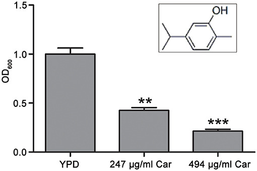
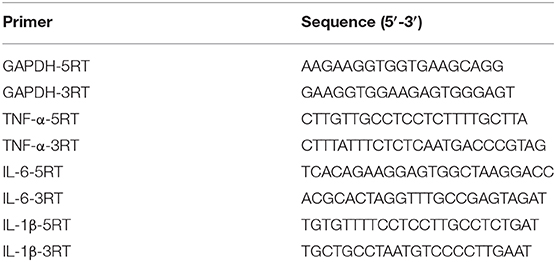
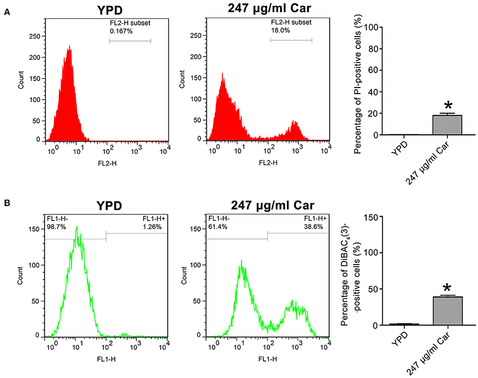
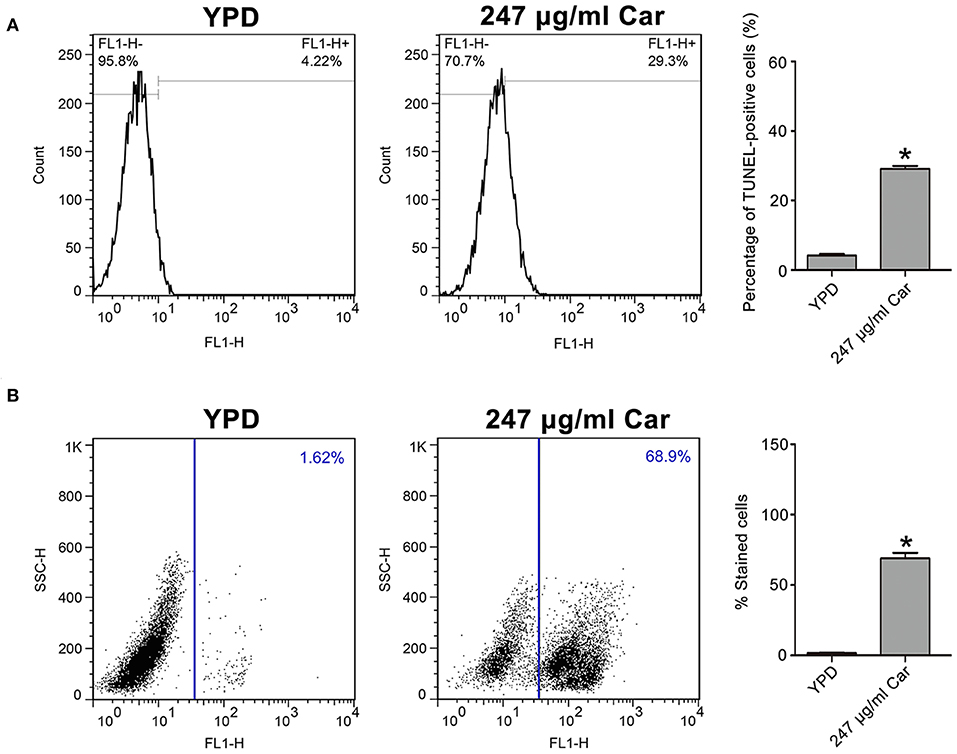
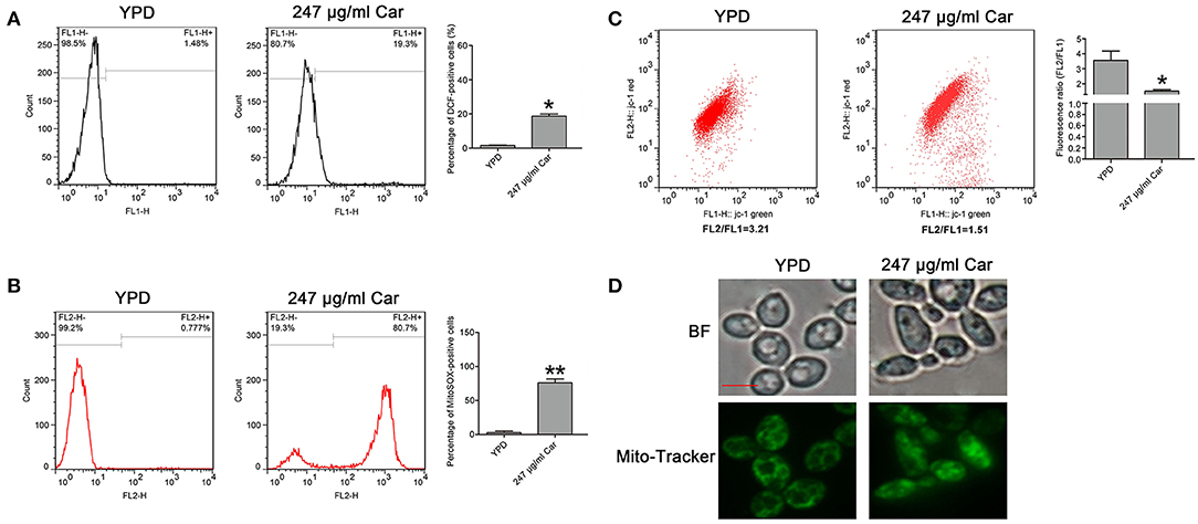
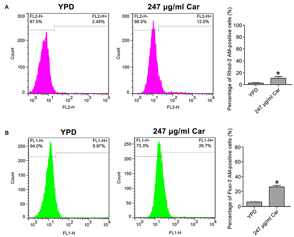
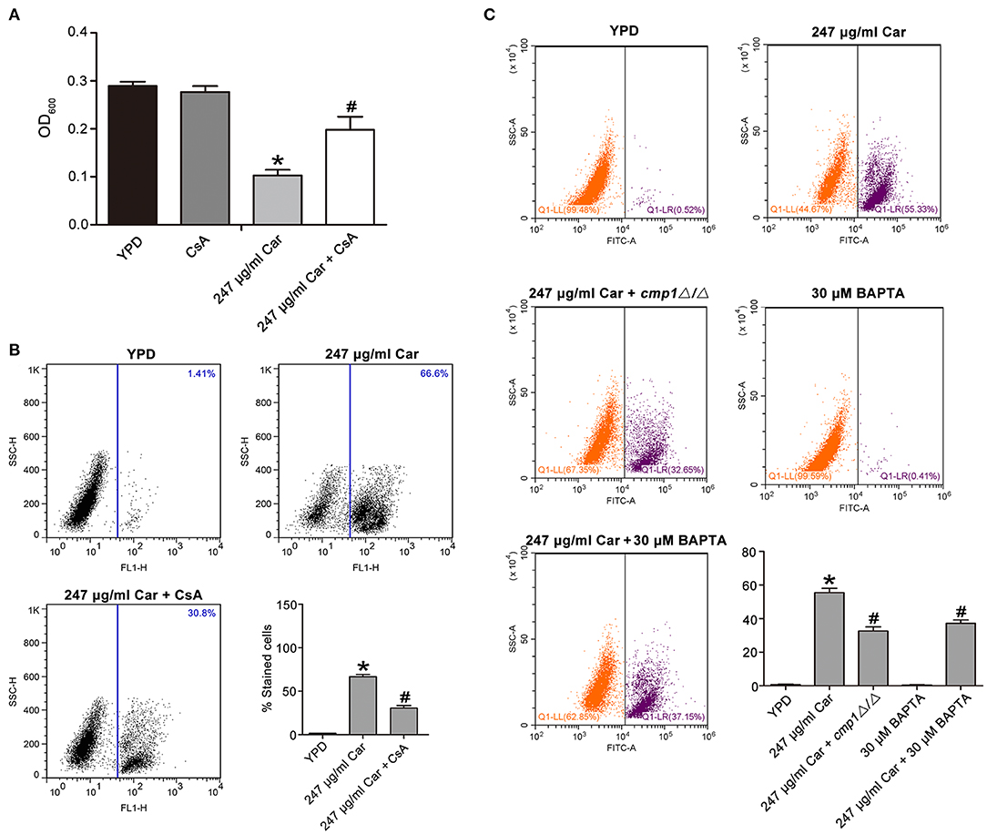
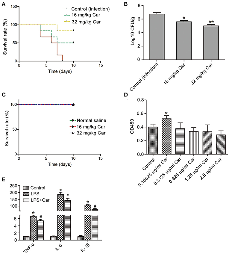





 Jun Chen


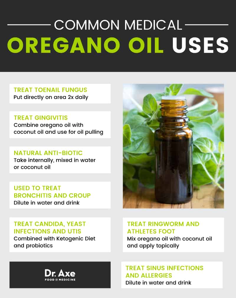


 CHARGED WITH SUN ENERGY
 PURE STEAM EXTRACTED
 SCIENTIFICALLY STUDIED
 HAND PICKED BY VILLAGERS

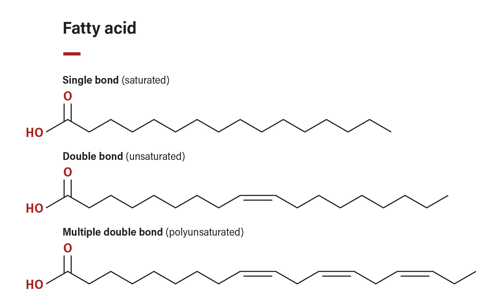


![Table 3 Different oxygenated compounds found in essential oils [15]](profile/S_M_Ashrafur_Rahman/publication/328700253/figure/tbl1/AS:722903939174418@1549365138766/Different-oxygenated-compounds-found-in-essential-oils-15_Q320.jpg)


This is seriously the best cure all for inflammation. I had a crazy toothache and I put some drops on it 3 x a day and it was gone in 2 days… I also use it on my toothbrush and it keeps my breath smelling and feeling fresh. Put a couple of drops with water to ward off a cold or illness…. beware it does burn, but to me feels so good, like it’s really working 🙂
Tanich2u
Athletes Foot – GONE! Cold Symptoms, caught in time – GONE! Sore throat? I just tip my head back & drip drops right down to the back of my throat! Mosquito bites, minor skin irritations, minor skin or wound infections – GONE! On cotton balls for earaches, drops, just a couple, on my kid’s earache/infections – GONE! I use for allergy symptoms, too! ACCEPT NO SUBSTITUTES! This brand and its MEDITERRANEAN Oregano components, the P-73 (or whatever it is that does the work), IT is the KIND! I’ve tried others with WAY less curative effects! Make your own capsules straight up, I don’t bother with measure, just fill half capsule up ‘n swallow w/beverage. I’ve seen reviews concerned with effects w/beneficial bacteria or whatever, personally, I’ve used this product for almost 20 years or so and have never had any problems, only benefits! So whats a big deal about a strong oregano burp, anyways!? I must say, I absolutely do consume a complete, well rounded diet with plenty of fruits & vegetables & work a physically demanding job. I’m almost 61 and plenty healthy so far!
thejuandrr
Oreganol is a product I use daily for my overall health. I have come to believe that I need this in today’s unhealthy world environment to combat any ugly bacteria, virus, or fungus. This is the best non-gmo, organic oregano oil on the market and the only one with the P73 enzyme.
M.D. Morris
This was recommended by the Crohn’s Foundation. I have colitis that has been out of control for years. After 2 weeks of taking 2 drops in a glass of water 1x a day, I am off of my colitis meds altogether. The taste and smell are a bit hard to handle, but my health has shown such an unexpected and pleasant improvement, I can hold my nose while drinking it!!!
jh
Not as effective as I thought it would be on the warts on my skin. Have been taking it orally and applying it directly to the wart.
Marsha Clarke
As a school nurse, I’m exposed to illness daily. Recently when I notice a slight sore throat, I take a few drops of Oregano P73 or gargle with a few drops in water and it’s gone by morning. Love it!
Laura Collinwood