Does Hydroxychloroquine work for COVID or not? The Overwhelming Evidence says: YES!!!
Does Hydroxychloroquine really work to treat COVID-19?
I'm not a doctor or scientist, but all the research I have done says: YES! And if you look at all the studies, information and testimonials on this page, you'll see why.
There are SO many doctors that swear Hydroxychloroquine works, it is really sad to see how many of these doctors are getting censored!
What you need to know about hydroxychloroquine:
1. It was used in 2005 to effectively treat SARS (See study)
2. MANY doctors insist it is a VERY effective, inexpensive treatment (see their stories and testimonials in the videos below). Some have treated thousands of patients with a 100% success rate (or close to it) when it is used early.
3. It's commonly used in countries where people have easy access to hydroxychloroquine and similar drugs, because they are are used to having to treat malaria. If you look at the countries that have the lowest cases of COVID per million, you'll see that it tends to be the poorer countries with the highest cases of malaria.... this is likely because they have easy access to Hydroxychloroquine!
4. The rumors that have been spread about hydroxychloroquine causing heart problems are extremely misleading and a threat to public health and safety. Hydroxychloroquine only affects people negatively if you take it in VERY HIGH DOSES - MUCH higher than the doctors who are having great success with it.
Dr. Zelenko has used Hydroxychloroquine with GREAT SUCCESS. He's treated over 7,000 patients, and hasn't lost a single patient in over 9 months. In the video below, you can see him speaking to fellow Israeli ministers, and one of them actually thanks him, because he used his protocol and it saved him from COVID.
DR. VLADIMIR 'ZEV' ZELENKO INTERVIEW ON THE HIGHWIRE WITH DEL BIGTREE
THE HIGHWIRE WITH DEL BIGTREE - ZELENKO: HCQ DENIERS ARE “GUILTY OF MASS MURDER”
Why do Asian countries use hydroxychloroquine for Covid-19 despite Western rejection?
Dr Christina Lin
August 2020
Analysis
In the competition between East and West over global governance models, it seems Asian countries may be
emerging as the winners when it comes to health governance.
On the issue of pandemic management, currently East Asian and South Asian countries enjoy comparative lower
mortality rate for Covid-19 than western countries.1
Taiwan, now touted as the gold standard for pandemic
management, has a population of 23.7 million people but only seven deaths.2
Vietnam has registered zero death
with a population of 95.5 million, while South Korea (population: 51 million) has lost 300 and Japan (population:
126 million) has lost 1001.
In contrast, the US (population: 328 million) has lost 154,000 while the UK (population: 66 million) has lost
45,961. Italy (population 60 million) has lost 35,121, Spain (population: 47 million) has lost 28,443 while France
(population: 67 million) has lost 30,238 and Germany (population: 83 million) has lost 9,217.
In order to control for population variance across countries, as measured by case-fatality-rate (CFR) per 100,000
people, Asian countries still rank lower in the CFR vis-à-vis Western countries.3
According to Johns Hopkins
University, European countries have the highest CFR rate in double digits with UK at 15.2% followed by Belgium
(14.6%), Italy (14.2%), France (13.7%), Spain (10.1%), and Germany and US at 4.4% and 3.4% respectively.
How Did Vietnam and Cambodia Contain COVID-19 With Few Resources?
Manisha MirchandaniDirector of Strategy at Atlantic 57
This piece — published on August 7 — was BRINK’s best-performing article of 2020. Strategist Manisha Mirchandani explored how Vietnam and Cambodia were handling the pandemic so successfully despite little access to resources.
As the coronavirus has ravaged its way across the world, some countries in Asia have shown a remarkable ability to evade its worst effects. As of early August, Cambodia had zero deaths and Vietnam had just six recorded deaths from the coronavirus. [since the piece was published, Vietnam has recorded a total of 35 deaths, while Cambodia has continued to have zero deaths].
This is in spite of these countries having far fewer resources to respond to the threat, compared to wealthy nations such as the United States and Italy, where the virus has exacted a terrible price.
Not all of Southeast Asia has been as successful in containing the virus’ spread: Both Indonesia and the Philippines register amongst the highest deaths in Asia outside of China. So why have Cambodia and Vietnam been so successful? Vietnam has recorded only 500 cases for a population of nearly 100 million (five per million) as of the end of July, while Cambodia has recorded just 15 cases per million at this point.
A Younger Population?
Experts have pointed to a younger demographic in these countries as being a possible inoculating factor, but the virus has not spared the youthful populations of Indonesia and the Philippines. Others say that low testing rates and possible underreporting of cases is hiding the true extent of COVID-19 in these countries. But to date, there has been no risk of health systems in Vietnam or Cambodia becoming overwhelmed by the severity of outbreaks, as we have seen in other countries.
The early success of these countries in responding to the coronavirus shows that an effective response is not dependent on resource availability. Much has been made of the importance of testing to contain the disease, as exemplified by South Korea’s strategy to “test, test, test.” But lacking the resources to mobilize an expansive testing infrastructure, Vietnam and Cambodia have relied on a combination of tactics to “box in” the virus.
Source: Southeast Asia COVID-19 Tracker, Center for Strategic & International Studies, as of July 31, 2020
Boxing in the Virus
One tactic has been targeted testing, where health authorities have focused on high-risk individuals or homed in on buildings or neighborhoods where positive cases have emerged. This was combined with extensive contact tracing: In Vietnam, people up to three degrees removed from an infected person were identified and tested for infection.
This was enabled by national guidelines that empowered members of communities — including members of the public, teachers, religious and community leaders — to report public health events. Official tracing efforts were complemented by a mobile app that allows for the reporting of health status and suspected cases and another that notifies users of possible exposure.
Local-level surveillance and the participation of communities have helped early identification of possible outbreaks in Vietnam, as did an approach based on risk of exposure regardless of whether a person exhibited symptoms. This may have been critical in containing COVID-19, given the emerging evidence that asymptomatic people are able to transmit the virus to others.
Once identified, Vietnam had set up the infrastructure to quarantine infected persons and international travelers, minimizing household exposure to the virus; some 200,000 people spent time in a quarantine facility between January and May of 2020. In Cambodia, a network of 2,900 health care workers was trained in January and February to assist detection and contact tracing at the community level.
A Population Used to Combating Epidemics
Having experienced the SARS epidemic in 2003 and bouts of the avian flu between 2004 and 2010, the Vietnamese and Cambodian authorities took the threat of COVID-19 seriously from the beginning. Health was prioritized above economic concerns as each country quickly implemented border controls and enforced the wearing of masks and social distancing. Past experience in combating epidemics may have led to greater public comfort with such measures, and an understanding of the need for decisive action to contain the pandemic. In a March 2020 survey, most Vietnamese (62%) agreed that the government’s reaction was “the right amount,” indicating a high degree of public support for aggressive measures.
Compliance is also a function of an authoritarian style of governance in both countries, which has allowed for the quick implementation of containment measures and the enforcement of them. In this environment, privacy concerns related to surveillance and contact tracing are given little credence, nor the individual rights of people who were forced into quarantine or had their freedom of movement restricted.
Risk of a Surveillance State
Indeed, there is a risk of the government’s public surveillance infrastructure being strengthened and normalized during this period. A state of emergency lawbeing passed in Cambodia will allow for unlimited telecommunications surveillance and the right for the government to control the press and social media, and restrict freedom of movement.
Measures taken so far have worked to control the spread of the coronavirus, but both countries are still vulnerable to subsequent waves. The discovery of the first locally transmitted case of COVID-19 in 100 days in Danang has prompted the evacuationof 80,000 people from the Vietnamese town.
The scale of the response by authorities and ongoing vigilance augurs well for the country to manage any outbreaks. Meanwhile, the health system has stocked up on supplies and equipment needed to withstand a surge of sick patients in case of an outbreak.
Targeted testing and the involvement of the community are critical in responding to COVID-19 where resources are more limited. What is less certain is the extent to which citizen concerns about privacy and individual liberties could become impediments to public trust and their willingness to participate in the response. So far, however, Vietnam and Cambodia have proved to be great examples of how countries with limited resources can successfully hold back the tide of coronavirus.
Wondering about the safety or efficacy of Hydroxychloroquine?
Call your own doctor and find out for yourself:
1) Is Hydroxychloroquine an FDA-approved drug? YES
2) Is it safe YES, it is a SAFE DRUG
3) If you go to a country where hydroxychloroquine is prevalent, is your doctor likely to prescribe hydroxychloroquine to you? Doctors routinely prescribe it for Malaria
4) How long has this drug been around? 65 years
I never before seen a retracted study in my life, and I've looked at hundreds of studies over the last 7 years. This one about Hydroxychloroquine is the first!
Severe acute respiratory syndrome (SARS) is caused by a newly discovered coronavirus (SARS-CoV). No effective prophylactic or post-exposure therapy is currently available.
Results
We report, however, that chloroquine has strong antiviral effects on SARS-CoV infection of primate cells. These inhibitory effects are observed when the cells are treated with the drug either before or after exposure to the virus, suggesting both prophylactic and therapeutic advantage. In addition to the well-known functions of chloroquine such as elevations of endosomal pH, the drug appears to interfere with terminal glycosylation of the cellular receptor, angiotensin-converting enzyme 2. This may negatively influence the virus-receptor binding and abrogate the infection, with further ramifications by the elevation of vesicular pH, resulting in the inhibition of infection and spread of SARS CoV at clinically admissible concentrations.
Severe acute respiratory syndrome (SARS) is caused by a newly discovered coronavirus (SARS-CoV). No effective prophylactic or post-exposure therapy is currently available.
Results
We report, however, that chloroquine has strong antiviral effects on SARS-CoV infection of primate cells. These inhibitory effects are observed when the cells are treated with the drug either before or after exposure to the virus, suggesting both prophylactic and therapeutic advantage. In addition to the well-known functions of chloroquine such as elevations of endosomal pH, the drug appears to interfere with terminal glycosylation of the cellular receptor, angiotensin-converting enzyme 2. This may negatively influence the virus-receptor binding and abrogate the infection, with further ramifications by the elevation of vesicular pH, resulting in the inhibition of infection and spread of SARS CoV at clinically admissible concentrations.
Conclusion
Chloroquine is effective in preventing the spread of SARS CoV in cell culture. Favorable inhibition of virus spread was observed when the cells were either treated with chloroquine prior to or after SARS CoV infection. In addition, the indirect immunofluorescence assay described herein represents a simple and rapid method for screening SARS-CoV antiviral compounds.
Keywords: severe acute respiratory syndrome coronavirus, chloroquine, inhibition, therapy
Severe acute respiratory syndrome (SARS) is an emerging disease that was first reported in Guangdong Province, China, in late 2002. The disease rapidly spread to at least 30 countries within months of its first appearance, and concerted worldwide efforts led to the identification of the etiological agent as SARS coronavirus (SARS-CoV), a novel member of the family Coronaviridae [1]. Complete genome sequencing of SARS-CoV [2,3] confirmed that this pathogen is not closely related to any of the previously established coronavirus groups. Budding of the SARS-CoV occurs in the Golgi apparatus [4] and results in the incorporation of the envelope spike glycoprotein into the virion. The spike glycoprotein is a type I membrane protein that facilitates viral attachment to the cellular receptor and initiation of infection, and angiotensin-converting enzyme-2 (ACE2) has been identified as a functional cellular receptor of SARS-CoV [5]. We have recently shown that the processing of the spike protein was effected by furin-like convertases and that inhibition of this cleavage by a specific inhibitor abrogated cytopathicity and significantly reduced the virus titer of SARS-CoV [6].
Due to the severity of SARS-CoV infection, the potential for rapid spread of the disease, and the absence of proven effective and safe in vivo inhibitors of the virus, it is important to identify drugs that can effectively be used to treat or prevent potential SARS-CoV infections. Many novel therapeutic approaches have been evaluated in laboratory studies of SARS-CoV: notable among these approaches are those using siRNA [7], passive antibody transfer [8], DNA vaccination [9], vaccinia or parainfluenza virus expressing the spike protein [10,11], interferons [12,13], and monoclonal antibody to the S1-subunit of the spike glycoprotein that blocks receptor binding [14]. In this report, we describe the identification of chloroquine as an effective pre- and post-infection antiviral agent for SARS-CoV. Chloroquine, a 9-aminoquinoline that was identified in 1934, is a weak base that increases the pH of acidic vesicles. When added extracellularly, the non-protonated portion of chloroquine enters the cell, where it becomes protonated and concentrated in acidic, low-pH organelles, such as endosomes, Golgi vesicles, and lysosomes. Chloroquine can affect virus infection in many ways, and the antiviral effect depends in part on the extent to which the virus utilizes endosomes for entry. Chloroquine has been widely used to treat human diseases, such as malaria, amoebiosis, HIV, and autoimmune diseases, without significant detrimental side effects [15]. Together with data presented here, showing virus inhibition in cell culture by chloroquine doses compatible with patient treatment, these features suggest that further evaluation of chloroquine in animal models of SARS-CoV infection would be warranted as we progress toward finding effective antivirals for prevention or treatment of the disease.
Preinfection chloroquine treatment renders Vero E6 cells refractory to SARS-CoV infection
In order to investigate if chloroquine might prevent SARS-CoV infection, permissive Vero E6 cells [1] were pretreated with various concentrations of chloroquine (0.1–10 μM) for 20–24 h prior to virus infection. Cells were then infected with SARS-CoV, and virus antigens were visualized by indirect immunofluorescence as described in Materials and Methods. Microscopic examination (Fig. (Fig.1A)1A) of the control cells (untreated, infected) revealed extensive SARS-CoV-specific immunostaining of the monolayer. A dose-dependant decrease in virus antigen-positive cells was observed starting at 0.1 μM chloroquine, and concentrations of 10 μM completely abolished SARS-CoV infection. For quantitative purposes, we counted the number of cells stained positive from three random locations on a slide. The average number of positively stained control cells was scored as 100% and was compared with the number of positive cells observed under various chloroquine concentrations (Fig. (Fig.1B).1B). Pretreatment with 0.1, 1, and 10 μM chloroquine reduced infectivity by 28%, 53%, and 100%, respectively. Reproducible results were obtained from three independent experiments. These data demonstrated that pretreatment of Vero E6 cells with chloroquine rendered these cells refractory to SARS-CoV infection.
Prophylactic effect of chloroquine. Vero E6 cells pre-treated with chloroquine for 20 hrs. Chloroquine-containing media were removed and the cells were washed with phosphate buffered saline before they were infected with SARS-CoV (0.5 multiplicity of infection) for 1 h. in the absence of chloroquine. Virus was then removed and the cells were maintained in Opti-MEM (Invitrogen) for 16–18 h in the absence of chloroquine. SARS-CoV antigens were stained with virus-specific HMAF, followed by FITC-conjugated secondary antibodies. (A) The concentration of chloroquine used is indicated on the top of each panel. (B) SARS-CoV antigen-positive cells at three random locations were captured by using a digital camera, the number of antigen-positive cells was determined, and the average inhibition was calculated. Percent inhibition was obtained by considering the untreated control as 0% inhibition. The vertical bars represent the range of SEM.
Postinfection chloroquine t
Postinfection chloroquine treatment is effective in preventing the spread of SARS-CoV infection
In order to investigate the antiviral properties of chloroquine on SARS-CoV after the initiation of infection, Vero E6 cells were infected with the virus and fresh medium supplemented with various concentrations of chloroquine was added immediately after virus adsorption. Infected cells were incubated for an additional 16–18 h, after which the presence of virus antigens was analyzed by indirect immunofluorescence analysis. When chloroquine was added after the initiation of infection, there was a dramatic dose-dependant decrease in the number of virus antigen-positive cells (Fig. (Fig.2A).2A). As little as 0.1–1 μM chloroquine reduced the infection by 50% and up to 90–94% inhibition was observed with 33–100 μM concentrations (Fig. (Fig.2B).2B). At concentrations of chloroquine in excess of 1 μM, only a small number of individual cells were initially infected, and the spread of the infection to adjacent cells was all but eliminated. A half-maximal inhibitory effect was estimated to occur at 4.4 ± 1.0 μM chloroquine (Fig. (Fig.2C).2C). These data clearly show that addition of chloroquine can effectively reduce the establishment of infection and spread of SARS-CoV if the drug is added immediately following virus adsorption.
Post-infection chloroquine treatment reduces SARS-CoV infection and spread. Vero E6 cells were seeded and infected as described for Fig. 1 except that chloroquine was added only after virus adsorption. Cells were maintained in Opti-MEM (Invitrogen) containing chloroquine for 16–18 h, after which they were processed for immunofluorescence. (A) The concentration of chloroquine is indicated on the top. (B) Percent inhibition and SEM were calculated as in Fig. 1B. (C) The effective dose (ED50) was calculated using commercially available software (Grafit, version 4, Erithacus Software).
pH-dependent entry of severe acute respiratory syndrome coronavirus is mediated by the spike glycoprotein and enhanced by dendritic cell transfer through DC-SIGN
The severe acute respiratory syndrome coronavirus (SARS-CoV) synthesizes several putative viral envelope proteins, including the spike (S), membrane (M), and small envelope (E) glycoproteins. Although these proteins likely are essential for viral replication, their specific roles in SARS-CoV entry have not been defined. In this report, we show that the SARS-CoV S glycoprotein mediates viral entry through pH-dependent endocytosis. Further, we define its cellular tropism and demonstrate that virus transmission occurs through cell-mediated transfer by dendritic cells. The S glycoprotein was used successfully to pseudotype replication-defective retroviral and lentiviral vectors that readily infected Vero cells as well as primary pulmonary and renal epithelial cells from human, nonhuman primate, and, to a lesser extent, feline species. The tropism of this reporter virus was similar to that of wild-type, replication-competent SARS-CoV, and binding of purified S to susceptible target cells was demonstrated by flow cytometry. Although myeloid dendritic cells were able to interact with S and to bind virus, these cells could not be infected by SARS-CoV. However, these cells were able to transfer the virus to susceptible target cells through a synapse-like structure. Both cell-mediated infection and direct infection were inhibited by anti-S antisera, indicating that strategies directed toward this gene product are likely to confer a therapeutic benefit for antiviral drugs or the development of a SARS vaccine.
Gorshkov K, Chen CZ, Bostwick R, Rasmussen L, Tran BN, Cheng YS, Xu M, Pradhan M, Henderson M, Zhu W, Oh E, Susumu K, Wolak M, Shamim K, Huang W, Hu X, Shen M, Klumpp-Thomas C, Itkin Z, Shinn P, Carlos de la Torre J, Simeonov A, Michael SG, Hall MD, Lo DC, Zheng W.ACS Infect Dis. 2020 Dec 21:acsinfecdis.0c00349. doi: 10.1021/acsinfecdis.0c00349. Online ahead of print.PMID: 33346633Free PMC article.
This manuscript addresses the physicochemical behavior of an antimalarial drug Chloroquine Diphosphate (CLQ) as well as its interaction with anionic surfactants and Human Serum Albumin (HSA). Surface tension and specific conductivity were employed to detect the critical micelle concentration (CMC) and thus its surface and thermodynamic parameters were calculated. Solubilization of this drug within micelles of anionic surfactant sodium dodecyl sulfate (SDS) has also been studied. UV/Visible spectroscopy was used to calculate partition coefficient (Kx), free energy of partition and number of drug molecules per micelle. The complexation of drug with HSA at physiological conditions (pH 7.4) has also been analyzed by using UV/Visible and fluorescence spectroscopy. The values of drug-protein binding constant, number of binding sites and free energy of binding were calculated.
► Free energy of adsorption is more negative than free energy of micellization. ► Shifts in UV/Visible spectra in presence of SDS indicated interaction of CLQ with SDS. ► The decrease in fluorescence intensity of HSA by CLQ shows its binding with HSA.
THE FRONTLINE FIGHT AGAINST MEDICAL DISCRIMINATION
FRONTLINE DOCTORS MAKE URGENT PLEA HYDROXYCHLOROQUINE WITH ZINC CURES COVID IN D C
THE CURE FOR COVID-19: HYDROXYCHLOROQUINE - AMERICAN FRONTLINE DOCTORS
COVID PATIENT ON DEATH'S DOORSTEP IN ICU: A FLORIDA MIRACLE CURE WITH HCQ
pinion on Hydroxychloroquine plus Zinc: Yes, it works. And for a very simple reason: It makes your endosomal pH more alkaline (see studies on this page that show how it changes the pH of your endosomes - credit goes to Raphaela Laurean for pointing this out - you can see her video on this page). I believe this is why baking soda and lime juice seems to have been so helpful for people who've contractted the SARS-2 Coronavirus or influenza (both are enveloped viruses, with a lipid bilayer that requires an acidic environment in order for viral fusion to take place).
I've done a good amount of research on Hydroxychloroquine, and had gone back and forth on my opinion that it works. I remember seeing Elon Musk had tweeted about it, and somehow that made it sound more credible. Then, I thought Remdesivir must be the better thing. I heard a story about how a little girl died from taking it after she found an old prescription bottle in a bathroom drawer. When I posted that on Facebook, a friend of mine (who works for the company that makes it) pointed out that the story was misleading, and it's safe. I did realize that kids are not supposed to take drugs made for adults and kids could die from MANY drugs that are safe for adults, if they take too much. The news stories keep changing.
I've realized that a lot of the mainstream news outlets are reporting that it doesn't work, but these are also outlets that get funded by pharmaceutical companies, and there is hardly any profit to be made from Hydroxychloroquine, because it's a really cheap, affordable drug.
So I had to ask myself the question:
Why would all these doctors (in the video below) put their jobs on the line, as they did (at least one was fired) unless they were really passionate about helping people to keep from getting sick? If there's no profit to be made from Hydroxychloroquine, why would they lie about it?
You can see what many physicians have to say, about Hydroxychloroquine, in this video:
KEEP IN MIND - these articles that are circulating about chloroquine misleading the public are misleading the public because doctors that find it effective and safe DO NOT use it in the way that has been causing heart problems. The subjects in these studies are using unusually high doses.
Small Chloroquine Study Halted Over Risk of Fatal Heart Complications
A research trial of coronavirus patients in Brazil ended after patients taking a higher dose of chloroquine, one of the drugs President Trump has promoted, developed irregular heart rates.
(NOTE how they say the patients were taking a HIGHER DOSE). They were also using azythromycin, an antibiotic
A small study in Brazil was halted early for safety reasons after coronavirus patients taking a higher dose of chloroquine developed irregular heart rates that increased their risk of a potentially fatal heart arrhythmia.
Despite its limitations, infectious disease doctors and drug safety experts said the study provided further evidence that chloroquine and hydroxychloroquine, which are both used to treat malaria, can pose significant harm to some patients, specifically the risk of a fatal heart arrhythmia. Patients in the trial were also given the antibiotic azithromycin, which carries the same heart risk. Hospitals in the United States are also using azithromycin to treat coronavirus patients, often in combination with hydroxychloroquine.
“To me, this study conveys one useful piece of information, which is that chloroquine causes a dose-dependent increase in an abnormality in the ECG that could predispose people to sudden cardiac death,” said Dr. David Juurlink, an internist and the head of the division of clinical pharmacology at the University of Toronto, referring to an electrocardiogram, which reads the heart’s electrical activity.
It also seems kind of odd that WebMD's page about Azithromycin says it won't work for viral infections. I don't understand why they felt the need to use it at all.
Azithromycin is used to treat a wide variety of bacterial infections. It is a macrolide-type antibiotic. It works by stopping the growth of bacteria.
This medication will not work for viral infections (such as common cold, flu). Unnecessary use or misuse of any antibiotic can lead to its decreased effectiveness.
Remember that your doctor has prescribed this medication because he or she has judged that the benefit to you is greater than the risk of side effects. Many people using this medication do not have serious side effects.
Tell your doctor right away if any of these unlikely but serious side effects occur: hearing changes (such as decreased hearing, deafness), eye problems (such as drooping eyelids, blurred vision), difficulty speaking/swallowing, muscle weakness, signs of liver problems (such as unusual tiredness, persistent nausea/vomiting, severe stomach/abdominal pain, yellowing eyes/skin, dark urine).
Get medical help right away if any of these rare but serious side effects occur: fast/irregular heartbeat, severe dizziness, fainting.
This medication may rarely cause a severe intestinal condition (Clostridium difficile-associated diarrhea) due to a resistant bacteria. This condition may occur during treatment or weeks to months after treatment has stopped. Do not use anti-diarrhea or opioidmedications if you have any of the following symptoms because these products may make them worse. Tell your doctor right away if you develop: persistent diarrhea, abdominal or stomach pain/cramping, blood/mucus in your stool.
Small Chloroquine Study Halted Over Risk of Fatal Heart Complications
A research trial of coronavirus patients in Brazil ended after patients taking a higher dose of chloroquine, one of the drugs President Trump has promoted, developed irregular heart rates.
Packets of Nivaquine tablets containing chloroquine, and Plaquenil tablets containing hydroxychloroquine. Both drugs have been promoted by President Trump as a coronavirus treatment.Credit...Gerard Julien/Agence France-Presse — Getty Images
Roughly half of the study participants were given a low dose of chloroquine (450 milligrams of chloroquine for five days, with two doses on the first day), while the rest were prescribed a high dose (600 milligrams twice daily for 10 days). Within three days, researchers started noticing heart arrhythmias in patients taking the higher dose. By the sixth day of treatment, 11 patients had died, leading to an immediate end to the high-dose segment of the trial.
The researchers said the study did not have enough patients in the lower-dose portion of the trial to conclude if chloroquine was effective in patients with severe disease. More studies evaluating the drug earlier in the course of the disease are “urgently needed,” the researchers said.
Several clinical trials for chloroquine and hydroxychloroquine are testing low doses for shorter periods of time in coronavirus patients. But the Health Commission of Guangdong Province in China had initially recommended those sick with the virus be treated with 500 milligrams of chloroquine twice daily for 10 days.
CORONAVIRUS SCHOOLS BRIEFING: It’s back to school — or is it?
One of the authors of the Brazilian study, Dr. Marcus Lacerda, said in an email on Sunday that his study found that “the high dosage that the Chinese were using is very toxic and kills more patients.”
“That is the reason this arm of the study was halted early,” he said, adding that the manuscript was being reviewed by the journal Lancet Global Health.
A small study in Brazil was halted early for safety reasons after coronavirus patients taking a higher dose of chloroquine developed irregular heart rates that increased their risk of a potentially fatal heart arrhythmia.
Chloroquine is closely related to the more widely used drug hydroxychloroquine. President Trump has enthusiastically promoted them as a potential treatment for the novel coronavirus despite little evidence that they work, and despite concerns from some of his top health officials. Last month, the Food and Drug Administration granted emergency approval to allow hospitals to use chloroquine and hydroxychloroquine from the national stockpile if clinical trials were not feasible. Companies that manufacture both drugs are ramping up production.
The Brazilian study involved 81 hospitalized patients in the city of Manaus and was sponsored by the Brazilian state of Amazonas. It was posted on Saturday at medRxiv, an online server for medical articles, before undergoing peer review by other researchers. Because Brazil’s national guidelines recommend the use of chloroquine in coronavirus patients, the researchers said including a placebo in their trial — considered the best way to evaluate a drug — was an “impossibility.”
Despite its limitations, infectious disease doctors and drug safety experts said the study provided further evidence that chloroquine and hydroxychloroquine, which are both used to treat malaria, can pose significant harm to some patients, specifically the risk of a fatal heart arrhythmia. Patients in the trial were also given the antibiotic azithromycin, which carries the same heart risk. Hospitals in the United States are also using azithromycin to treat coronavirus patients, often in combination with hydroxychloroquine.
Coronavirus: Influential study on hydroxychloroquine withdrawn
An influential article that found hydroxychloroquine increases the risk of death in coronavirus patients has been retracted over data concerns.
Three of the study's authors said they could not longer vouch for its veracity because Surgisphere, a healthcare firm behind the data, would not allow an independent review of its dataset.
Its findings led the WHO to suspend its testing on the anti-malaria drug.
But leaders including US President Donald Trump continue to tout its use.
Surgisphere chief executive Sapan Desai, the study's fourth author, told The Guardian newspaper he would co-operate with an independent audit but said transferring the data would "violate client agreements and confidentiality requirements".
What did the study say?
Research for the article, published last month in medical journal The Lancet, involved 96,000 coronavirus patients across 671 hospitals worldwide. Nearly 15,000 were given hydroxychloroquine - or a related form, chloroquine - either alone or with an antibiotic.
It concluded that the drug showed no benefits against coronavirus and increased the risk patients developing irregular heart rhythms and dying.
Mandeep Mehra, a Harvard University professor who led the study, together with Frank Ruschitzka of the University Hospital Zurich and Amit Patel of the University of Utah, said in a statement that they had tried to arrange for a third-party peer review of the data, but Surgisphere had refused to co-operate.
"We deeply apologize to you, the editors, and the journal readership for any embarrassment or inconvenience that this may have caused," the group added.
Facebook Twitter Flipboard Email May 21, 20207:01 AM ET
WILL STONE
Hydroxychloroquine, a drug used to prevent malaria and treat certain autoimmune conditions, is being studied to treat or prevent COVID-19.
George Frey/AFP via Getty Images
Publicity around the drug hydroxychloroquine spiked this week when President Trump revealed that he's taking it to prevent COVID-19.
All the attention on the drug in recent months is increasingly spilling into science and making it harder for some researchers to actually study whether the drug has potential for COVID-19. Doctors have used hydroxychloroquine for decades to treat autoimmune conditions and to prevent malaria.
While the Food and Drug Administration has given emergency use authorization for doctors to try the treatment on COVID-19 patients, the agency has also cautioned about possible deadly side effects.
On Wednesday, the executive director of the World Health Organization's emergencies program, Dr. Mike Ryan, echoed other leading medical experts, saying at a press briefing that hydroxychloroquine and chloroquine have "yet to be found effective in the treatment of COVID-19" or for preventing the disease.
Because of potential side effects, Ryan says the WHO advises the drug "be reserved" for use in the context of clinical trials, which are underway in many places.
But for some researchers, running such trials is becoming more difficult because of the controversy around the drug.Article continues after sponsor message
Missed opportunity
Dr. Jon Giles didn't expect to have any problems drumming up interest in his clinical trial testing if hydroxychloroquine can help prevent COVID-19.
"We were getting calls all the time from people who were interested," says Giles, an epidemiologist and rheumatologist at Columbia University.
For his study, Giles planned to give a short course of the medication to people who were "household contacts" of COVID-19 patients. Like many of the ongoing trials, it would be randomized with a control group.
By the end of April, Giles was ready to start enrolling people. But his team ran into a problem when they started calling potential participants.
"Pretty much everybody said, 'Well that's the drug that's dangerous to your heart,' or, 'I talked to my friends and they said don't take it,' or that 'I saw on TV it's dangerous,' " says Giles. Just a month earlier, he says demand for the drug was soaring. Some patients with autoimmune conditions couldn't even get their prescriptions filled, after Trump spoke enthusiastically about the drug.
In April, the FDA issued a warning about using the drug for COVID-19 patients without strict medical supervision in a hospital or as part of a clinical trial. The agency had received reports of serious heart-related adverse events and death in patients with COVID-19 receiving hydroxychloroquine and chloroquine.
But the back-and-forth headlines and the ongoing political wrangling seemed to make people wary of the medication, Giles says, even in the context of a carefully run clinical trial. "It became almost impossible to get anyone interested," says Giles.
Giles says the committee overseeing his study added a new requirement: that study participants needed to have had an electrocardiogram within the last year.
As a rheumatologist, Giles knows the medication better than most doctors because he prescribes it to many of his patients. "It's a very, very safe drug; it's been used for over 75 years," he says. "When I give someone hydroxychloroquine, I don't get an ECG or do blood monitoring."
Giles was planning to enroll otherwise healthy people and screen out anyone who could be at risk of heart problems. But that new requirement, plus the negative press, made it too difficult to find study subjects.
He gave up on doing the study. He says it's a missed opportunity. "It's not unreasonable to think that a short course of this drug might have some protective ability," he says.Risks and potential
The concern over cardiac risk for hospitalized patients is real, says Dr. Mark Poznansky, director of the Vaccine and Immunotherapy Center in the Infectious Disease Division of Massachusetts General Hospital. Even though the drug has been used safely to prevent malaria, he says, "that is very different from using the drug in acutely ill patients with COVID-19."
He recently published a review of the evidence for using hydroxychloroquine to treat COVID-19 that casts doubt on its effectiveness.
"The data up to date ... doesn't make one entirely optimistic that we are going to find something different in a clinical trial," he says.
Poznansky notes a recent study of nearly 1,400 people with moderate to severe COVID-19 found no benefit and concluded that the drug should not be routinely used on patients. He says the drug should only be used in the context of clinical trials "or under strict compassionate use, in order to do no harm." Still, Poznansky doesn't rule out that further well-designed trials could turn up better results. 'The virus doesn't care about politics'
At Henry Ford Health System in Detroit, cardiologist Dr. William O'Neill says enrollment for their clinical trial studying hydroxychloroquine to prevent COVID-19 plummeted in late April.
"It really caused a huge problem for us," O'Neill says. "It set us back probably a month."
O'Neill attributes the drop in enrollment to the FDA warning and a highly publicized study of over 300 hospitalized veterans with COVID-19. The authors of that study found the drug did not reduce the need for a ventilator and even linked hydroxychloroquine to higher death rates. However, the study wasn't a rigorous controlled trial and the results weren't peer-reviewed.
"That was interpreted as a warning that the drug is dangerous," says O'Neill. But he says, using it for prevention is a different matter than treating already sick patients. "Everything that we see about hydroxychloroquine suggests that the earlier you use the drug, the more likely it is to be effective."
O'Neill says the fact that President Trump is touting this drug means it now has become a political flashpoint. "It has made people absolutely committed to proving him wrong," he says.
"The problem with that is this is not politics, this is life and death," he says. "We're talking about a treatment. Who would be rooting for us not to find a therapy, for God's sakes?" Multiple clinical trials involving hydroxychloroquine are underway at the University of Washington in Seattle. But Dr. Christine Johnston says recruitment has become a major challenge.
"We are hearing now from some participants that the study and the drug feel too political and they just don't want to participate at all," says Johnston. "As physicians, we would not do something that we thought was harmful."
"A clinical trial is the only way that we will be able to answer this question quickly and with high confidence," she says. Researchers at Duke University are also testing whether hydroxychloroquine can be a new tool for prevention during the pandemic. But Duke professor of medicine Dr. Susanna Naggie says they need to be able to recruit enough people. "I do worry that there are many well-controlled trials that are underway where we won't get an answer," says Naggie. "That, I think, would be a shame."
Another researcher conducting large trials, Dr. Wesley Self, says the coverage of hydroxychloroquine has felt a bit like an "emotional roller coaster," which has led to more questions from patients.
"I actually find it helpful," says Self, an emergency physician at Vanderbilt University Medical Center. "We're giving people relatively low doses of hydroxychloroquine, which we believe are quite safe. And we're monitoring for side effects very closely." Self is the lead investigator of the ORCHID trial that focuses on hydroxychloroquine in treating hospitalized COVID-19 patients. "This is a perfect situation for a clinical trial to really understand what the balance of those benefits and risks are," he says.
Another large clinical trial is looking at whether hydroxychloroquine and the antibiotic azithromycin can keep people with COVID-19 from being hospitalized or dying.
"We think the way that this medication works, it would be better to take it early, rather than later, especially when somebody is really really sick already," says Dr. Davey Smith, head of Infectious Diseases and Global Public Health at University of California San Diego School of Medicine.
Surfactant protein C (SP-C) is a hydrophobic protein synthesized and secreted exclusively by alveolar type II cells through proteolysis of a 21-kDa propeptide (SP-C21) to produce the 3.7-kDa surface active form. Previous studies from this laboratory have demonstrated that early processing of proSP-C involves extensive intracellular proteolysis of the COOH terminus of proSP-C21 in subcellular compartments, which include the acidic type II cell-specific subcellular organelle, the lamellar body. (Beers, M. F., Kim, C. Y., Dodia, C., and Fisher, A. B.(1994) J. Biol. Chem. 269, 20318-20328). The role of intracellular pH gradients in SP-C processing was studied in freshly isolated rat type II cells. Using vital fluorescence microscopy, the pH indicator acridine orange (AO) identified intense fluorescence staining of acidic cytoplasmic vesicles within fresh type II cells. The AO vesicular staining pattern was similar in cells labeled with the lamellar body marker phosphine 3R and the phospholipid dye nile red. AO fluorescence was quenched by the addition of a membrane-permeable weak base, methylamine. Immunoprecipitation of cell lysates with anti-proSP-C antisera following pulse-chase labeling (0-2 h) with 35S-Translabel demonstrated rapid synthesis of 35S-proSP-C21 with a time-dependent appearance of 16- and 6-kDa intermediates (SP-C16 and SP-C6). Tricine polyacrylamide gel electrophoresis analysis of organic extracts of cell lysates showed time-dependent appearance of mature SP-C3.7. The addition of 5 mM methylamine significantly blocked the post-translational processing of proSP-C resulting in disruption of normal precursor-product relationships and inhibition of SP-C3.7 formation. Methylamine-treated cells exhibited slow accumulation of SP-C16 and SP-C6, a persistence of SP-C21, and an absence of SP-C3.7 for the duration of the chase period. The lysosomotropic agent chloroquine, the proton ionophore monensin, and bafilomycin A1, a specific vacuolar H+-ATPase inhibitor, each caused inhibition of proSP-C processing in a similar manner. These results demonstrate that normal post-translational proteolysis of proSP-C occurs in acidic intracellular compartments, which include the lamellar body, and that complete processing to SP-C3.7 is dependent upon maintenance of transmembrane pH gradients by a vacuolar H+-ATPase.
Chloroquine (CQ) is a widely used drug and its administration has been reported to increase surfactant- associated phospholipids in lungs. We have used histomorphometric techniques to study the effects of CQ on foetal lung maturation in rats. CQ (40mg/kg BW) or saline was administered to pregnant rats on days 20 and 21 of gestation. A third group received the same dose of CQ on days 20 and 21, and in addition, hydrocortisone (HD; 25 mg/kg BW) on day 21 of pregnancy. Foetuses were delivered by hysterotomy on day 22. The lungs were weighed, fixed in Bouin's fixative and embedded in paraffin. Morphometric studies were performed on 5 microns-thick sections. The lung weight/100g BW, the volume density of lung saccular spaces, and the cross-sectional area and volume of the saccular spaces were reduced but the numerical density of the saccules was not decreased in foetuses exposed to CQ. With the exception of lung weights, which were lower in the foetuses exposed to CQ and HD, there were no other differences between this group and that exposed to CQ only. The results suggest that CQ attenuates the expansion of saccular spaces which occurs in preparation for post-natal gaseous exchange, and thus CQ retards foetal lung maturation. Although HD further reduced lung weights as expected from its reported action on foetal lungs, it did not reverse the CQ-induced retardation in lung maturation.
It really seems like there is a smear campaign against Chloroquine and Hydroxychloroquine.
Across the world, scientists are searching for ways to fight COVID-19. What might prevent people from catching the coronavirus that causes the disease? And in people who are infected, what treatments might reduce symptoms or slow COVID-19’s spread?
After early studies showed promise, the FDA issued an emergency ruling that would allow doctors to use chloroquine and hydroxychloroquine (a less-toxic derivative of chloroquine) in people who were in the hospital with COVID-19. But the agency later revoked the ruling amid serious concerns about the drugs’ safety and how well they worked against the virus.
What Is It?
Chloroquine is a low-cost drug that has been in use for decades. First discovered in 1934, it is the synthetic version of quinine, an antimalarial drug derived from the bark of the cinchona tree. Chloroquine is taken as a pill that you swallow. It’s already approved by the FDA to treat malaria as well as lupus and rheumatoid arthritis.
How Could It Help COVID-19 Patients?
Doctors thought chloroquine might reduce how long people with mild-to-moderate COVID-19 symptoms feel sick. They also hoped it would shorten the time when infected people are contagious.
What Does the Science Say?
As far back as the late 1960s, scientists have known that chloroquine could kill viruses in a petri dish. Chloroquine is also commonly taken to prevent and treat malaria infection in areas where the disease is widespread.
ADVERTISEMENT
But what about people who have infections other than malaria? That’s less clear. Human studies in influenza and dengue have shown no effect, either good or bad. And in chikungunya, though chloroquine did well in lab tests, later research showed it might make the illness worse.
Researchers around the world studied hydroxychloroquine’s effects in people with COVID-19. Some found early evidence of an effect against the new coronavirus. But many of those trials were stopped when they failed to show results or found serious side effects.
Side Effects
Chloroquine can upset your stomach, so people take it with food. Other side effects include headache, poor appetite, diarrhea, stomach pain, skin rash or itching, hair loss, and mood changes. Most of these problems are mild and temporary.
More serious side effects include eye problems (light flashes or streaks, blurred vision, difficulty reading), ear problems (ringing, trouble hearing), muscle weakness, drowsiness, vomiting, convulsions (sudden, unusual movement), irregular heartbeat, and breathing problems.
Be aware: If you take too much chloroquine, it can cause heart problems that can be life-threatening. It can also suppress the immune system. So only take this medicine under a doctor’s supervision.
This is what you will see, when you look up chloroquine on Google:
Seems like everything on the first page gives the impression that it's dangerous.
The people who have had a great experience with it are convenitntly found on much later pages.
Oct 1, 2020 — Chloroquine is used to prevent and treat malaria. It is also used to treat ... This is a decision you and your doctor will make. For this medicine ...
Chloroquine is a medication primarily used to prevent and treat malaria in areas where malaria ... Chloroquine is a member of the drug class 4-aminoquinoline. ... This could be avoided by "taste-masked and controlled release" formulations such as ... present five months after the drug was cleared from the rest of the body.
When a virus infects cells in your body, it tricks the cells into using their own machinery to make copies of the virus instead of doing all the things your cells would ...
If you have other side effects, be sure to tell your doctor. Do not take chloroquine that is strictly intended for veterinary use – such as to treat fish in aquariums or ...
a history of vision changes or damage to your retina caused by an anti-malaria medication. To make sure chloroquine is safe for you, tell your doctor if you have ...
Chloroquine is not recommended for coronavirus infection, also known as COVID-19, unless you are enrolled in a study. Talk to your doctor about the risks and ...
Jul 7, 2020 — Chloroquine is taken as a pill that you swallow. ... chloroquine did well in lab tests, later research showed it might make the illness worse.
Mar 20, 2020 — Now that chloroquine is in the news everywhere, I thought it might be ... no how, was he going to make quinine – but the purple dye he did produce made ... You might wonder how an antiparasitical drug might do that, but the ...
Mar 28, 2020 — There is limited information known about the safety and effectiveness (whether this will make you better) of using chloroquine phosphate for ...
Jun 21, 2019 — Chloroquine is used to protect against malaria when taken alongside insect ... If you are taking chloroquine to protect against malaria, it is important that ... Do not take two doses on the same day to make up for a missed dose.
Endocytosis is a cellular process in which substances are brought into the cell. The material to be internalized is surrounded by an area of cell membrane, which then buds off inside the cell to form a vesicle containing the ingested material. Endocytosis includes pinocytosis and phagocytosis. Wikipedia
Nov 29, 2016 — We demonstrate that chloroquine reduces the number of ZIKV-infected cells in vitro, and inhibits virus production and cell death promoted by ZIKV ...
Severe acute respiratory syndrome (SARS) is caused by a newly discovered coronavirus (SARS-CoV). No effective prophylactic or post-exposure therapy is currently available.
Results
We report, however, that chloroquine has strong antiviral effects on SARS-CoV infection of primate cells. These inhibitory effects are observed when the cells are treated with the drug either before or after exposure to the virus, suggesting both prophylactic and therapeutic advantage. In addition to the well-known functions of chloroquine such as elevations of endosomal pH, the drug appears to interfere with terminal glycosylation of the cellular receptor, angiotensin-converting enzyme 2. This may negatively influence the virus-receptor binding and abrogate the infection, with further ramifications by the elevation of vesicular pH, resulting in the inhibition of infection and spread of SARS CoV at clinically admissible concentrations.
Conclusion
Chloroquine is effective in preventing the spread of SARS CoV in cell culture. Favorable inhibition of virus spread was observed when the cells were either treated with chloroquine prior to or after SARS CoV infection. In addition, the indirect immunofluorescence assay described herein represents a simple and rapid method for screening SARS-CoV antiviral compounds.
Keywords: severe acute respiratory syndrome coronavirus, chloroquine, inhibition, therapy
Severe acute respiratory syndrome (SARS) is an emerging disease that was first reported in Guangdong Province, China, in late 2002. The disease rapidly spread to at least 30 countries within months of its first appearance, and concerted worldwide efforts led to the identification of the etiological agent as SARS coronavirus (SARS-CoV), a novel member of the family Coronaviridae [1]. Complete genome sequencing of SARS-CoV [2,3] confirmed that this pathogen is not closely related to any of the previously established coronavirus groups. Budding of the SARS-CoV occurs in the Golgi apparatus [4] and results in the incorporation of the envelope spike glycoprotein into the virion. The spike glycoprotein is a type I membrane protein that facilitates viral attachment to the cellular receptor and initiation of infection, and angiotensin-converting enzyme-2 (ACE2) has been identified as a functional cellular receptor of SARS-CoV [5]. We have recently shown that the processing of the spike protein was effected by furin-like convertases and that inhibition of this cleavage by a specific inhibitor abrogated cytopathicity and significantly reduced the virus titer of SARS-CoV [6].
Due to the severity of SARS-CoV infection, the potential for rapid spread of the disease, and the absence of proven effective and safe in vivo inhibitors of the virus, it is important to identify drugs that can effectively be used to treat or prevent potential SARS-CoV infections. Many novel therapeutic approaches have been evaluated in laboratory studies of SARS-CoV: notable among these approaches are those using siRNA [7], passive antibody transfer [8], DNA vaccination [9], vaccinia or parainfluenza virus expressing the spike protein [10,11], interferons [12,13], and monoclonal antibody to the S1-subunit of the spike glycoprotein that blocks receptor binding [14]. In this report, we describe the identification of chloroquine as an effective pre- and post-infection antiviral agent for SARS-CoV. Chloroquine, a 9-aminoquinoline that was identified in 1934, is a weak base that increases the pH of acidic vesicles. When added extracellularly, the non-protonated portion of chloroquine enters the cell, where it becomes protonated and concentrated in acidic, low-pH organelles, such as endosomes, Golgi vesicles, and lysosomes. Chloroquine can affect virus infection in many ways, and the antiviral effect depends in part on the extent to which the virus utilizes endosomes for entry. Chloroquine has been widely used to treat human diseases, such as malaria, amoebiosis, HIV, and autoimmune diseases, without significant detrimental side effects [15]. Together with data presented here, showing virus inhibition in cell culture by chloroquine doses compatible with patient treatment, these features suggest that further evaluation of chloroquine in animal models of SARS-CoV infection would be warranted as we progress toward finding effective antivirals for prevention or treatment of the disease.
Preinfection chloroquine treatment renders Vero E6 cells refractory to SARS-CoV infection
In order to investigate if chloroquine might prevent SARS-CoV infection, permissive Vero E6 cells [1] were pretreated with various concentrations of chloroquine (0.1–10 μM) for 20–24 h prior to virus infection. Cells were then infected with SARS-CoV, and virus antigens were visualized by indirect immunofluorescence as described in Materials and Methods. Microscopic examination (Fig. (Fig.1A)1A) of the control cells (untreated, infected) revealed extensive SARS-CoV-specific immunostaining of the monolayer. A dose-dependant decrease in virus antigen-positive cells was observed starting at 0.1 μM chloroquine, and concentrations of 10 μM completely abolished SARS-CoV infection. For quantitative purposes, we counted the number of cells stained positive from three random locations on a slide. The average number of positively stained control cells was scored as 100% and was compared with the number of positive cells observed under various chloroquine concentrations (Fig. (Fig.1B).1B). Pretreatment with 0.1, 1, and 10 μM chloroquine reduced infectivity by 28%, 53%, and 100%, respectively. Reproducible results were obtained from three independent experiments. These data demonstrated that pretreatment of Vero E6 cells with chloroquine rendered these cells refractory to SARS-CoV infection.
Prophylactic effect of chloroquine. Vero E6 cells pre-treated with chloroquine for 20 hrs. Chloroquine-containing media were removed and the cells were washed with phosphate buffered saline before they were infected with SARS-CoV (0.5 multiplicity of infection) for 1 h. in the absence of chloroquine. Virus was then removed and the cells were maintained in Opti-MEM (Invitrogen) for 16–18 h in the absence of chloroquine. SARS-CoV antigens were stained with virus-specific HMAF, followed by FITC-conjugated secondary antibodies. (A) The concentration of chloroquine used is indicated on the top of each panel. (B) SARS-CoV antigen-positive cells at three random locations were captured by using a digital camera, the number of antigen-positive cells was determined, and the average inhibition was calculated. Percent inhibition was obtained by considering the untreated control as 0% inhibition. The vertical bars represent the range of SEM.
Postinfection chloroquine treatment is effective in preventing the spread of SARS-CoV infection
In order to investigate the antiviral properties of chloroquine on SARS-CoV after the initiation of infection, Vero E6 cells were infected with the virus and fresh medium supplemented with various concentrations of chloroquine was added immediately after virus adsorption. Infected cells were incubated for an additional 16–18 h, after which the presence of virus antigens was analyzed by indirect immunofluorescence analysis. When chloroquine was added after the initiation of infection, there was a dramatic dose-dependant decrease in the number of virus antigen-positive cells (Fig. (Fig.2A).2A). As little as 0.1–1 μM chloroquine reduced the infection by 50% and up to 90–94% inhibition was observed with 33–100 μM concentrations (Fig. (Fig.2B).2B). At concentrations of chloroquine in excess of 1 μM, only a small number of individual cells were initially infected, and the spread of the infection to adjacent cells was all but eliminated. A half-maximal inhibitory effect was estimated to occur at 4.4 ± 1.0 μM chloroquine (Fig. (Fig.2C).2C). These data clearly show that addition of chloroquine can effectively reduce the establishment of infection and spread of SARS-CoV if the drug is added immediately following virus adsorption.
Post-infection chloroquine treatment reduces SARS-CoV infection and spread. Vero E6 cells were seeded and infected as described for Fig. 1 except that chloroquine was added only after virus adsorption. Cells were maintained in Opti-MEM (Invitrogen) containing chloroquine for 16–18 h, after which they were processed for immunofluorescence. (A) The concentration of chloroquine is indicated on the top. (B) Percent inhibition and SEM were calculated as in Fig. 1B. (C) The effective dose (ED50) was calculated using commercially available software (Grafit, version 4, Erithacus Software).
Electron microscopic analysis indicated the appearance of significant amounts of extracellular virus particles 5–6 h after infection [16]. Since we observed antiviral effects by chloroquine immediately after virus adsorption, we further extended the analysis by adding chloroquine 3 and 5 h after virus adsorption and examined for the presence of virus antigens after 20 h. We found that chloroquine was still significantly effective even when added 5 h after infection (Fig. (Fig.3);3); however, to obtain equivalent antiviral effect, a higher concentration of chloroquine was required if the drug was added 3 or 5 h after adsorption.
Timed post-infection treatment with chloroquine. This experiment is similar to that depicted in Fig. 2 except that cells were infected at 1 multiplicity of infection, and chloroquine (10, 25, and 50 μM) was added 3 or 5 h after infection.
Ammonium chloride inhibits SARS-CoV infection of Vero E6 cells
Since chloroquine inhibited SARS-CoV infection when added before or after infection, we hypothesized that another common lysosomotropic agent, NH4Cl, might also function in a similar manner. Ammonium chloride has been widely used in studies addressing endosome-mediated virus entry. Coincidently, NH4Cl was recently shown to reduce the transduction of pseudotype viruses decorated with SARS-CoV spike protein [17,18]. In an attempt to examine if NH4Cl functions similarly to chloroquine, we performed infection analyses in Vero E6 cells before (Fig. (Fig.4A)4A) and after (Fig. (Fig.4B)4B) they were treated with various concentrations of NH4Cl. In both cases, we observed a 93–99% inhibition with NH4Cl at ≥ 5 mM. These data indicated that NH4Cl (≥ 5 mM) and chloroquine (≥ 10 μM) are very effective in reducing SARS-CoV infection. These results suggest that effects of chloroquine and NH4Cl in controlling SARS CoV infection and spread might be mediated by similar mechanism(s).
NH4Cl inhibits SARS-CoV during pre or post infection treatment. NH4Cl was added to the cells either before (A) or after (B) infection, similar to what was done for chloroquine in Figs 1 and 2. Antigen-positive cells were counted, and the results were presented as in Fig. 1B.
Effect of chloroquine and NH4Cl on cell surface expression of ACE2
We performed additional experiments to elucidate the mechanism of SARS-CoV inhibition by chloroquine and NH4Cl. Since intra-vesicular acidic pH regulates cellular functions, including N-glycosylation trimming, cellular trafficking, and various enzymatic activities, it was of interest to characterize the effect of both drugs on the processing, glycosylation, and cellular sorting of SARS-CoV spike glycoprotein and its receptor, ACE2. Flow cytometry analysis was performed on Vero E6 cells that were either untreated or treated with highly effective anti-SARS-CoV concentrations of chloroquine or NH4Cl. The results revealed that neither drug caused a significant change in the levels of cell-surface ACE2, indicating that the observed inhibitory effects on SARS-CoV infection are not due to the lack of available cell-surface ACE2 (Fig. (Fig.5A).5A). We next analyzed the molecular forms of endogenous ACE2 in untreated Vero E6 cells and in cells that were pre-incubated for 1 h with various concentrations of either NH4Cl (2.5–10 mM) or chloroquine (1 and 10 μM) and labeled with 35S-(Met) for 3 h in the presence or absence of the drugs (Fig. (Fig.5B5B and and5C).5C). Under normal conditions, we observed two immunoreactive ACE2 forms, migrating at ~105 and ~113 kDa, respectively (Fig. (Fig.5B,5B, lane 1). The ~105-kDa protein is endoglycosidase H sensitive, suggesting that it represents the endoplasmic reticulum (ER) localized form, whereas the ~113-kDa protein is endoglycosidase H resistant and represents the Golgi-modified form of ACE2 [19]. The specificity of the antibody was confirmed by displacing the immunoreactive protein bands with excess cold-soluble human recombinant ACE2 (+ rhACE2; Fig. Fig.5B,5B, lane 2). When we analyzed ACE2 forms in the presence of NH4Cl, a clear stepwise increase in the migration of the ~113-kDa protein was observed with increasing concentrations of NH4Cl, with a maximal effect observed at 10 mM NH4Cl, resulting in only the ER form of ACE2 being visible on the gel (Fig. (Fig.5B,5B, compare lanes 3–5). This suggested that the trimming and/or terminal modifications of the N-glycosylated chains of ACE2 were affected by NH4Cl treatment. In addition, at 10 mM NH4Cl, the ER form of ACE2 migrated with slightly faster mobility, indicating that NH4Cl at that concentration might also affect core glycosylation. We also examined the terminal glycosylation status of ACE2 when the cells were treated with chloroquine (Fig. (Fig.5C).5C). Similar to NH4Cl, a stepwise increase in the electrophoretic mobility of ACE2 was observed with increasing concentrations of chloroquine. At 25 μM chloroquine, the faster electrophoretic mobility of the Golgi-modified form of ACE2 was clearly evident. On the basis of the flow cytometry and immunoprecipitation analyses, it can be inferred that NH4Cl and chloroquine both impaired the terminal glycosylation of ACE2, while NH4Cl resulted in a more dramatic effect. Although ACE2 is expressed in similar quantities at the cell surface, the variations in its glycosylation status might render the ACE2-SARS-CoV interaction less efficient and inhibit virus entry when the cells are treated with NH4Cl and chloroquine.
Effect of lysomotropic agents on the cell-surface expression and biosynthesis of ACE2. (A) Vero E6 cells were cultured for 20 h in the absence (control) or presence of chloroquine (10 μM) or NH4Cl (20 mM). Cells were labeled with anti-ACE2 (grey histogram) or with a secondary antibody alone (white histogram). (B) Biosynthesis of ACE2 in untreated cells or in cells treated with NH4Cl. Vero E6 cells were pulse-labeled for 3 h with 35S-Met, and the cell lysates were immunoprecipitated with an ACE2 antibody (lane 1). Preincunbation of the antibody with recombinant human ACE2 (rhACE2) completely abolished the signal (lane 2). The positions of the endoglycosidase H-sensitive ER form and the endoglycosidase H-resistant Golgi form of ACE2 are emphasized. Note that the increasing concentration of NH4Cl resulting in the decrease of the Golgi form of ACE2. (C) A similar experiment was performed in the presence of the indicated concentrations of chloroquine. Note the loss of terminal glycans with increasing concentrations of chloroquine. (D) The terminal glycosidic modification of ACE2 was evaluated by neuraminidase treatment of immunoprecipitated ACE2. Here cells were treated with 1–25 μM concentrations of chloroquine during starvation, pulse, and 3-h chase.
To confirm that ACE2 undergoes terminal sugar modifications and that the terminal glycosylation is affected by NH4Cl or chloroquine treatment, we performed immunopreipitation of 35S-labeled ACE2 and subjected the immunoprecipitates to neuraminidase digestion. Proteins were resolved using SDS-PAGE (Fig (Fig5D).5D). It is evident from the slightly faster mobility of the Golgi form of ACE2 after neuraminidase treatment (Fig (Fig5D,5D, compare lanes 1 and 2), that ACE2 undergoes terminal glycosylation; however, the ER form of ACE2 was not affected by neuraminidase. Cells treated with 10 μM chloroquine did not result in a significant shift; whereas 25 μM chloroquine caused the Golgi form of ACE2 to resolve similar to the neuraminidase-treated ACE2 (Fig (Fig5D,5D, compare lanes 5 and 6). These data provide evidence that ACE2 undergoes terminal glycosylation and that chloroquine at anti-SARS-CoV concentrations abrogates the process.
Effect of chloroquine and NH4Cl on the biosynthesis and processing of SARS-CoV spike protein
We next addressed whether the lysosomotropic drugs (NH4Cl and chloroquine) affect the biosynthesis, glycosylation, and/or trafficking of the SARS-CoV spike glycoprotein. For this purpose, Vero E6 cells were infected with SARS-CoV for 18 h. Chloroquine or ammonium chloride was added to these cells during while they were being starved (1 h), labeled (30 min) or chased (3 h). The cell lysates were analyzed by immunoprecipitation with the SARS-specific polyclonal antibody (HMAF). The 30-min pulse results indicated that pro-spike (proS) was synthesized as a ~190-kDa precursor (proS-ER) and processed into ~125-, ~105-, and ~80-kDa proteins (Fig. (Fig.6A,6A, lane 2), a result identical to that in our previous analysis [6]. Except for the 100 μM chloroquine (Fig. (Fig.6A,6A, lane 3), there was no significant difference in the biosynthesis or processing of the virus spike protein in untreated or chloroquine-treated cells (Fig. (Fig.6A,6A, lanes 4–6). It should be noted that chloroquine at 100 μM resulted in an overall decrease in biosynthesis and in the levels of processed virus glycoprotein. In view of the lack of reduction in the biosynthesis and processing of the spike glycoprotein in the presence of chloroquine concentrations (10 and 50 μM) that caused large reductions in SARS-CoV replication and spread, we conclude that the antiviral effect is probably not due to alteration of virus glycoprotein biosynthesis and processing. Similar analyses were performed with NH4Cl, and the data suggested that the biosynthesis and processing of the spike protein were also not negatively affected by NH4Cl (Fig. (Fig.6A,6A, lanes 7–12). Consistent with our previous analysis [6], we observed the presence of a larger protein, which is referred to here as oligomers. Recently, Song et al. [20] provided evidence that these are homotrimers of the SARS-CoV spike protein and were incorporated into the virions. Interestingly, the levels of the homotrimers in cells treated with 100 μM chloroquine and 40 and 20 mM NH4Cl (Fig. (Fig.6A,6A, lanes 3, 9, and 10) were slightly lower than in control cells or cells treated with lower drug concentrations.
Effects of NH4Cl and chloroquine (CQ) on the biosynthesis, processing, and glycosylation of SARS-CoV spike protein. Vero E6 cells were infected with SARS-CoV as described in Fig. 2. CQ or NH4Cl was added during the periods of starvation (1 h) and labeling (30 min) with 35S-Cys and followed by chase for 3 h in the presence of unlabeled medium. Cells were lysed in RIPA buffer and immunoprecipitated with HMAF. Virus proteins were resolved using 3–8% NuPAGE gel (Invitrogen). The cells presented were labeled for 30 min (A) and chased for 3 h (B). The migration positions of the various spike molecular forms are indicated at the right side, and those of the molecular standards are shown to the left side. proS-ER and proS-Golgi are the pro-spike of SARS-Co in the ER and Golgi compartments, respectively and proS-ungly is the unglycosylated pro-spike ER.
The data obtained from a 30-min pulse followed by a 3-h chase (Fig. (Fig.6B,6B, lanes 2 and 8) confirmed our earlier observation that the SARS-CoV spike protein precursor (proS-ER) acquires Golgi-specific modifications (proS-Golgi) resulting in a ~210-kDa protein [6]. Chloroquine at 10, 25, and 50 μM had no substantial negative impact on the appearance of the Golgi form (Fig. (Fig.6B,6B, compare lane 2 to lanes 4–6). Only at 100 μM chloroquine was a reduction in the level of the Golgi-modified pro-spike observed (lane 3). On the other hand, NH4Cl abrogated the appearance of Golgi-modified forms at ≥10 mM (compare lane 8 with 9–11) and had a milder effect at 1 mM (lane 12). These data clearly demonstrate that the biosynthesis and proteolytic processing of SARS-CoV spike protein are not affected at chloroquine (25 and 50 μM) and NH4Cl (1 mM) doses that cause virus inhibitory effects. In addition, with 40, 20, and 10 mM NH4Cl, there was an increased accumulation of proS-ER with a concomitant decrease in the amount of oligomers (Fig. (Fig.6B,6B, lanes 9–11). When we examined the homotrimers, we found that chloroquine at 100 μM and NH4Cl at 40 and 20 mM resulted in slightly faster mobility of the trimers (Fig. (Fig.6B,6B, lanes 3, 9, and 10), but lower drug doses, which did exhibit significant antiviral effects, did not result in appreciable differences. These data suggest that the newly synthesized intracellular spike protein may not be a major target for chloroquine and NH4Cl antiviral action. The faster mobility of the trimer at certain higher concentration of the drugs might be due the effect of these drugs on the terminal glycosylation of the trimers.
We have identified chloroquine as an effective antiviral agent for SARS-CoV in cell culture conditions, as evidenced by its inhibitory effect when the drug was added prior to infection or after the initiation and establishment of infection. The fact that chloroquine exerts an antiviral effect during pre- and post-infection conditions suggest that it is likely to have both prophylactic and therapeutic advantages. Recently, Keyaerts et al. [21] reported the antiviral properties of chloroquine and identified that the drug affects SARS-CoV replication in cell culture, as evidenced by quantitative RT-PCR. Taken together with the findings of Keyaerts et al. [21], our analysis provides further evidence that chloroquine is effective against SARS-CoV Frankfurt and Urbani strains. We have provided evidence that chloroquine is effective in preventing SARS-CoV infection in cell culture if the drug is added to the cells 24 h prior to infection. In addition, chloroquine was significantly effective even when the drug was added 3–5 h after infection, suggesting an antiviral effect even after the establishment of infection. Since similar results were obtained by NH4Cl treatment of Vero E6 cells, the underlying mechanism(s) of action of these drugs might be similar.
Apart from the probable role of chloroquine on SARS-CoV replication, the mechanisms of action of chloroquine on SARS-CoV are not fully understood. Previous studies have suggested the elevation of pH as a mechanism by which chloroquine reduces the transduction of SARS-CoV pseudotype viruses [17,18]. We examined the effect of chloroquine and NH4Cl on the SARS-CoV spike proteins and on its receptor, ACE2. Immunoprecipitation results of ACE2 clearly demonstrated that effective anti-SARS-CoV concentrations of chloroquine and NH4Cl also impaired the terminal glycosylation of ACE2. However, the flow cytometry data demonstrated that there are no significant differences in the cell surface expression of ACE2 in cells treated with chloroquine or NH4Cl. On the basis of these results, it is reasonable to suggest that the pre-treatment with NH4Cl or chloroquine has possibly resulted in the surface expression of the under-glycosylated ACE2. In the case of chloroquine treatment prior to infection, the impairment of terminal glycosylation of ACE2 may result in reduced binding affinities between ACE2 and SARS-CoV spike protein and negatively influence the initiation of SARS-CoV infection. Since the biosynthesis, processing, Golgi modification, and oligomerization of the newly synthesized spike protein were not appreciably affected by anti-SARS-CoV concentrations of either chloroquine or NH4Cl, we conclude that these events occur in the cell independent of the presence of the drugs. The potential contribution of these drugs in the elevation of endosomal pH and its impact on subsequent virus entry or exit could not be ruled out. A decrease in SARS-CoV pseudotype transduction in the presence of NH4Cl was observed and was attributed to the effect on intracellular pH [17,18]. When chloroquine or NH4Cl are added after infection, these agents can rapidly raise the pH and subvert on-going fusion events between virus and endosomes, thus inhibiting the infection.
In addition, the mechanism of action of NH4Cl and chloroquine might depend on when they were added to the cells. When added after the initiation of infection, these drugs might affect the endosome-mediated fusion, subsequent virus replication, or assembly and release. Previous studies of chloroquine have demonstrated that it has multiple effects on mammalian cells in addition to the elevation of endosomal pH, including the prevention of terminal glycosyaltion of immunoglobulins [22]. When added to virus-infected cells, chloroquine inhibited later stages in vesicular stomatitis virus maturation by inhibiting the glycoprotein expression at the cell surface [23], and it inhibited the production of infectious HIV-1 particles by interfering with terminal glycosylation of the glycoprotein [24,25]. On the basis of these properties, we suggest that the cell surface expression of under-glycosylated ACE2 and its poor affinity to SARS-CoV spike protein may be the primary mechanism by which infection is prevented by drug pretreatment of cells prior to infection. On the other hand, rapid elevation of endosomal pH and abrogation of virus-endosome fusion may be the primary mechanism by which virus infection is prevented under post-treatment conditions. More detailed SARS CoV spike-ACE2 binding assays in the presence or absence of chloroquine will be performed to confirm our findings. Our studies indicate that the impact of NH4Cl and chloroquine on the ACE2 and spike protein profiles are significantly different. NH4Cl exhibits a more pronounced effect than does chloroquine on terminal glycosylation, highlighting the novel intricate differences between chloroquine and ammonium chloride in affecting the protein transport or glycosylation of SARS-CoV spike protein and its receptor, ACE2, despite their well-established similar effects of endosomal pH elevation.
The infectivity of coronaviruses other than SARS-CoV are also affected by chloroquine, as exemplified by the human CoV-229E [15]. The inhibitory effects observed on SARS-CoV infectivity and cell spread occurred in the presence of 1–10 μM chloroquine, which are plasma concentrations achievable during the prophylaxis and treatment of malaria (varying from 1.6–12.5 μM) [26] and hence are well tolerated by patients. It recently was speculated that chloroquine might be effective against SARS and the authors suggested that this compound might block the production of TNFα, IL6, or IFNγ [15]. Our data provide evidence for the possibility of using the well-established drug chloroquine in the clinical management of SARS.
Chloroquine, a relatively safe, effective and cheap drug used for treating many human diseases including malaria, amoebiosis and human immunodeficiency virus is effective in inhibiting the infection and spread of SARS CoV in cell culture. The fact that the drug has significant inhibitory antiviral effect when the susceptible cells were treated either prior to or after infection suggests a possible prophylactic and therapeutic use.
SARS-CoV infection, immunofluorescence, and immunoprecipitation analyses
Vero E6 cells (an African green monkey kidney cell line) were infected with SARS-CoV (Urbani strain) at a multiplicity of infection of 0.5 for 1 h. The cells were washed with PBS and then incubated in OPTI-MEM (Invitrogen) medium with or without various concentrations of either chloroquine or NH4Cl (both from Sigma). Immunofluorescence staining was performed with SARS-CoV-specific hyperimmune mouse ascitic fluid (HMAF) [8] followed by anti-mouse fluorescein-coupled antibody.
Eighteen hours after infection, the virus-containing supernatants were removed, and the cells were pulsed with 35S-(Cys) for 30 min and chased for 3 h before lysis in RIPA buffer. Clarified cell lysates and media were incubated with HMAF, and immunoprecipitated proteins were separated by 3–8% NuPAGE gel (Invitrogen); proteins were visualized by autoradiography. In some experiments, cells were chased for 3 h with isotope-free medium. Clarified cell supernatants were also immunoprecipitated with SARS-CoV-specific HMAF.
ACE2 flow cytometry analysis and biosynthesis
Vero E6 cells were seeded in Dulbecco's modified Eagle medium (Invitrogen) supplemented with 10% fetal bovine serum. The next day, the cells were incubated in Opti-MEM (Invitrogen) in the presence or absence of 10 μM chloroquine or 20 mM NH4Cl. To analyze the levels of ACE2 at the cell surface, cells were incubated on ice with 10 μg/mL affinity-purified goat anti-ACE2 antibody (R&D Systems) and then incubated with FITC-labeled swine anti-goat IgG antibody (Caltag Laboratories). Labeled cells were analyzed by flow cytometry with a FACSCalibur flow cytometer (BD Biosciences). For ACE2 biosynthesis studies, Vero E6 cells were pulsed with 250 μCi 35S-(Met) (Perkin Elmer) for 3 h with the indicated concentrations of chloroquine or NH4Cl and then lysed in RIPA buffer. Clarified lysates were immunoprecipitated with an affinity-purified goat anti-ACE2 antibody (R&D systems), and the immunoprecipitated proteins were separated by SDS-polyacrylamide gel electrophoresis.
MV did all the experiments pertaining to SARS CoV infection and coordinated the drafting of the manuscript. EB and SB performed experiments on ACE2 biosynthesis and FACS analysis. BE performed data acquisition from the immunofluorescence experiments. PR and TK provided critical reagents and revised the manuscript critically. NS and SN along with MV and EB participated in the planning of the experiments, review and interpretation of data and critical review of the manuscript. All authors read and approved the content of the manuscript.
We thank Claudia Chesley and Jonathan Towner for critical reading of the manuscript. This work was supported by a Canadian PENCE grant (T3), CIHR group grant #MGC 64518, and CIHR grant #MGP-44363 (to NGS).
Ksiazek TG, Erdman D, Goldsmith CS, Zaki SR, Peret T, Emery S, Tong S, Urbani C, Comer JA, Lim W, Rollin PE, Dowell SF, Ling AE, Humphrey CD, Shieh WJ, Guarner J, Paddock CD, Rota PB, Fields B, DeRisi J, Yang JY, Cox N, Hughes J, LeDuc JW, Bellini WJ, Anderson LJ, SARS Working Group A novel coronavirus associated with severe acute respiratory syndrome. N Engl J Med. 2003;348:1953–1966. doi: 10.1056/NEJMoa030781. [PubMed] [CrossRef] [Google Scholar]
Marra MA, Jones SJ, Astell CR, Holt RA, Brooks-Wilson A, Butterfield YS, Khattra J, Asano JK, Barber SA, Chan SY, Cloutier A, Coughlin SM, Freeman D, Girn N, Griffith OL, Leach SR, Mayo , McDonald H, Montgomery SB, Pandoh PK, Petrescu AS, Robertson AG, Schein JE, Siddiqui A, Smailus DE, Stott JM, Yang GS, Plummer F, Andonov A, Artsob H, Bastien N, Bernard K, Booth TF, Bowness D, Czub M, Drebot M, Fernando L, Flick R, Garbutt M, Gray M, Grolla A, Jones S, Feldmann H, Meyers A, Kabani A, Li Y, Normand S, Stroher U, Tipples GA, Tyler S, Vogrig R, Ward D, Watson B, Brunham RC, Krajden M, Petric M, Skowronski DM, Upton C, Roper RL. The Genome sequence of the SARS-associated coronavirus. Science. 2003;300:1399–1404. doi: 10.1126/science.1085953. [PubMed] [CrossRef] [Google Scholar]
Rota PA, Oberste MS, Monroe SS, Nix WA, Campagnoli R, Icenogle JP, Penaranda S, Bankamp B, Maher K, Chen MH, Tong S, Tamin A, Lowe L, Frace M, DeRisi JL, Chen Q, Wang D, Erdman DD, Peret TC, Burns C, Ksiazek TG, Rollin PE, Sanchez A, Liffick S, Holloway B, Limor J, McCaustland K, Olsen Rasmussen M, Fouchier R, Gunther S, Osterhaus AS, Drosten C, Pallansch MA, Anderson LJ, Bellini WJ. Characterization of a novel coronavirus associated with severe acute respiratory syndrome. Science. 2003;300:1394–1399. doi: 10.1126/science.1085952. [PubMed] [CrossRef] [Google Scholar]
Ng ML, Tan SH, See EE, Ooi EE, Ling AE. Proliferative growth of SARS coronavirus in Vero E6 cells. J Gen Virol. 2003;84:3291–3303. doi: 10.1099/vir.0.19505-0. [PubMed] [CrossRef] [Google Scholar]
Li M, Moore WJ, Vasilieva N, Sui J, Wong SK, Berne MA, Somasundaran M, Sullivan JL, Luzuriaga K, Greenough TC, Choe H, Farzan M. Angiotensin-converting enzyme 2 is a functional receptor for the SARS coronavirus. Nature. 2003;426:450–454. doi: 10.1038/nature02145. [PMC free article] [PubMed] [CrossRef] [Google Scholar]
Bergeron E, Vincent MJ, Wickham L, Hamelin J, Basak A, Nichol ST, Chrétien M, NG Seidah. Implication of proprotein convertases in the processing and spread of severe acute respiratory syndrome coronavirus. Biochem Biophys Res Comm. 2005;326:554–563. doi: 10.1016/j.bbrc.2004.11.063. [PMC free article] [PubMed] [CrossRef] [Google Scholar]
Zhang Y, Li T, Fu L, Yu C, Li Y, Xu X, Wang Y, Ning H, Zhang S, Chen W, Babiuk LA, Chang Z. Silencing SARS-CoV spike protein expression in cultured cells by RNA interference. FEBS Lett. 2004;560:141–146. doi: 10.1016/S0014-5793(04)00087-0. [PMC free article] [PubMed] [CrossRef] [Google Scholar]
Subbarao K, McAuliffe J, Vogel L, Fahle G, Fischer S, Tatti K, Packard M, Shieh WJ, Zaki S, Murphy B. Prior infection and passive transfer of neutralizing antibody prevent replication of severe acute respiratory syndrome coronavirus in the respiratory tract of mice. J Virol. 2004;78:3572–3577. doi: 10.1128/JVI.78.7.3572-3577.2004. [PMC free article] [PubMed] [CrossRef] [Google Scholar]
Yang ZY, Kong WP, Huang Y, Roberts A, Murphy BR, Subbarao K, Nabel GJ. A DNA vaccine induces SARS coronavirus neutralization and protective immunity in mice. Nature. 2004;428:561–564. doi: 10.1038/nature02463. [PMC free article] [PubMed] [CrossRef] [Google Scholar]
Bisht H, Roberts A, Vogel L, Bukreyev A, Collins PL, Murphy BR, Subbarao K, Moss B. Severe acute respiratory syndrome coronavirus spike protein expressed by attenuated vaccinia virus protectively immunizes mice. Proc Natl Acad Sci USA. 2004;101:6641–6646. doi: 10.1073/pnas.0401939101. [PMC free article] [PubMed] [CrossRef] [Google Scholar]
Bukreyev A, Lamirande EW, Buchholz UJ, Vogel LN, Elkins WR, St. Claire M, Murphy BR, Subbarao K, Collins PL. Mucosal immunization of African green monkeys (Cercopithecus aethiops) with an attenuated parainfluenza virus expressing the SARS coronavirus spike protein for the prevention of SARS. Lancet. 2004;363:2122–2127. doi: 10.1016/S0140-6736(04)16501-X. [PMC free article] [PubMed] [CrossRef] [Google Scholar]
Sainz B, Jr, Mossel EC, Peters CJ, Garry RF. Interferon-beta and interferon-gamma synergistically inhibit the replication of severe acute respiratory syndrome-associated coronavirus (SARS-CoV) Virology. 2004;329:11–17. doi: 10.1016/j.virol.2004.08.011. [PMC free article] [PubMed] [CrossRef] [Google Scholar]
Stroher U, DiCaro A, Li Y, Strong JE, Aoki F, Plummer F, Jones SM, Feldmann H. Severe acute respiratory syndrome-related coronavirus is inhibited by interferon- alpha. J Infect Dis. 2004;189:1164–1167. doi: 10.1086/382597. [PMC free article] [PubMed] [CrossRef] [Google Scholar]
Sui J, Li W, Murakami A, Tamin A, Matthews LJ, Wong SK, Moore MJ, Tallarico AS, Olurinde M, Choe H, Anderson LJ, Bellini WJ, Farzan M, Marasco WA. Potent neutralization of severe acute respiratory syndrome (SARS) coronavirus by a human mAb to S1 protein that blocks receptor association. Proc Natl Acad Sci USA. 2004;101:2536–2541. doi: 10.1073/pnas.0307140101. [PMC free article] [PubMed] [CrossRef] [Google Scholar]
Savarino A, Boelaert JR, Cassone A, Majori G, Cauda R. Effects of chloroquine on viral infections: an old drug against today's diseases? Lancet Infect Dis. 2003;3:722–727. doi: 10.1016/S1473-3099(03)00806-5. [PMC free article] [PubMed] [CrossRef] [Google Scholar]
Ng ML, Tan SH, See EE, Ooi EE, Ling AE. Early events of SARS coronavirus infection in vero cells. J Med Virol. 2003;71:323–331. doi: 10.1002/jmv.10499. [PMC free article] [PubMed] [CrossRef] [Google Scholar]
Simmons G, Reeves JD, Rennekamp AJ, Amberg SM, Piefer AJ, Bates P. Characterization of severe acute respiratory syndrome-associated coronavirus (SARS-CoV) spike glycoprotein-mediated viral entry. Proc Natl Acad Sci USA. 2004;101:4240–4245. doi: 10.1073/pnas.0306446101. [PMC free article] [PubMed] [CrossRef] [Google Scholar]
Yang ZY, Huang Y, Ganesh L, Leung K, Kong WP, Schwartz O, Subbarao K, Nabel GJ. pH-dependent entry of severe acute respiratory syndrome coronavirus is mediated by the spike glycoprotein and enhanced by dendritic cell transfer through DC-SIGN. J Virol. 2004;78:5642–5650. doi: 10.1128/JVI.78.11.5642-5650.2004. [PMC free article] [PubMed] [CrossRef] [Google Scholar]
Tipnis SR, Hooper NM, Hyde R, Karran E, Christie G, Turner AJ. A human homolog of angiotensin-converting enzyme. Cloning and functional expression as a captopril-insensitive carboxypeptidase. J Biol Chem. 2000;275:33238–33243. doi: 10.1074/jbc.M002615200. [PubMed] [CrossRef] [Google Scholar]
Song HC, Seo MY, Stadler K, Yoo BJ, Choo QL, Coates SR, Uematsu Y, Harada T, Greer CE, Polo JM, Pileri P, Eickmann M, Rappuoli R, Abrignani S, Houghton M, Han JH. Synthesis and characterization of a native, oligomeric form of recombinant severe acute respiratory syndrome coronavirus spike glycoprotein. J Virol. 2004;78:10328–10335. doi: 10.1128/JVI.78.19.10328-10335.2004. [PMC free article] [PubMed] [CrossRef] [Google Scholar]
Keyaerts E, Vijgen L, Maes P, Neyts J, Ranst MV. In vitro inhibition of severe acute respiratory syndrome coronavirus by chloroquine. Biochem Biophys Res Commun. 2004;323:264–268. doi: 10.1016/j.bbrc.2004.08.085. [PMC free article] [PubMed] [CrossRef] [Google Scholar]
Thorens B, Vassalli P. Chloroquine and ammonium chloride prevent terminal glycosylation of immunoglobulins in plasma cells without affecting secretion. Nature. 1986;321:618–620. doi: 10.1038/321618a0. [PubMed] [CrossRef] [Google Scholar]
Dille BJ, Johnson TC. Inhibition of vesicular stomatitis virus glycoprotein expression by chloroquine. J Gen Virol. 1982;62:91–103. [PubMed] [Google Scholar]
Tsai WP, Nara PL, Kung HF, Oroszlan S. Inhibition of human immunodeficiency virus infectivity by chloroquine. AIDS Res Hum Retroviruses. 1990;6:481–489. [PubMed] [Google Scholar]
Savarino A, Lucia MB, Rastrelli E, Rutella S, Golotta C, Morra E, Tamburrini E, Perno CF, Boelaert JR, Sperber K, Cauda RC. Anti-HIV effects of chloroquine: inhibition of viral particle glycosylation and synergism with protease inhibitors. J Acquir Immune Defic Syndr. 2004;35:223–232. [PubMed] [Google Scholar]
Ducharme J, Farinotti R. Clinical pharmacokinetics and metabolism of chloroquine. Focus on recent advancements. Clin Pharmacokinet. 1996;31:257–274. [PubMed] [Google Scholar]
No comments:
Post a Comment
Note: Only a member of this blog may post a comment.


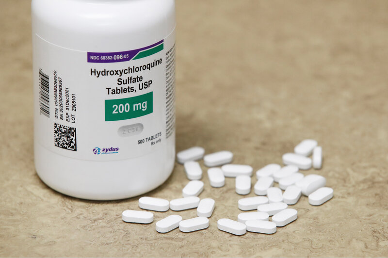


No comments:
Post a Comment
Note: Only a member of this blog may post a comment.